<?xml version="1.0" encoding="ISO-8859-1"?><article xmlns:mml="http://www.w3.org/1998/Math/MathML" xmlns:xlink="http://www.w3.org/1999/xlink" xmlns:xsi="http://www.w3.org/2001/XMLSchema-instance">
<front>
<journal-meta>
<journal-id>0075-5222</journal-id>
<journal-title><![CDATA[Kasmera]]></journal-title>
<abbrev-journal-title><![CDATA[Kasmera]]></abbrev-journal-title>
<issn>0075-5222</issn>
<publisher>
<publisher-name><![CDATA[Universidad del Zulia]]></publisher-name>
</publisher>
</journal-meta>
<article-meta>
<article-id>S0075-52222005000200003</article-id>
<title-group>
<article-title xml:lang="en"><![CDATA[Identification of Cryptococcus neoformans isolates using Staib agar without creatinine]]></article-title>
<article-title xml:lang="es"><![CDATA[Identificación de Aislados de Cryptococcus neoformans Usando Agar Staib sin Creatinina]]></article-title>
</title-group>
<contrib-group>
<contrib contrib-type="author">
<name>
<surname><![CDATA[Nardelli]]></surname>
<given-names><![CDATA[Vanessa]]></given-names>
</name>
<xref ref-type="aff" rid="A01"/>
</contrib>
<contrib contrib-type="author">
<name>
<surname><![CDATA[Pérez]]></surname>
<given-names><![CDATA[Celina]]></given-names>
</name>
<xref ref-type="aff" rid="A01"/>
</contrib>
<contrib contrib-type="author">
<name>
<surname><![CDATA[Mata-Essayag]]></surname>
<given-names><![CDATA[Sofía]]></given-names>
</name>
<xref ref-type="aff" rid="A01"/>
</contrib>
<contrib contrib-type="author">
<name>
<surname><![CDATA[Colella]]></surname>
<given-names><![CDATA[María Teresa]]></given-names>
</name>
<xref ref-type="aff" rid="A01"/>
</contrib>
<contrib contrib-type="author">
<name>
<surname><![CDATA[Roselló]]></surname>
<given-names><![CDATA[Arantza]]></given-names>
</name>
<xref ref-type="aff" rid="A01"/>
</contrib>
<contrib contrib-type="author">
<name>
<surname><![CDATA[Hartung de Capriles]]></surname>
<given-names><![CDATA[Claudia]]></given-names>
</name>
<xref ref-type="aff" rid="A01"/>
</contrib>
<contrib contrib-type="author">
<name>
<surname><![CDATA[Landaeta]]></surname>
<given-names><![CDATA[María Eugenia]]></given-names>
</name>
<xref ref-type="aff" rid="A02"/>
</contrib>
<contrib contrib-type="author">
<name>
<surname><![CDATA[Olaizola]]></surname>
<given-names><![CDATA[Carolina]]></given-names>
</name>
<xref ref-type="aff" rid="A01"/>
</contrib>
<contrib contrib-type="author">
<name>
<surname><![CDATA[Magaldi]]></surname>
<given-names><![CDATA[Sylvia]]></given-names>
</name>
<xref ref-type="aff" rid="A01"/>
</contrib>
</contrib-group>
<aff id="A01">
<institution><![CDATA[,UCV Instituto de Medicina Tropical Sección de Micología Médica]]></institution>
<addr-line><![CDATA[Caracas ]]></addr-line>
</aff>
<aff id="A02">
<institution><![CDATA[,Universidad Central de Venezuela Escuela de Medicina Luis Razetti Cátedra de Microbiología]]></institution>
<addr-line><![CDATA[Caracas ]]></addr-line>
<country>Venezuela</country>
</aff>
<pub-date pub-type="pub">
<day>00</day>
<month>07</month>
<year>2005</year>
</pub-date>
<pub-date pub-type="epub">
<day>00</day>
<month>07</month>
<year>2005</year>
</pub-date>
<volume>33</volume>
<numero>2</numero>
<fpage>102</fpage>
<lpage>108</lpage>
<copyright-statement/>
<copyright-year/>
<self-uri xlink:href="http://ve.scielo.org/scielo.php?script=sci_arttext&amp;pid=S0075-52222005000200003&amp;lng=en&amp;nrm=iso"></self-uri><self-uri xlink:href="http://ve.scielo.org/scielo.php?script=sci_abstract&amp;pid=S0075-52222005000200003&amp;lng=en&amp;nrm=iso"></self-uri><self-uri xlink:href="http://ve.scielo.org/scielo.php?script=sci_pdf&amp;pid=S0075-52222005000200003&amp;lng=en&amp;nrm=iso"></self-uri><abstract abstract-type="short" xml:lang="es"><p><![CDATA[En micología, el cultivo es la mejor manera de realizar el diagnóstico definitivo de una micosis. Muchos tipos de medios han sido desarrollados y modificados para tal fin, utilizando las propiedades bioquímicas de los hongos para reconocerlos. Los medios más comunes son Sabouraud, agar Malta, agar maíz, agar Papa-dextrosa, y agar Staib. Este último ha sido ampliamente usado para la identificación de levaduras del género Cryptococcus y en otros hongos. El objetivo de este estudio fue demostrar la utilidad del agar Staib y de un medio de Staib modificado, sin creatinina, en el aislamiento e identificación de hongos patógenos. Cuarenta y seis aislados de Cryptococcus neoformans más un control fueron identificados por criterios morfológicos y bioquímicos, usando la prueba de ureasa (agar urea de Christensen) y cultivo en medio Sablac. Las cepas fueron transferidas a placas de agar Staib estandar y de agar Staib sin creatinina. Las placas fueron incubadas a temperatura ambiente (26-28°C) durante una semana y luego fueron evaluadas las características morfológicas de las colonias y producción de fenoloxidasa. En 43 (91.5%) de las cepas estudiadas, se observó producción de fenoloxidasa (presencia de colonias marrones) tanto en el agar estandar como en el agar sin creatinina. Conclusión: el agar Staib sin creatinina es un medio excelente para la identificación de C. neoformans, con las ventajas de un bajo costo y fácil preparación.]]></p></abstract>
<abstract abstract-type="short" xml:lang="en"><p><![CDATA[In mycology, culture is the best way to demonstrate and identify pathogenic fungi. Many kinds of media have been developed and modified, using fungal biochemical properties to recognize them. The most common media are Sabouraud, Malt Agar, Cornmeal agar, Potato-dextrose agar, and Staib agar. Staib agar has been widely used for identification of yeasts from the genus Cryptococcus and other fungi. The aim of this study was to demonstrate the usefulness of Staib agar, and of a modified Staib medium without creatinine, in the isolation and identification of fungi. Forty-six Cryptococcus neoformans strains plus one control strain were identified by morphological and biochemical criteria, using urease test(Christensen urea agar) and culture in Sablac medium. The strains were then transferred into standard Staib and Staib without creatinine agar plates. Morphological features and phenoloxidase production were evaluated after one week incubation at room temperature (26-28°C). Culture in standard and modified Staib agar yielded brown color colonies (phenoloxidase production) in both media in 43 (91.5%) cases. Staib agar without creatinine is an excellent medium for identification of C. neoformans, with the advantage of a low cost and easy preparation.]]></p></abstract>
<kwd-group>
<kwd lng="es"><![CDATA[Agar Staib]]></kwd>
<kwd lng="es"><![CDATA[Guizotia abyssinica]]></kwd>
<kwd lng="es"><![CDATA[Cryptococcus neoformans]]></kwd>
<kwd lng="es"><![CDATA[creatinina]]></kwd>
<kwd lng="en"><![CDATA[Staib agar]]></kwd>
<kwd lng="en"><![CDATA[Guizotia abyssinica]]></kwd>
<kwd lng="en"><![CDATA[Cryptococcus neoformans]]></kwd>
<kwd lng="en"><![CDATA[creatinine]]></kwd>
</kwd-group>
</article-meta>
</front><body><![CDATA[  <BASEFONT SIZE="3">     <P ALIGN="center"><b><FONT COLOR="000000" size="3" face="Verdana"> Identification of Cryptococcus neoformans isolates using Staib agar without  creatinine </FONT></b></P>      <P ALIGN="center"><b><FONT COLOR="000000" size="3" face="Verdana"> Identificación de Aislados de Cryptococcus neoformans Usando Agar Staib  sin Creatinina </FONT></b></P>      <P ALIGN="center"><font size="2" face="Verdana"> <B><FONT COLOR="000000"> Nardelli, </FONT> </B><FONT COLOR="000000"> <B>Vanessa</B><SUP><B>1</B></SUP><B>; Pérez, Celina</B><SUP><B>1</B></SUP><B>; Mata-Essayag, Sofía</B><SUP><B>1</B></SUP><B>; Colella, María  Teresa</B><SUP><B>1</B></SUP><B>;  Roselló, Arantza</B><SUP><B>1</B></SUP><B>; Hartung de Capriles, Claudia</B><SUP><B>1</B></SUP><B>;  Landaeta,  María Eugenia</B><SUP><B>2</B></SUP><B>; Olaizola, Carolina</B><SUP><B>1</B></SUP><B> y  Magaldi, Sylvia</B><SUP><B>1</B></SUP> </FONT></font></P>      <P ALIGN="justify"><font size="2" face="Verdana"> <SUP><FONT COLOR="000000"> 1 </FONT></SUP> <FONT COLOR="000000">  Sección de Micología Médica, Instituto de Medicina Tropical, UCV, Caracas.  <SUP>2</SUP>Cátedra de Microbiología, Escuela de Medicina Luis Razetti, Universidad  Central de Venezuela. Caracas, Venezuela. E-mail: somae50@hotmail.com </FONT></font></P>      <P ALIGN="justify"><B><FONT COLOR="000000" size="2" face="Verdana"> Resumen </FONT></B></P>      <P ALIGN="justify"><FONT COLOR="000000" size="2" face="Verdana"> En micología, el cultivo es la mejor manera de realizar el diagnóstico  definitivo de una micosis. Muchos tipos de medios han sido desarrollados  y modificados para tal fin, utilizando las propiedades bioquímicas de los  hongos para reconocerlos. Los medios más comunes son Sabouraud, agar Malta,  agar maíz, agar Papa-dextrosa, y agar Staib. Este último ha sido ampliamente  usado para la identificación de levaduras del género <I>Cryptococcus</I> y en  otros hongos. El objetivo de este estudio fue demostrar la utilidad del  agar Staib y de un medio de Staib modificado, sin creatinina, en el aislamiento  e identificación de hongos patógenos. Cuarenta y seis aislados de <I>Cryptococcus  neoformans</I> más un control fueron identificados por criterios morfológicos  y bioquímicos, usando la prueba de ureasa (agar urea de Christensen) y  cultivo en medio Sablac. Las cepas fueron transferidas a placas de agar  Staib estandar y de agar Staib sin creatinina. Las placas fueron incubadas  a temperatura ambiente (26-28°C) durante una semana y luego fueron evaluadas  las características morfológicas de las colonias y producción de fenoloxidasa.  En 43 (91.5%) de las cepas estudiadas, se observó producción de fenoloxidasa  (presencia de colonias marrones) tanto en el agar estandar como en el agar  sin creatinina. Conclusión: el agar Staib sin creatinina es un medio excelente  para la identificación de <I>C. neoformans</I>, con las ventajas de un bajo costo  y fácil preparación. </FONT></P>      <P ALIGN="justify"><B><FONT COLOR="000000" size="2" face="Verdana"> Palabras clave:</FONT></B> <FONT COLOR="000000" size="2" face="Verdana">Agar Staib, <I>Guizotia abyssinica</I>, <I>Cryptococcus neoformans, </I>creatinina.</FONT></P>      <P ALIGN="justify"> <B><FONT COLOR="000000" size="2" face="Verdana"> Abstract </FONT></B> </P>      <P ALIGN="justify"><FONT COLOR="000000" size="2" face="Verdana"> In mycology, culture is the best way to demonstrate and identify pathogenic  fungi. Many kinds of media have been developed and modified, using fungal  biochemical properties to recognize them. The most common media are Sabouraud,  Malt Agar, Cornmeal agar, Potato-dextrose agar, and Staib agar. Staib agar  has been widely used for identification of yeasts from the genus <I>Cryptococcus</I>  and other fungi. The aim of this study was to demonstrate the usefulness  of Staib agar, and of a modified Staib medium without creatinine, in the  isolation and identification of fungi. Forty-six <I>Cryptococcus neoformans</I>  strains plus one control strain were identified by morphological and biochemical  criteria, using urease test<SUP> </SUP>(Christensen urea agar) and culture in Sablac  medium. The strains were then transferred into standard Staib and Staib  without creatinine agar plates. Morphological features and phenoloxidase  production were evaluated after one week incubation at room temperature  (26-28°C). Culture in standard and modified Staib agar yielded brown color  colonies (phenoloxidase production) in both media in 43 (91.5%) cases.  Staib agar without creatinine is an excellent medium for identification  of <I>C. neoformans</I>, with the advantage of a low cost and easy preparation. </FONT></P>      ]]></body>
<body><![CDATA[<P ALIGN="justify"><B><FONT COLOR="000000" size="2" face="Verdana"> Key words:</FONT></B> <FONT COLOR="000000" size="2" face="Verdana">Staib agar, <I>Guizotia abyssinica</I>, <I>Cryptococcus neoformans, </I>creatinine. </FONT></P>      <P ALIGN="justify"><font color="#000000" face="Verdana" size="2">Recibido: 15-07-05 / Aceptado: 27-09-05</font></P>      <P ALIGN="justify"><B><FONT COLOR="000000" size="2" face="Verdana"> Introduction </FONT></B> </P>      <P ALIGN="justify"><FONT COLOR="000000" size="2" face="Verdana"> Staib agar medium has been widely used for isolation and identification  of yeasts from the genus <I>Cryptococcus</I>. It was developed in 1962, by the  German mycologist Friedrich Staib, who thought of a culture medium made  of <I>Guizotia abyssinica</I>, an oval black seed used for bird feeding. It is  also used in India and Ethiopia for oil production (1, 2). </FONT></P>      <P ALIGN="justify"><FONT COLOR="000000" size="2" face="Verdana"> In his first studies, Staib employed powdered canary droppings, which is  used by <I>Cryptococcus</I> <I>neoformans</I> as a nitrogen source. Thus, Staib observed  a color change in the colonies, which grew in this medium, turning into  a brown color after 4 days of incubation (3). </FONT></P>      <P ALIGN="justify"><FONT COLOR="000000" size="2" face="Verdana"> Later, Staib found that the color variation was not only dependent on the  canary droppings, but on the food these birds consumed, principally made  of <I>Guizotia abyssinica </I>seeds<SUP> </SUP>(3). </FONT></P>      <P ALIGN="justify"><FONT COLOR="000000" size="2" face="Verdana"> These seeds contain cafeinic acid, among other phenolic compounds, in which  O-diphenol undergoes an oxidation process by action of the phenoloxidase  enzyme, produced by <I>C. neoformans</I>, originating a melanin pigment which  covers the yeast wall, thus turning the colonies growing in culture to  a dark brown color. This feature differentiates <I>C. neoformans</I> from other  yeasts. This reaction was named “Brown color effect” (BCE) by this author  (2, 4). Then, BCE depends on the enzyme diphenoloxidase, an antioxidant,  which helps the yeast to survive inside the host by the production of melanin  (5). </FONT></P>      <P ALIGN="justify"><FONT COLOR="000000" size="2" face="Verdana"> This medium is prepared with glucose, creatinine and potassium phosphate  (6). Creatinine is assimilated by <I>C. neoformans</I>; this is possible only  in the presence of thiamine or other compounds like thiazol and pirimidine,  which are in the seeds. Creatinine is used by the yeast as nitrogen source,  its assimilation is especially important in serotypes B and C, but not  A and D. This assimilation has been demonstrated by the presence of a green  diffusible pigment (1, 2, 3, 7). </FONT></P>      <P ALIGN="justify"><FONT COLOR="000000" size="2" face="Verdana"> Staib medium is used routinely since the beginning of the AIDS epidemic,  which increased the frequency of opportunistic infections, including fungal.  Culture of samples (especially sputum) from AIDS patients directly onto  this medium shows brown colonies of <I>C. neoformans</I> (5, 7-13). </FONT></P>      <P ALIGN="justify"><FONT COLOR="000000" size="2" face="Verdana"> For these reasons, we started to use this medium in our medical mycology  laboratory, for the isolation and identification of <I>C. neoformans</I> in clinical  samples from AIDS patients. </FONT></P>      ]]></body>
<body><![CDATA[<P ALIGN="justify"><FONT COLOR="000000" size="2" face="Verdana"> Initially, Staib agar was being prepared with the traditional method, but  further on in the study there was a failure in the supply of some of the  compounds, especially creatinine. For this reason the medium was modified,  and prepared without creatinine. Surprisingly, <I>C. neoformans</I> colonies showed  a deeper brown color than in the original Staib agar. We decided to continue  using this medium with excellent results. </FONT></P>      <P ALIGN="justify"><FONT COLOR="000000" size="2" face="Verdana"> The aim of this study was to assess the usefulness of Staib agar without  creatinine for the identification of <I>C. neoformans</I>, by the observation  of the presence of pigment, macro and microscopic morphological development  and time of growth, using strains from the Medical Mycology Department  Culture Collection. </FONT></P>      <P ALIGN="justify"><B><FONT COLOR="000000" size="2" face="Verdana"> Materials and Methods </FONT></B> </P>      <P ALIGN="justify"><FONT COLOR="000000" size="2" face="Verdana"> After certifying their pureness and viability, forty-six <I>C. neoformans</I>  strains from the Medical Mycology Department Culture Collection were identified  by morphological and biochemical criteria, using urease test<SUP> </SUP>(Christensen  urea agar) (14)<SUP> </SUP>and culture in Sablac medium, which allows better capsule  development. They were also identified with the Microscan™ Dade Behring  Automated System. </FONT></P>      <P ALIGN="justify"><font size="2" face="Verdana"> <I><FONT COLOR="000000"> C. neoformans </FONT></I> <FONT COLOR="000000">  (WC 1400 IMT) and <I>Candida albicans</I> (ATCC 90028) were used  as urease positive and negative control isolates, respectively. </FONT></font></P>      <P ALIGN="justify"><FONT COLOR="000000" size="2" face="Verdana"> Additionally 5 isolates of <I>Candida dubliniensis</I> (S2-5, S2-6, S2-14, S636  and S645) from the Health Science Center, University of Texas (supplied  by Dr. Anette Fothergill) were used to assess their development in Staib  agar with and without creatinine. </FONT></P>      <P ALIGN="justify"><FONT COLOR="000000" size="2" face="Verdana"> All strains were then transferred into standard Staib and Staib without  creatinine agar plates (two isolates per plate), they were incubated at  room temperature (26-28°C) for one week, and then the morphological features  were evaluated. </FONT></P>      <P ALIGN="justify"><FONT COLOR="000000" size="2" face="Verdana"> Canavanine-glycine-bromotimol blue (CGB) medium (15) was used to identify  <I>Cryptococcus</I> isolates. All isolates were inoculated in this medium and  incubated at room temperature for 5 days. Interpretation of results: yellow  color: <I>C. neoformans var neoformans</I>, dark blue color: <I>C. neoformans var  gattii</I>. </FONT></P>      <P ALIGN="justify"><FONT COLOR="000000" size="2" face="Verdana"> Descriptive statistics were used to analyze data obtained, using frequency  tables, and percentage and sensitivity determination. </FONT></P>      <P ALIGN="justify"><B><FONT COLOR="000000" size="2" face="Verdana"> Results </FONT></B> </P>      ]]></body>
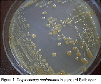
<body><![CDATA[<P ALIGN="justify"><FONT COLOR="000000" size="2" face="Verdana"> During the course of this study we were forced to use Staib medium without  one of its main components, creatinine. The experiments were continued  using the modified medium. Later in the study, we were able to find the  creatinine and nevertheless decided to use both kinds of Staib medium,  the original and the modified. </FONT></P>      <P ALIGN="justify"><FONT COLOR="000000" size="2" face="Verdana"> Forty-seven <I>C. neoformans </I>strains used for the study were tested for urease  production, being all positive. The same strains were tested for phenoloxidase  production, culturing in standard and modified Staib agar. They yielded  brown color colonies in both media in 43 (91.5%) cases (<a href="#f1">Figura 1</a>, <a href="#f2">2</a>). In  4 (8.5%) of the studied strains, the colonies remained cream color, in  spite of being urease positive and identified by automated Microscan™ method (<a href="#Table 1">Table 1</a>). </FONT></P>      <P ALIGN="center"><a name="f1"><img border="0" src="/img/fbpe/km/v33n2/art03img01.gif" width="341" height="284"></a></P>      
<P ALIGN="center"><a name="f2"><img border="0" src="/img/fbpe/km/v33n2/art03img02.gif" width="347" height="300"></a></P>  <basefont>     
<p align="center"><b><font color="#000000" face="Verdana" size="2"><a name="Table 1">Table 1</a>.</font></b> <font color="#000000" face="Verdana" size="2">Phenotypic differences of <i>Cryptococcus neoformans</i> in Staib agar with and without creatinine.</font></p>     <div align="center">       <center>   <table width="580" border="1" cellspacing="1">     <tbody>       <tr>         <td vAlign="top">               <p align="center"><font color="#000000" face="Verdana" size="2">Media</font></p>         </td>         <td vAlign="top">               <p align="center"><font size="2"><b><font color="#000000" face="Verdana">“</font></b><font color="#000000" face="Verdana">Diffusible           Pigment”</font></font></p>         </td>         <td vAlign="top">               <p align="center"><font color="#000000" face="Verdana" size="2">Colony    ]]></body>
<body><![CDATA[<br>           Color</font></p>         </td>         <td vAlign="top">               <p align="center"><font color="#000000" face="Verdana" size="2">Strains</font></p>         </td>         <td vAlign="top">               <p align="center"><font color="#000000" face="Verdana" size="2">Microscopic           Features</font></p>         </td>         <td vAlign="top">               <p align="center"><font color="#000000" face="Verdana" size="2">Growth           Time</font> <font color="#000000" face="Verdana" size="2">(Hours)</font></td>         <td vAlign="top">               <p align="center"><font color="#000000" face="Verdana" size="2">Brown           color colonies (Days)</font></p>         </td>         <td vAlign="top">               <p align="center"><font color="#000000" face="Verdana" size="2">Growth           Temperature (°C)</font></p>         </td>       </tr>       <tr>         <td vAlign="top">               <p align="center"><font face="Verdana" size="2">&nbsp;</font></p>         </td>         <td vAlign="top">               <p align="center"><font color="#000000" face="Verdana" size="2">No</font></p>         </td>         <td vAlign="top">               <p align="center"><font color="#000000" face="Verdana" size="2">Light           brown</font></p>         </td>         <td vAlign="top">               <p align="center"><font color="#000000" face="Verdana" size="2">33</font></p>         </td>         <td vAlign="top">               ]]></body>
<body><![CDATA[<p align="center"><font color="#000000" face="Verdana" size="2">Encapsulated           yeasts</font></p>         </td>         <td vAlign="top">               <p align="center"><font color="#000000" face="Verdana" size="2">48-72</font></p>         </td>         <td vAlign="top">               <p align="center"><font color="#000000" face="Verdana" size="2">5-8</font></p>         </td>         <td vAlign="top">               <p align="center"><font color="#000000" face="Verdana" size="2">26-28</font></p>         </td>       </tr>       <tr>         <td vAlign="top">               <p align="center"><font color="#000000" face="Verdana" size="2">Standard</font></p>         </td>         <td vAlign="top">               <p align="center"><font face="Verdana" size="2">&nbsp;</font></p>         </td>         <td vAlign="top">               <p align="center"><font color="#000000" face="Verdana" size="2">Cream</font></p>         </td>         <td vAlign="top">               <p align="center"><font color="#000000" face="Verdana" size="2">–</font></p>         </td>         <td vAlign="top">               <p align="center"><font color="#000000" face="Verdana" size="2">–</font></p>         </td>         <td vAlign="top">               <p align="center"><font color="#000000" face="Verdana" size="2">–</font></p>         </td>         <td vAlign="top">               ]]></body>
<body><![CDATA[<p align="center"><font color="#000000" face="Verdana" size="2">–</font></p>         </td>         <td vAlign="top">               <p align="center"><font color="#000000" face="Verdana" size="2">–</font></p>         </td>       </tr>       <tr>         <td vAlign="top">               <p align="center"><font color="#000000" face="Verdana" size="2">Staib</font></p>         </td>         <td vAlign="top">               <p align="center"><font face="Verdana" size="2">&nbsp;</font></p>         </td>         <td vAlign="top">               <p align="center"><font color="#000000" face="Verdana" size="2">Light           brown</font></p>         </td>         <td vAlign="top">               <p align="center"><font color="#000000" face="Verdana" size="2">10</font></p>         </td>         <td vAlign="top">               <p align="center"><font color="#000000" face="Verdana" size="2">Encapsulated           yeasts</font></p>         </td>         <td vAlign="top">               <p align="center"><font color="#000000" face="Verdana" size="2">24-48</font></p>         </td>         <td vAlign="top">               <p align="center"><font color="#000000" face="Verdana" size="2">5-8</font></p>         </td>         <td vAlign="top">               <p align="center"><font color="#000000" face="Verdana" size="2">26-28</font></p>         </td>       </tr>       <tr>         <td vAlign="top"><font face="Verdana" size="2">&nbsp;&nbsp;</font></td>         <td vAlign="top">               ]]></body>
<body><![CDATA[<p align="center"><font color="#000000" face="Verdana" size="2">Green</font></p>         </td>         <td vAlign="top">               <p align="center"><font color="#000000" face="Verdana" size="2">Cream</font></p>         </td>         <td vAlign="top">               <p align="center"><font color="#000000" face="Verdana" size="2">4</font></p>         </td>         <td vAlign="top">               <p align="center"><font color="#000000" face="Verdana" size="2">Encapsulated           yeasts</font></p>         </td>         <td vAlign="top">               <p align="center"><font color="#000000" face="Verdana" size="2">24-48</font></p>         </td>         <td vAlign="top">               <p align="center"><font color="#000000" face="Verdana" size="2">–</font></p>         </td>         <td vAlign="top">               <p align="center"><font color="#000000" face="Verdana" size="2">26-28</font></p>         </td>       </tr>       <tr>         <td vAlign="top">               <p align="center"><font color="#000000" face="Verdana" size="2">Staib           without</font></p>         </td>         <td vAlign="top">               <p align="center"><font color="#000000" face="Verdana" size="2">No</font></p>         </td>         <td vAlign="top">               <p align="center"><font color="#000000" face="Verdana" size="2">Dark           brown</font></p>         </td>         <td vAlign="top">               ]]></body>
<body><![CDATA[<p align="center"><font color="#000000" face="Verdana" size="2">43</font></p>         </td>         <td vAlign="top">               <p align="center"><font color="#000000" face="Verdana" size="2">Encapsulated           yeasts</font></p>         </td>         <td vAlign="top">               <p align="center"><font color="#000000" face="Verdana" size="2">48-72</font></p>         </td>         <td vAlign="top">               <p align="center"><font color="#000000" face="Verdana" size="2">5-8</font></p>         </td>         <td vAlign="top">               <p align="center"><font color="#000000" face="Verdana" size="2">26-28</font></p>         </td>       </tr>       <tr>         <td vAlign="top">               <p align="center"><font color="#000000" face="Verdana" size="2">Creatinine</font></p>         </td>         <td vAlign="top">               <p align="center"><font face="Verdana" size="2">&nbsp;</font></p>         </td>         <td vAlign="top">               <p align="center"><font color="#000000" face="Verdana" size="2">Cream</font></p>         </td>         <td vAlign="top">               <p align="center"><font color="#000000" face="Verdana" size="2">4</font></p>         </td>         <td vAlign="top">               <p align="center"><font color="#000000" face="Verdana" size="2">Encapsulated           yeasts</font></p>         </td>         <td vAlign="top">               ]]></body>
<body><![CDATA[<p align="center"><font color="#000000" face="Verdana" size="2">48-72</font></p>         </td>         <td vAlign="top">               <p align="center"><font color="#000000" face="Verdana" size="2">–</font></p>         </td>         <td vAlign="top">               <p align="center"><font color="#000000" face="Verdana" size="2">26-28</font></p>         </td>       </tr>     </tbody>   </table>   </center> </div>      <P ALIGN="justify"><FONT COLOR="000000" face="Verdana" size="2">From the 47 isolates inoculated in CGB medium, only one showed dark blue  color, which suggested C. <I>neoformans var. gattii</I>. This isolate came from  a HIV seronegative patient. In standard Staib agar, colonies were light  brown color, without green diffusible pigment; in Staib without creatinine,  colonies were dark brown.</FONT></P>      <P ALIGN="justify"><FONT COLOR="000000" size="2" face="Verdana"> Five <I>C. dubliniensis</I> strains were inoculated on standard Staib agar and  Staib agar without creatinine. In standard Staib agar, all 5 isolates showed  the same characteristics: rough colonies, and formation of chlamydoconidea.  On the other hand, on Staib agar without creatinine, neither of the isolates  showed these features. Instead, colonies were smooth, and no chlamydoconidea  were formed. </FONT></P>      <P ALIGN="justify"><B><FONT COLOR="000000" face="Verdana" size="2">Discussion </FONT></B> </P>      <P ALIGN="justify"><FONT COLOR="000000" face="Verdana" size="2">In mycology, culture is the best method used to demonstrate the presence  of pathogenic fungi. This is obtained by culturing a clinical sample in  a nutrient rich medium, which “feeds” the fungus, in favorable temperature  and time conditions, until it can be seen and identified.  </FONT></P>      <P ALIGN="justify"><FONT COLOR="000000" face="Verdana" size="2">During the history of mycology dozens of culture media have been developed,  which have been made to highlight the morphological properties of different  pathogenic fungi. The most common are Sabouraud agar, Malt agar, Cornmeal  agar, Potato-dextrose agar, and Staib agar (6, 16, 17). </FONT></P>      <P ALIGN="justify"><FONT COLOR="000000" face="Verdana" size="2">Staib in 1962, developed Staib agar, using <I>Guizotia abyssinica</I> seeds (1).  This medium has been very useful in the identification of <I>Cryptococcus</I>  strains, and moreover since the beginning of the AIDS epidemic, which has  provoked a rise in fungal opportunistic infections. </FONT></P>      <P ALIGN="justify"><FONT COLOR="000000" size="2" face="Verdana"> Forty-three (91.5%) out of 47 <I>C. neoformans </I>strains, showed light brown  colonies on the standard Staib medium and dark brown colonies on the modified  Staib medium (without creatinine). There is no explanation for the different  brown colors observed. </FONT></P>      ]]></body>
<body><![CDATA[<P ALIGN="justify"><FONT COLOR="000000" size="2" face="Verdana"> The other 4 (8.5%) strains showed cream color colonies on both media. Being  these isolates from a culture collection, there is no information about  their original color. Since these strains were 20 years old, and conserved  in Castellani medium (distilled water) <SUP>18</SUP>, maybe they could have lost the  capacity of production of phenoloxidase in time, which is necessary to  produce melanin (2, 5, 19-22). Melanin production is considered one of  the main pathogenic factors of <I>C. neoformans </I>(22).</FONT></P>      <P ALIGN="justify"><FONT COLOR="000000" size="2" face="Verdana"> On the other hand, Staib, in 1962 determined that serotypes B and C, but  not A and D assimilate creatinine from the medium, evidenced by the presence  of a green diffusible pigment (1). In our study we found 14 strains that  produced green diffusible pigment. We initially presumed that these were  serotype B and C strains. However, when the isolates were inoculated in  CGB medium, only one showed the characteristically dark blue color of <I>C.  neoformans var gattii</I>. CGB medium is very specific for identification of  this variety (15), and it is also useful for epidemiological studies of  Cryptococcal disease.</FONT></P>      <P ALIGN="justify"><FONT COLOR="000000" size="2" face="Verdana"> Bennett et al. (1978) also found that certain <I>C. neoformans&nbsp;var gattii </I>(serotypes  B and C) produced a green diffusible pigment in Staib medium. This phenomenon  did not occur with <I>C. neoformans var neoformans </I>(serotypes A and D), which  have a very slow assimilation of this compound. They explained that when  serotypes B and C assimilate creatinine, it is metabolized producing ammonium,  turning the medium to an alkaline pH and green color<SUP> </SUP>(7, 23, 24). For this  reason the term “diffusible green pigment” used in published papers, actually  refers to a color change in the medium provoked by the pH variation, and  not to production of pigment. Since we didn’t find similar results, we  concluded that Staib medium is not a sensitive or specific way of identifying  B and C serotypes. </FONT></P>      <P ALIGN="justify"><FONT COLOR="000000" size="2" face="Verdana"> Bennett et al in 1978, reported that not all <I>C. neoformans&nbsp;</I>strains assimilate  creatinine in the same way. <I>C. neoformans&nbsp;</I>Serotype A grew more slowly (48-72  hours) in this medium<SUP> </SUP>(7), which also made creatinine assimilation slower.  In our study, strains that assimilated creatinine between 48 and 72 hours  of observation could correspond to serotype A (<a href="#Table 1">Table 1</a>). This will be verified  in future studies of strain serotyping.</FONT></P>      <P ALIGN="justify"><FONT COLOR="000000" size="2" face="Verdana"> In our revision of international literature, we found that in 1978, Paliwal  and Randhawa (21) prepared a simplified <I>Guizotia abyssinica</I> seed medium,  eliminating glucose, creatinine, and phosphate. This was evaluated for  the isolation and presumptive identification of <I>C. neoformans</I>. The results  of this study demonstrated that the simplified or modified <I>G. abyssinica</I>  seed agar was superior to the complete medium for the rapid development  of the brown pigment by <I>C. neoformans</I>. This finding is very similar to  our results. </FONT></P>      <P ALIGN="justify"><font size="2" face="Verdana"><FONT COLOR="000000">In the case of <I>C. dubliniensis</I>, Staib agar without creatinine was not useful  in identifying these strains, as distinctive features were not present  in this medium (rough colonies, presence of chlamydoconidea). For this  reason, standard Staib agar continues to be the best medium to identify <I>C.</I> <I>dubliniensis</I>, as well as differentiating it from <I>C. albicans </I>(28)</FONT><FONT COLOR="000080">.</FONT></font></P>      <P ALIGN="justify"><FONT COLOR="000000" size="2" face="Verdana"> Therefore Staib agar without creatinine can be used as a cheaper option,  for the identification of <I>C. neoformans</I>, especially in laboratories with  restricted economic resources.</FONT></P>      <P ALIGN="justify"><FONT COLOR="000000" size="2" face="Verdana"> It can be used for culture of clinical samples from patients with or without  immune suppression, or AIDS patients, when cryptococcal disease is suspected,  as recommended by other authors in the literature (5, 8, 10-12, 25, 26,  27, 29-31). </FONT></P>      <P ALIGN="justify"><FONT COLOR="000000" face="Verdana" size="2">This medium was not useful in the morphological identification of <I>C. dubliniensis  </I>or recognition of <I>C. neoformans</I> serotypes. </FONT></P>      <P ALIGN="justify"><B><FONT COLOR="000000" size="2" face="Verdana"> References </FONT></B> </P>      ]]></body>
<body><![CDATA[<!-- ref --><P ALIGN="justify"><FONT COLOR="000000" size="2" face="Verdana"> (1)</FONT> <FONT COLOR="000000" size="2" face="Verdana"> Staib, F. <I>Cryptococcus neoformans</I> und <I>Guizotia abyssinica</I> (syn. G. Oleifera  D. C.).<I> Z Hyg. Infekt.-Kr. </I>1962; 148: 466-475.</FONT>&nbsp;&nbsp;&nbsp;&nbsp;&nbsp;&nbsp;&nbsp;&nbsp;[&#160;<a href="javascript:void(0);" onclick="javascript: window.open('/scielo.php?script=sci_nlinks&ref=1330279&pid=S0075-5222200500020000300001&lng=','','width=640,height=500,resizable=yes,scrollbars=1,menubar=yes,');">Links</a>&#160;]<!-- end-ref --><!-- ref --><P ALIGN="justify"><FONT COLOR="000000" size="2" face="Verdana"> (2)</FONT> <FONT COLOR="000000" size="2" face="Verdana"> Nardelli, V.; Pérez, C.; Colella M.T.; Mata S.; Hartung C.; Roselló A.;  Olaizola C., Landaeta ME. Identificación de diferentes hongos patógenos  al hombre mediante el uso del medio Agar Staib. <I>Antib. Infecc</I>. 2004; 12  (1-2) (In press)</FONT>&nbsp;&nbsp;&nbsp;&nbsp;&nbsp;&nbsp;&nbsp;&nbsp;[&#160;<a href="javascript:void(0);" onclick="javascript: window.open('/scielo.php?script=sci_nlinks&ref=1330280&pid=S0075-5222200500020000300002&lng=','','width=640,height=500,resizable=yes,scrollbars=1,menubar=yes,');">Links</a>&#160;]<!-- end-ref --><!-- ref --><P ALIGN="justify"><FONT COLOR="000000" size="2" face="Verdana"> (3)</FONT> <FONT COLOR="000000" size="2" face="Verdana"> Staib, F. New concepts in the occurrence and identification of <I>Cryptococcus  neoformans. Mycopathol Mycol Appl</I>.1963;19:143-145.</FONT>&nbsp;&nbsp;&nbsp;&nbsp;&nbsp;&nbsp;&nbsp;&nbsp;[&#160;<a href="javascript:void(0);" onclick="javascript: window.open('/scielo.php?script=sci_nlinks&ref=1330281&pid=S0075-5222200500020000300003&lng=','','width=640,height=500,resizable=yes,scrollbars=1,menubar=yes,');">Links</a>&#160;]<!-- end-ref --><!-- ref --><P ALIGN="justify"><FONT COLOR="000000" size="2" face="Verdana"> (4)</FONT> <FONT COLOR="000000" size="2" face="Verdana"> Staib, F. The green colour effect (GCE) of the killer strain <I>Cryptococcus  laurentii</I> CBS 139 on Staib agar. <I>Mycoses</I> 1999; 42: 103-6.</FONT>&nbsp;&nbsp;&nbsp;&nbsp;&nbsp;&nbsp;&nbsp;&nbsp;[&#160;<a href="javascript:void(0);" onclick="javascript: window.open('/scielo.php?script=sci_nlinks&ref=1330282&pid=S0075-5222200500020000300004&lng=','','width=640,height=500,resizable=yes,scrollbars=1,menubar=yes,');">Links</a>&#160;]<!-- end-ref --><!-- ref --><P ALIGN="justify"><FONT COLOR="000000" size="2" face="Verdana"> (5)</FONT> <FONT COLOR="000000" size="2" face="Verdana"> Golubev, W.I. and Staib, F. Green and brown colour effects in tremellaceous  yeast fungi on Staib agar. <I>Mycoses</I> 2000; 43:1-5.</FONT>&nbsp;&nbsp;&nbsp;&nbsp;&nbsp;&nbsp;&nbsp;&nbsp;[&#160;<a href="javascript:void(0);" onclick="javascript: window.open('/scielo.php?script=sci_nlinks&ref=1330283&pid=S0075-5222200500020000300005&lng=','','width=640,height=500,resizable=yes,scrollbars=1,menubar=yes,');">Links</a>&#160;]<!-- end-ref --><!-- ref --><P ALIGN="justify"><FONT COLOR="000000" size="2" face="Verdana"> (6)</FONT> <FONT COLOR="000000" size="2" face="Verdana"> Shields A.B. and Ajello L., Medium for selective isolation of <I>Cryptococcus</I>  <I>neoformans</I>. <I>Science</I> 1966;151:208-209.</FONT>&nbsp;&nbsp;&nbsp;&nbsp;&nbsp;&nbsp;&nbsp;&nbsp;[&#160;<a href="javascript:void(0);" onclick="javascript: window.open('/scielo.php?script=sci_nlinks&ref=1330284&pid=S0075-5222200500020000300006&lng=','','width=640,height=500,resizable=yes,scrollbars=1,menubar=yes,');">Links</a>&#160;]<!-- end-ref --><!-- ref --><P ALIGN="justify"><FONT COLOR="000000" size="2" face="Verdana"> (7)</FONT> <FONT COLOR="000000" size="2" face="Verdana"> Bennett J.E.; Kwon-Chung K.J. and Theodore T.S. Biochemical differences  between serotypes of <I>Cryptococcus neoformans. Sabouraudia</I>.<I> </I>1978; 16:167-174.</FONT>&nbsp;&nbsp;&nbsp;&nbsp;&nbsp;&nbsp;&nbsp;&nbsp;[&#160;<a href="javascript:void(0);" onclick="javascript: window.open('/scielo.php?script=sci_nlinks&ref=1330285&pid=S0075-5222200500020000300007&lng=','','width=640,height=500,resizable=yes,scrollbars=1,menubar=yes,');">Links</a>&#160;]<!-- end-ref --><!-- ref --><P ALIGN="justify"><FONT COLOR="000000" size="2" face="Verdana"> (8)</FONT> <FONT COLOR="000000" size="2" face="Verdana"> Bava, A.J., Robles, A.M., Negroni, R., Arechavala, A., Bíarichi, M. Estudio  de algunos aspectos epidemiológicos de 253 casos de criptococosis. <I>Rev  Iberoam Micol</I> 1997; 14:111-114.</FONT>&nbsp;&nbsp;&nbsp;&nbsp;&nbsp;&nbsp;&nbsp;&nbsp;[&#160;<a href="javascript:void(0);" onclick="javascript: window.open('/scielo.php?script=sci_nlinks&ref=1330286&pid=S0075-5222200500020000300008&lng=','','width=640,height=500,resizable=yes,scrollbars=1,menubar=yes,');">Links</a>&#160;]<!-- end-ref --><!-- ref --><P ALIGN="justify"><FONT COLOR="000000" size="2" face="Verdana"> (9)</FONT> <FONT COLOR="000000" size="2" face="Verdana"> Fernández, O.; Costa, T.; Costa, M.; Soares, A. et al. <I>Cryptococcus neoformans  </I>isolados de pacientes com AIDS. <I>Rev Soc Bras Med Trop</I>. 2000; 33:75-78</FONT>&nbsp;&nbsp;&nbsp;&nbsp;&nbsp;&nbsp;&nbsp;&nbsp;[&#160;<a href="javascript:void(0);" onclick="javascript: window.open('/scielo.php?script=sci_nlinks&ref=1330287&pid=S0075-5222200500020000300009&lng=','','width=640,height=500,resizable=yes,scrollbars=1,menubar=yes,');">Links</a>&#160;]<!-- end-ref --><!-- ref --><P ALIGN="justify"><FONT COLOR="000000" size="2" face="Verdana"> (10)</FONT> <FONT COLOR="000000" size="2" face="Verdana"> Helou, S.; Robles, A.M.;<I>, </I>Arechavala, A.; Bianchi, M.; Negroni R. Criptococosis  respiratoria en pacientes VIH positivos. <I>Rev Iberoam Micol</I>. 1999; 16: 126-129.</FONT>&nbsp;&nbsp;&nbsp;&nbsp;&nbsp;&nbsp;&nbsp;&nbsp;[&#160;<a href="javascript:void(0);" onclick="javascript: window.open('/scielo.php?script=sci_nlinks&ref=1330288&pid=S0075-5222200500020000300010&lng=','','width=640,height=500,resizable=yes,scrollbars=1,menubar=yes,');">Links</a>&#160;]<!-- end-ref --><!-- ref --><P ALIGN="justify"><FONT COLOR="000000" size="2" face="Verdana"> (11)</FONT> <FONT COLOR="000000" size="2" face="Verdana"> Vidotto, V.; Defina, N.; Pugliese, A.; Aoki, S.; Nakamura, K.; Takeo K.  Effect of different K+ concentrations on <I>cryptococcus neoformans </I>phenoloxidase  activity<I> Mycopathologia </I>2002;156: 171–176.</FONT>&nbsp;&nbsp;&nbsp;&nbsp;&nbsp;&nbsp;&nbsp;&nbsp;[&#160;<a href="javascript:void(0);" onclick="javascript: window.open('/scielo.php?script=sci_nlinks&ref=1330289&pid=S0075-5222200500020000300011&lng=','','width=640,height=500,resizable=yes,scrollbars=1,menubar=yes,');">Links</a>&#160;]<!-- end-ref --><!-- ref --><P ALIGN="justify"><FONT COLOR="000000" size="2" face="Verdana"> (12)</FONT> <FONT COLOR="000000" size="2" face="Verdana"> Mitchel, T.; Perfect, J. Cryptococosis in the era of AIDS -100 years after  the discovery of <I>Cryptococcus neoformans. Clin Microbiol Rev</I>. 1995; 8:  515-548.</FONT>&nbsp;&nbsp;&nbsp;&nbsp;&nbsp;&nbsp;&nbsp;&nbsp;[&#160;<a href="javascript:void(0);" onclick="javascript: window.open('/scielo.php?script=sci_nlinks&ref=1330290&pid=S0075-5222200500020000300012&lng=','','width=640,height=500,resizable=yes,scrollbars=1,menubar=yes,');">Links</a>&#160;]<!-- end-ref --><!-- ref --><P ALIGN="justify"><FONT COLOR="000000" size="2" face="Verdana"> (13)</FONT> <FONT COLOR="000000" size="2" face="Verdana"> Staib, F.; Seibold, M.; Antweiler, E.; Frohlich, B.; Weber, S.; Blisse  A. The brown color effect (BCE) of Cryptococcus<I> neoformans</I> in the diagnosis,  control and epidemiology of <I>C. neoformans </I>infections in AIDS patients.<I>  Zhl. Bakt. Hyg. I. Abt. Orig. A.</I> 1987;<I> </I>266: 167-77.</FONT>&nbsp;&nbsp;&nbsp;&nbsp;&nbsp;&nbsp;&nbsp;&nbsp;[&#160;<a href="javascript:void(0);" onclick="javascript: window.open('/scielo.php?script=sci_nlinks&ref=1330291&pid=S0075-5222200500020000300013&lng=','','width=640,height=500,resizable=yes,scrollbars=1,menubar=yes,');">Links</a>&#160;]<!-- end-ref --><!-- ref --><P ALIGN="justify"><FONT COLOR="000000" size="2" face="Verdana"> (14)</FONT> <FONT COLOR="000000" size="2" face="Verdana"> Staib, F.; Blisse, A.; Randhawa H.S. Creatine and creatinine assimilation  by <I>Sporothrix schenckii. Zhl. Bakt. Hyg. I. Abt. Orig. A., </I>1972; 221:<B> </B>94-99.</FONT>&nbsp;&nbsp;&nbsp;&nbsp;&nbsp;&nbsp;&nbsp;&nbsp;[&#160;<a href="javascript:void(0);" onclick="javascript: window.open('/scielo.php?script=sci_nlinks&ref=1330292&pid=S0075-5222200500020000300014&lng=','','width=640,height=500,resizable=yes,scrollbars=1,menubar=yes,');">Links</a>&#160;]<!-- end-ref --><!-- ref --><P ALIGN="justify"><FONT COLOR="000000" size="2" face="Verdana"> (15)</FONT> <FONT COLOR="000000" size="2" face="Verdana"> Kwon-Chung, K.J.; Polacheck, I. Bennett JE. Improved diagnostic medium  for separation of <I>Cryptococcus neoformans var. neoformans</I> (serotypes A  and D) <I>Cryptococcus neoformans var. gatii</I> (serotypes B and C). <I>J. Clin.  Microbiol</I>. 1982; 15(3):535-537</FONT>&nbsp;&nbsp;&nbsp;&nbsp;&nbsp;&nbsp;&nbsp;&nbsp;[&#160;<a href="javascript:void(0);" onclick="javascript: window.open('/scielo.php?script=sci_nlinks&ref=1330293&pid=S0075-5222200500020000300015&lng=','','width=640,height=500,resizable=yes,scrollbars=1,menubar=yes,');">Links</a>&#160;]<!-- end-ref --><!-- ref --><P ALIGN="justify"><FONT COLOR="000000" size="2" face="Verdana"> (16)</FONT> <FONT COLOR="000000" size="2" face="Verdana"> Borelli, D. Medios caseros para micología. <I>Arch Venez Med Trop Parasitol  Med</I> 1962; 4: 301-309.</FONT>&nbsp;&nbsp;&nbsp;&nbsp;&nbsp;&nbsp;&nbsp;&nbsp;[&#160;<a href="javascript:void(0);" onclick="javascript: window.open('/scielo.php?script=sci_nlinks&ref=1330294&pid=S0075-5222200500020000300016&lng=','','width=640,height=500,resizable=yes,scrollbars=1,menubar=yes,');">Links</a>&#160;]<!-- end-ref --><!-- ref --><P ALIGN="justify"><FONT COLOR="000000" size="2" face="Verdana"> (17)</FONT> <FONT COLOR="000000" size="2" face="Verdana"> Lacaz, CD.; Porto, E.; Costa, J. <I>Micologia medica</I>. Chapter 36. 8th ed.  Sarvier. Sao Paulo 1991: 557-575</FONT>&nbsp;&nbsp;&nbsp;&nbsp;&nbsp;&nbsp;&nbsp;&nbsp;[&#160;<a href="javascript:void(0);" onclick="javascript: window.open('/scielo.php?script=sci_nlinks&ref=1330295&pid=S0075-5222200500020000300017&lng=','','width=640,height=500,resizable=yes,scrollbars=1,menubar=yes,');">Links</a>&#160;]<!-- end-ref --><!-- ref --><P ALIGN="justify"><FONT COLOR="000000" size="2" face="Verdana"> (18)</FONT> <FONT COLOR="000000" size="2" face="Verdana"> Hartung, C.; Mata, S. et al. Preservation of fungi in water: 20 years.  <I>Mycopathologia</I> 106: 73-79, 1989</FONT>&nbsp;&nbsp;&nbsp;&nbsp;&nbsp;&nbsp;&nbsp;&nbsp;[&#160;<a href="javascript:void(0);" onclick="javascript: window.open('/scielo.php?script=sci_nlinks&ref=1330296&pid=S0075-5222200500020000300018&lng=','','width=640,height=500,resizable=yes,scrollbars=1,menubar=yes,');">Links</a>&#160;]<!-- end-ref --><!-- ref --><P ALIGN="justify"><FONT COLOR="000000" size="2" face="Verdana"> (19)</FONT> <FONT COLOR="000000" size="2" face="Verdana"> Hartung, C.; Navas, T.; Dolande, M.; Guaimare, G.; Reviakina V. Patogenicidad  del <I>Cryptococcus neoformans</I> conservado en agua, según Castellani. <I>Rev Inst  Nac Hig</I> <I>Rafael Rangel</I>. 1991; 22:19-22.</FONT>&nbsp;&nbsp;&nbsp;&nbsp;&nbsp;&nbsp;&nbsp;&nbsp;[&#160;<a href="javascript:void(0);" onclick="javascript: window.open('/scielo.php?script=sci_nlinks&ref=1330297&pid=S0075-5222200500020000300019&lng=','','width=640,height=500,resizable=yes,scrollbars=1,menubar=yes,');">Links</a>&#160;]<!-- end-ref --><!-- ref --><P ALIGN="justify"><FONT COLOR="000000" size="2" face="Verdana"> (20)</FONT> <FONT COLOR="000000" size="2" face="Verdana"> Hopfer, R.; Groschel, D. Six-hour pigmentation test for the identification  of <I>Cryptococcus neoformans. J Clin Microbiol</I>. 1975; 2: 96-98.</FONT>&nbsp;&nbsp;&nbsp;&nbsp;&nbsp;&nbsp;&nbsp;&nbsp;[&#160;<a href="javascript:void(0);" onclick="javascript: window.open('/scielo.php?script=sci_nlinks&ref=1330298&pid=S0075-5222200500020000300020&lng=','','width=640,height=500,resizable=yes,scrollbars=1,menubar=yes,');">Links</a>&#160;]<!-- end-ref --><!-- ref --><P ALIGN="justify"><FONT COLOR="000000" size="2" face="Verdana"> (21)</FONT> <FONT COLOR="000000" size="2" face="Verdana"> Paliwal, D.K.; Randhawa, HS. Evaluation of a simplified <I>Guizotia abyssinica  </I>seed medium for differentiation of <I>Cryptococcus neoformans</I>. <I>J Clin Microbiol</I>  1978; 7: 346-8.</FONT>&nbsp;&nbsp;&nbsp;&nbsp;&nbsp;&nbsp;&nbsp;&nbsp;[&#160;<a href="javascript:void(0);" onclick="javascript: window.open('/scielo.php?script=sci_nlinks&ref=1330299&pid=S0075-5222200500020000300021&lng=','','width=640,height=500,resizable=yes,scrollbars=1,menubar=yes,');">Links</a>&#160;]<!-- end-ref --><!-- ref --><P ALIGN="justify"><FONT COLOR="000000" size="2" face="Verdana"> (22)</FONT> <FONT COLOR="000000" size="2" face="Verdana"> Polak, A. Melanin as a virulence factor in pathogenic fungi. Mycoses 1989;  33: 215-224.</FONT>&nbsp;&nbsp;&nbsp;&nbsp;&nbsp;&nbsp;&nbsp;&nbsp;[&#160;<a href="javascript:void(0);" onclick="javascript: window.open('/scielo.php?script=sci_nlinks&ref=1330300&pid=S0075-5222200500020000300022&lng=','','width=640,height=500,resizable=yes,scrollbars=1,menubar=yes,');">Links</a>&#160;]<!-- end-ref --><!-- ref --><P ALIGN="justify"><FONT COLOR="000000" size="2" face="Verdana"> (23)</FONT> <FONT COLOR="000000" size="2" face="Verdana"> Bennett, J.E.; Kwon-Chung K.J.; Howard DH. Epidemiologic differences among  serotypes of <I>Cryptococcus neoformans. Am. J. Epidemiol</I>. 1977;105: 582-586</FONT>&nbsp;&nbsp;&nbsp;&nbsp;&nbsp;&nbsp;&nbsp;&nbsp;[&#160;<a href="javascript:void(0);" onclick="javascript: window.open('/scielo.php?script=sci_nlinks&ref=1330301&pid=S0075-5222200500020000300023&lng=','','width=640,height=500,resizable=yes,scrollbars=1,menubar=yes,');">Links</a>&#160;]<!-- end-ref --><!-- ref --><P ALIGN="justify"><FONT COLOR="000000" size="2" face="Verdana"> (24)</FONT> <FONT COLOR="000000" size="2" face="Verdana"> Kwon-Chung, K.J.; Polacheck, I. and Bennett, JE. Improved diagnostic medium  for separation of Cryptococcus<I> neoformans var neoformans </I>(Serotypes A and  D) and <I>Cryptococcus neoformans var gattii </I>(Serotypes B and C)<I>. J. Clin.  Microbiol</I>. 1982; 15:<B> </B>535-537.</FONT>&nbsp;&nbsp;&nbsp;&nbsp;&nbsp;&nbsp;&nbsp;&nbsp;[&#160;<a href="javascript:void(0);" onclick="javascript: window.open('/scielo.php?script=sci_nlinks&ref=1330302&pid=S0075-5222200500020000300024&lng=','','width=640,height=500,resizable=yes,scrollbars=1,menubar=yes,');">Links</a>&#160;]<!-- end-ref --><!-- ref --><P ALIGN="justify"><FONT COLOR="000000" size="2" face="Verdana"> (25)</FONT> <FONT COLOR="000000" size="2" face="Verdana"> Bava AJ, Negroni R, Arechavala A, Robles AM, Bianchi M. Cryptococcal associated  wíth AIDS in me Muñiz Hospital of Buenos Aires. <I>Mycopathologia</I>. 1997; 140:13-17.</FONT>&nbsp;&nbsp;&nbsp;&nbsp;&nbsp;&nbsp;&nbsp;&nbsp;[&#160;<a href="javascript:void(0);" onclick="javascript: window.open('/scielo.php?script=sci_nlinks&ref=1330303&pid=S0075-5222200500020000300025&lng=','','width=640,height=500,resizable=yes,scrollbars=1,menubar=yes,');">Links</a>&#160;]<!-- end-ref --><!-- ref --><P ALIGN="justify"><FONT COLOR="000000" size="2" face="Verdana"> (26)</FONT> <FONT COLOR="000000" size="2" face="Verdana"> Bava AJ, Negroni R. Características epidemiológicas de 105 casos de criptococosis  diagnosticados en la República Argentina entre 1981-1990. <I>Rev. Inst. Med.  Trop. Sao Paulo </I> 1992; 34: 335-340.</FONT>&nbsp;&nbsp;&nbsp;&nbsp;&nbsp;&nbsp;&nbsp;&nbsp;[&#160;<a href="javascript:void(0);" onclick="javascript: window.open('/scielo.php?script=sci_nlinks&ref=1330304&pid=S0075-5222200500020000300026&lng=','','width=640,height=500,resizable=yes,scrollbars=1,menubar=yes,');">Links</a>&#160;]<!-- end-ref --><!-- ref --><P ALIGN="justify"><FONT COLOR="000000" size="2" face="Verdana"> (27)</FONT> <FONT COLOR="000000" size="2" face="Verdana"> Tortorano AM<I>, </I>Vivianí MA, Rigoni AL, Cogfiati M, Roverselh A, <I>Pagano </I>A.  Prevalence of serotype D in <I>Cryptococcus neoformans </I>isolates from HIV positive  and HIV negative patients in ltaly. <I>Mycoses</I> 1997; 40: 397-302.</FONT>&nbsp;&nbsp;&nbsp;&nbsp;&nbsp;&nbsp;&nbsp;&nbsp;[&#160;<a href="javascript:void(0);" onclick="javascript: window.open('/scielo.php?script=sci_nlinks&ref=1330305&pid=S0075-5222200500020000300027&lng=','','width=640,height=500,resizable=yes,scrollbars=1,menubar=yes,');">Links</a>&#160;]<!-- end-ref --><!-- ref --><P ALIGN="justify"><FONT COLOR="000000" size="2" face="Verdana"> (28)</FONT> <FONT COLOR="000000" size="2" face="Verdana"> Mata-Essayag S, Hartung de Capriles C, Sánchez L, Gallardo S, Colella MT,  Magaldi S, Reyes H, Ontiveros J, Olaizola C, Suárez R. Aislamiento de <I>Candida</I>  <I>dubliniensis</I> en Venezuela. <I>Antib e Infecc</I>. 2002; 10(4): 165-170</FONT>&nbsp;&nbsp;&nbsp;&nbsp;&nbsp;&nbsp;&nbsp;&nbsp;[&#160;<a href="javascript:void(0);" onclick="javascript: window.open('/scielo.php?script=sci_nlinks&ref=1330306&pid=S0075-5222200500020000300028&lng=','','width=640,height=500,resizable=yes,scrollbars=1,menubar=yes,');">Links</a>&#160;]<!-- end-ref --><!-- ref --><P ALIGN="justify"><FONT COLOR="000000" size="2" face="Verdana"> (29)</FONT> <FONT COLOR="000000" size="2" face="Verdana"> Denning DW, Stevens DA, Hamilton JR. Comparison of <I>Guizotia abyssinica</I>  seed extract (birdseed) agar with conventional media for selective identification  of <I>Cryptococcus neoformans </I>in patients with acquired immunodeficiency syndrome.  <I>J Clin Microbiol</I>. 1990; 28:<B> </B>2565-7.</FONT>&nbsp;&nbsp;&nbsp;&nbsp;&nbsp;&nbsp;&nbsp;&nbsp;[&#160;<a href="javascript:void(0);" onclick="javascript: window.open('/scielo.php?script=sci_nlinks&ref=1330307&pid=S0075-5222200500020000300029&lng=','','width=640,height=500,resizable=yes,scrollbars=1,menubar=yes,');">Links</a>&#160;]<!-- end-ref --><!-- ref --><P ALIGN="justify"><FONT COLOR="000000" size="2" face="Verdana"> (30)</FONT> <FONT COLOR="000000" size="2" face="Verdana"> Rozembaum R, Rios AJ, Wanke B, Vieira W. <I>Cryptococcus neoformans var gattii  </I>in a Brazilian AIDS patient. <I>Mycopathologia</I>. 1990; 112: 33-34.</FONT>&nbsp;&nbsp;&nbsp;&nbsp;&nbsp;&nbsp;&nbsp;&nbsp;[&#160;<a href="javascript:void(0);" onclick="javascript: window.open('/scielo.php?script=sci_nlinks&ref=1330308&pid=S0075-5222200500020000300030&lng=','','width=640,height=500,resizable=yes,scrollbars=1,menubar=yes,');">Links</a>&#160;]<!-- end-ref --><!-- ref --><P ALIGN="justify"><FONT COLOR="000000" size="2" face="Verdana"> (31)</FONT> <FONT COLOR="000000" size="2" face="Verdana"> Speed B, Dunt D. Clinical and host differences between infections with  the two varieties of <I>Cryptococcus neoformans</I>. <I>Clin Infect Dis</I> 1995; 21:  28-34.</FONT>&nbsp;&nbsp;&nbsp;&nbsp;&nbsp;&nbsp;&nbsp;&nbsp;[&#160;<a href="javascript:void(0);" onclick="javascript: window.open('/scielo.php?script=sci_nlinks&ref=1330309&pid=S0075-5222200500020000300031&lng=','','width=640,height=500,resizable=yes,scrollbars=1,menubar=yes,');">Links</a>&#160;]<!-- end-ref --> ]]></body>
<back>
<ref-list>
<ref id="B1">
<label>1</label><nlm-citation citation-type="journal">
<person-group person-group-type="author">
<name>
<surname><![CDATA[Staib]]></surname>
<given-names><![CDATA[F]]></given-names>
</name>
</person-group>
<article-title xml:lang="en"><![CDATA[Cryptococcus neoformans und Guizotia abyssinica (syn. G. Oleifera D. C.)]]></article-title>
<source><![CDATA[Z Hyg. Infekt.-Kr]]></source>
<year>1962</year>
<volume>148</volume>
<page-range>466-475</page-range></nlm-citation>
</ref>
<ref id="B2">
<label>2</label><nlm-citation citation-type="journal">
<person-group person-group-type="author">
<name>
<surname><![CDATA[Nardelli]]></surname>
<given-names><![CDATA[V]]></given-names>
</name>
<name>
<surname><![CDATA[Pérez]]></surname>
<given-names><![CDATA[C]]></given-names>
</name>
<name>
<surname><![CDATA[Colella]]></surname>
<given-names><![CDATA[M.T]]></given-names>
</name>
<name>
<surname><![CDATA[Mata]]></surname>
<given-names><![CDATA[S]]></given-names>
</name>
<name>
<surname><![CDATA[Hartung]]></surname>
<given-names><![CDATA[C]]></given-names>
</name>
<name>
<surname><![CDATA[Roselló]]></surname>
<given-names><![CDATA[A]]></given-names>
</name>
<name>
<surname><![CDATA[Olaizola]]></surname>
<given-names><![CDATA[C]]></given-names>
</name>
<name>
<surname><![CDATA[Landaeta]]></surname>
<given-names><![CDATA[ME]]></given-names>
</name>
</person-group>
<article-title xml:lang="es"><![CDATA[Identificación de diferentes hongos patógenos al hombre mediante el uso del medio Agar Staib]]></article-title>
<source><![CDATA[Antib. Infecc]]></source>
<year>2004</year>
<volume>12</volume>
<numero>1-2</numero>
<issue>1-2</issue>
</nlm-citation>
</ref>
<ref id="B3">
<label>3</label><nlm-citation citation-type="journal">
<person-group person-group-type="author">
<name>
<surname><![CDATA[Staib]]></surname>
<given-names><![CDATA[F]]></given-names>
</name>
</person-group>
<article-title xml:lang="en"><![CDATA[New concepts in the occurrence and identification of Cryptococcus neoformans]]></article-title>
<source><![CDATA[Mycopathol Mycol Appl]]></source>
<year>1963</year>
<volume>19</volume>
<page-range>143-145</page-range></nlm-citation>
</ref>
<ref id="B4">
<label>4</label><nlm-citation citation-type="journal">
<person-group person-group-type="author">
<name>
<surname><![CDATA[Staib]]></surname>
<given-names><![CDATA[F]]></given-names>
</name>
</person-group>
<article-title xml:lang="en"><![CDATA[The green colour effect (GCE) of the killer strain Cryptococcus laurentii CBS 139 on Staib agar]]></article-title>
<source><![CDATA[Mycoses]]></source>
<year>1999</year>
<volume>42</volume>
<page-range>103-6</page-range></nlm-citation>
</ref>
<ref id="B5">
<label>5</label><nlm-citation citation-type="journal">
<person-group person-group-type="author">
<name>
<surname><![CDATA[Golubev]]></surname>
<given-names><![CDATA[W.I]]></given-names>
</name>
<name>
<surname><![CDATA[Staib]]></surname>
<given-names><![CDATA[F]]></given-names>
</name>
</person-group>
<article-title xml:lang="en"><![CDATA[Green and brown colour effects in tremellaceous yeast fungi on Staib agar]]></article-title>
<source><![CDATA[Mycoses]]></source>
<year>2000</year>
<volume>43</volume>
<page-range>1-5</page-range></nlm-citation>
</ref>
<ref id="B6">
<label>6</label><nlm-citation citation-type="journal">
<person-group person-group-type="author">
<name>
<surname><![CDATA[Shields]]></surname>
<given-names><![CDATA[A.B]]></given-names>
</name>
<name>
<surname><![CDATA[Ajello]]></surname>
<given-names><![CDATA[L]]></given-names>
</name>
</person-group>
<article-title xml:lang="en"><![CDATA[Medium for selective isolation of Cryptococcus neoformans]]></article-title>
<source><![CDATA[Science]]></source>
<year>1966</year>
<volume>151</volume>
<page-range>208-209</page-range></nlm-citation>
</ref>
<ref id="B7">
<label>7</label><nlm-citation citation-type="journal">
<person-group person-group-type="author">
<name>
<surname><![CDATA[Bennett]]></surname>
<given-names><![CDATA[J.E]]></given-names>
</name>
<name>
<surname><![CDATA[Kwon-Chung]]></surname>
<given-names><![CDATA[K.J]]></given-names>
</name>
<name>
<surname><![CDATA[Theodore]]></surname>
<given-names><![CDATA[T.S]]></given-names>
</name>
</person-group>
<article-title xml:lang="en"><![CDATA[Biochemical differences between serotypes of Cryptococcus neoformans]]></article-title>
<source><![CDATA[Sabouraudia]]></source>
<year>1978</year>
<volume>16</volume>
<page-range>167-174</page-range></nlm-citation>
</ref>
<ref id="B8">
<label>8</label><nlm-citation citation-type="journal">
<person-group person-group-type="author">
<name>
<surname><![CDATA[Bava]]></surname>
<given-names><![CDATA[A.J]]></given-names>
</name>
<name>
<surname><![CDATA[Robles]]></surname>
<given-names><![CDATA[A.M]]></given-names>
</name>
<name>
<surname><![CDATA[Negroni]]></surname>
<given-names><![CDATA[R]]></given-names>
</name>
<name>
<surname><![CDATA[Arechavala]]></surname>
<given-names><![CDATA[A]]></given-names>
</name>
<name>
<surname><![CDATA[Bíarichi]]></surname>
<given-names><![CDATA[M]]></given-names>
</name>
</person-group>
<article-title xml:lang="es"><![CDATA[Estudio de algunos aspectos epidemiológicos de 253 casos de criptococosis]]></article-title>
<source><![CDATA[Rev Iberoam Micol]]></source>
<year>1997</year>
<volume>14</volume>
<page-range>111-114</page-range></nlm-citation>
</ref>
<ref id="B9">
<label>9</label><nlm-citation citation-type="journal">
<person-group person-group-type="author">
<name>
<surname><![CDATA[Fernández]]></surname>
<given-names><![CDATA[O]]></given-names>
</name>
<name>
<surname><![CDATA[Costa]]></surname>
<given-names><![CDATA[T]]></given-names>
</name>
<name>
<surname><![CDATA[Costa]]></surname>
<given-names><![CDATA[M]]></given-names>
</name>
<name>
<surname><![CDATA[Soares]]></surname>
<given-names><![CDATA[A]]></given-names>
</name>
</person-group>
<article-title xml:lang="en"><![CDATA[Cryptococcus neoformans isolados de pacientes com AIDS]]></article-title>
<source><![CDATA[Rev Soc Bras Med Trop]]></source>
<year>2000</year>
<volume>33</volume>
<page-range>75-78</page-range></nlm-citation>
</ref>
<ref id="B10">
<label>10</label><nlm-citation citation-type="journal">
<person-group person-group-type="author">
<name>
<surname><![CDATA[Helou]]></surname>
<given-names><![CDATA[S]]></given-names>
</name>
<name>
<surname><![CDATA[Robles]]></surname>
<given-names><![CDATA[A.M]]></given-names>
</name>
<name>
<surname><![CDATA[Arechavala]]></surname>
<given-names><![CDATA[A]]></given-names>
</name>
<name>
<surname><![CDATA[Bianchi]]></surname>
<given-names><![CDATA[M]]></given-names>
</name>
<name>
<surname><![CDATA[Negroni]]></surname>
<given-names><![CDATA[R]]></given-names>
</name>
</person-group>
<article-title xml:lang="es"><![CDATA[Criptococosis respiratoria en pacientes VIH positivos]]></article-title>
<source><![CDATA[Rev Iberoam Micol]]></source>
<year>1999</year>
<volume>16</volume>
<page-range>126-129</page-range></nlm-citation>
</ref>
<ref id="B11">
<label>11</label><nlm-citation citation-type="journal">
<person-group person-group-type="author">
<name>
<surname><![CDATA[Vidotto]]></surname>
<given-names><![CDATA[V]]></given-names>
</name>
<name>
<surname><![CDATA[Defina]]></surname>
<given-names><![CDATA[N]]></given-names>
</name>
<name>
<surname><![CDATA[Pugliese]]></surname>
<given-names><![CDATA[A]]></given-names>
</name>
<name>
<surname><![CDATA[Aoki]]></surname>
<given-names><![CDATA[S]]></given-names>
</name>
<name>
<surname><![CDATA[Nakamura]]></surname>
<given-names><![CDATA[K]]></given-names>
</name>
<name>
<surname><![CDATA[Takeo]]></surname>
<given-names><![CDATA[K]]></given-names>
</name>
</person-group>
<article-title xml:lang="en"><![CDATA[Effect of different K+ concentrations on cryptococcus neoformans phenoloxidase activity]]></article-title>
<source><![CDATA[Mycopathologia]]></source>
<year>2002</year>
<volume>156</volume>
<page-range>171-176</page-range></nlm-citation>
</ref>
<ref id="B12">
<label>12</label><nlm-citation citation-type="journal">
<person-group person-group-type="author">
<name>
<surname><![CDATA[Mitchel]]></surname>
<given-names><![CDATA[T]]></given-names>
</name>
<name>
<surname><![CDATA[Perfect]]></surname>
<given-names><![CDATA[J]]></given-names>
</name>
</person-group>
<article-title xml:lang="en"><![CDATA[Cryptococosis in the era of AIDS -100 years after the discovery of Cryptococcus neoformans]]></article-title>
<source><![CDATA[Clin Microbiol Rev]]></source>
<year>1995</year>
<volume>8</volume>
<page-range>515-548</page-range></nlm-citation>
</ref>
<ref id="B13">
<label>13</label><nlm-citation citation-type="journal">
<person-group person-group-type="author">
<name>
<surname><![CDATA[Staib]]></surname>
<given-names><![CDATA[F]]></given-names>
</name>
<name>
<surname><![CDATA[Seibold]]></surname>
<given-names><![CDATA[M]]></given-names>
</name>
<name>
<surname><![CDATA[Antweiler]]></surname>
<given-names><![CDATA[E]]></given-names>
</name>
<name>
<surname><![CDATA[Frohlich]]></surname>
<given-names><![CDATA[B]]></given-names>
</name>
<name>
<surname><![CDATA[Weber]]></surname>
<given-names><![CDATA[S]]></given-names>
</name>
<name>
<surname><![CDATA[Blisse]]></surname>
<given-names><![CDATA[A]]></given-names>
</name>
</person-group>
<article-title xml:lang="en"><![CDATA[The brown color effect (BCE) of Cryptococcus neoformans in the diagnosis, control and epidemiology of C. neoformans infections in AIDS patients]]></article-title>
<source><![CDATA[Zhl. Bakt. Hyg. I. Abt. Orig. A]]></source>
<year>1987</year>
<volume>266</volume>
<page-range>167-77</page-range></nlm-citation>
</ref>
<ref id="B14">
<label>14</label><nlm-citation citation-type="journal">
<person-group person-group-type="author">
<name>
<surname><![CDATA[Staib]]></surname>
<given-names><![CDATA[F]]></given-names>
</name>
<name>
<surname><![CDATA[Blisse]]></surname>
<given-names><![CDATA[A]]></given-names>
</name>
<name>
<surname><![CDATA[Randhawa]]></surname>
<given-names><![CDATA[H.S]]></given-names>
</name>
</person-group>
<article-title xml:lang="en"><![CDATA[Creatine and creatinine assimilation by Sporothrix schenckii]]></article-title>
<source><![CDATA[Zhl. Bakt. Hyg. I. Abt. Orig. A]]></source>
<year>1972</year>
<volume>221</volume>
<page-range>94-99</page-range></nlm-citation>
</ref>
<ref id="B15">
<label>15</label><nlm-citation citation-type="journal">
<person-group person-group-type="author">
<name>
<surname><![CDATA[Kwon-Chung]]></surname>
<given-names><![CDATA[K.J]]></given-names>
</name>
<name>
<surname><![CDATA[Polacheck]]></surname>
<given-names><![CDATA[I]]></given-names>
</name>
<name>
<surname><![CDATA[Bennett]]></surname>
<given-names><![CDATA[JE]]></given-names>
</name>
</person-group>
<article-title xml:lang="en"><![CDATA[Improved diagnostic medium for separation of Cryptococcus neoformans var. neoformans (serotypes A and D) Cryptococcus neoformans var. gatii (serotypes B and C)]]></article-title>
<source><![CDATA[J. Clin. Microbiol]]></source>
<year>1982</year>
<volume>15</volume>
<numero>3</numero>
<issue>3</issue>
<page-range>535-537</page-range></nlm-citation>
</ref>
<ref id="B16">
<label>16</label><nlm-citation citation-type="journal">
<person-group person-group-type="author">
<name>
<surname><![CDATA[Borelli]]></surname>
<given-names><![CDATA[D]]></given-names>
</name>
</person-group>
<article-title xml:lang="es"><![CDATA[Medios caseros para micología]]></article-title>
<source><![CDATA[Arch Venez Med Trop Parasitol Med]]></source>
<year>1962</year>
<volume>4</volume>
<page-range>301-309</page-range></nlm-citation>
</ref>
<ref id="B17">
<label>17</label><nlm-citation citation-type="book">
<person-group person-group-type="author">
<name>
<surname><![CDATA[Lacaz]]></surname>
<given-names><![CDATA[CD]]></given-names>
</name>
<name>
<surname><![CDATA[Porto]]></surname>
<given-names><![CDATA[E]]></given-names>
</name>
<name>
<surname><![CDATA[Costa]]></surname>
<given-names><![CDATA[J]]></given-names>
</name>
</person-group>
<source><![CDATA[Micologia medica]]></source>
<year>1991</year>
<edition>8th</edition>
<page-range>557-575</page-range><publisher-loc><![CDATA[Sao Paulo ]]></publisher-loc>
<publisher-name><![CDATA[Sarvier]]></publisher-name>
</nlm-citation>
</ref>
<ref id="B18">
<label>18</label><nlm-citation citation-type="journal">
<person-group person-group-type="author">
<name>
<surname><![CDATA[Hartung]]></surname>
<given-names><![CDATA[C]]></given-names>
</name>
<name>
<surname><![CDATA[Mata]]></surname>
<given-names><![CDATA[S]]></given-names>
</name>
</person-group>
<article-title xml:lang="en"><![CDATA[Preservation of fungi in water: 20 years]]></article-title>
<source><![CDATA[Mycopathologia]]></source>
<year>1989</year>
<volume>106</volume>
<page-range>73-79</page-range></nlm-citation>
</ref>
<ref id="B19">
<label>19</label><nlm-citation citation-type="journal">
<person-group person-group-type="author">
<name>
<surname><![CDATA[Hartung]]></surname>
<given-names><![CDATA[C]]></given-names>
</name>
<name>
<surname><![CDATA[Navas]]></surname>
<given-names><![CDATA[T]]></given-names>
</name>
<name>
<surname><![CDATA[Dolande]]></surname>
<given-names><![CDATA[M]]></given-names>
</name>
<name>
<surname><![CDATA[Guaimare]]></surname>
<given-names><![CDATA[G]]></given-names>
</name>
<name>
<surname><![CDATA[Reviakina]]></surname>
<given-names><![CDATA[V]]></given-names>
</name>
</person-group>
<article-title xml:lang="es"><![CDATA[Patogenicidad del Cryptococcus neoformans conservado en agua, según Castellani]]></article-title>
<source><![CDATA[Rev Inst Nac Hig Rafael Rangel]]></source>
<year>1991</year>
<volume>22</volume>
<page-range>19-22</page-range></nlm-citation>
</ref>
<ref id="B20">
<label>20</label><nlm-citation citation-type="journal">
<person-group person-group-type="author">
<name>
<surname><![CDATA[Hopfer]]></surname>
<given-names><![CDATA[R]]></given-names>
</name>
<name>
<surname><![CDATA[Groschel]]></surname>
<given-names><![CDATA[D]]></given-names>
</name>
</person-group>
<article-title xml:lang="en"><![CDATA[Six-hour pigmentation test for the identification of Cryptococcus neoformans]]></article-title>
<source><![CDATA[J Clin Microbiol]]></source>
<year>1975</year>
<volume>2</volume>
<page-range>96-98</page-range></nlm-citation>
</ref>
<ref id="B21">
<label>21</label><nlm-citation citation-type="journal">
<person-group person-group-type="author">
<name>
<surname><![CDATA[Paliwal]]></surname>
<given-names><![CDATA[D.K]]></given-names>
</name>
<name>
<surname><![CDATA[Randhawa]]></surname>
<given-names><![CDATA[HS]]></given-names>
</name>
</person-group>
<article-title xml:lang="en"><![CDATA[Evaluation of a simplified Guizotia abyssinica seed medium for differentiation of Cryptococcus neoformans]]></article-title>
<source><![CDATA[J Clin Microbiol]]></source>
<year>1978</year>
<volume>7</volume>
<page-range>346-8</page-range></nlm-citation>
</ref>
<ref id="B22">
<label>22</label><nlm-citation citation-type="journal">
<person-group person-group-type="author">
<name>
<surname><![CDATA[Polak]]></surname>
<given-names><![CDATA[A]]></given-names>
</name>
</person-group>
<article-title xml:lang="en"><![CDATA[Melanin as a virulence factor in pathogenic fungi]]></article-title>
<source><![CDATA[Mycoses]]></source>
<year>1989</year>
<volume>33</volume>
<page-range>215-224</page-range></nlm-citation>
</ref>
<ref id="B23">
<label>23</label><nlm-citation citation-type="journal">
<person-group person-group-type="author">
<name>
<surname><![CDATA[Bennett]]></surname>
<given-names><![CDATA[J.E]]></given-names>
</name>
<name>
<surname><![CDATA[Kwon-Chung]]></surname>
<given-names><![CDATA[K.J]]></given-names>
</name>
<name>
<surname><![CDATA[Howard]]></surname>
<given-names><![CDATA[DH]]></given-names>
</name>
</person-group>
<article-title xml:lang="en"><![CDATA[Epidemiologic differences among serotypes of Cryptococcus neoformans]]></article-title>
<source><![CDATA[Am. J. Epidemiol]]></source>
<year>1977</year>
<volume>105</volume>
<page-range>582-586</page-range></nlm-citation>
</ref>
<ref id="B24">
<label>24</label><nlm-citation citation-type="journal">
<person-group person-group-type="author">
<name>
<surname><![CDATA[Kwon-Chung]]></surname>
<given-names><![CDATA[K.J]]></given-names>
</name>
<name>
<surname><![CDATA[Polacheck]]></surname>
<given-names><![CDATA[I]]></given-names>
</name>
<name>
<surname><![CDATA[Bennett]]></surname>
<given-names><![CDATA[JE]]></given-names>
</name>
</person-group>
<article-title xml:lang="en"><![CDATA[Improved diagnostic medium for separation of Cryptococcus neoformans var neoformans (Serotypes A and D) and Cryptococcus neoformans var gattii (Serotypes B and C)]]></article-title>
<source><![CDATA[J. Clin. Microbiol]]></source>
<year>1982</year>
<volume>15</volume>
<page-range>535-537</page-range></nlm-citation>
</ref>
<ref id="B25">
<label>25</label><nlm-citation citation-type="journal">
<person-group person-group-type="author">
<name>
<surname><![CDATA[Bava]]></surname>
<given-names><![CDATA[AJ]]></given-names>
</name>
<name>
<surname><![CDATA[Negroni]]></surname>
<given-names><![CDATA[R]]></given-names>
</name>
<name>
<surname><![CDATA[Arechavala]]></surname>
<given-names><![CDATA[A]]></given-names>
</name>
<name>
<surname><![CDATA[Robles]]></surname>
<given-names><![CDATA[AM]]></given-names>
</name>
<name>
<surname><![CDATA[Bianchi]]></surname>
<given-names><![CDATA[M]]></given-names>
</name>
</person-group>
<article-title xml:lang="en"><![CDATA[Cryptococcal associated wíth AIDS in me Muñiz Hospital of Buenos Aires]]></article-title>
<source><![CDATA[Mycopathologia]]></source>
<year>1997</year>
<volume>140</volume>
<page-range>13-17</page-range></nlm-citation>
</ref>
<ref id="B26">
<label>26</label><nlm-citation citation-type="journal">
<person-group person-group-type="author">
<name>
<surname><![CDATA[Bava]]></surname>
<given-names><![CDATA[AJ]]></given-names>
</name>
<name>
<surname><![CDATA[Negroni]]></surname>
<given-names><![CDATA[R]]></given-names>
</name>
</person-group>
<article-title xml:lang="es"><![CDATA[Características epidemiológicas de 105 casos de criptococosis diagnosticados en la República Argentina entre 1981-1990]]></article-title>
<source><![CDATA[Rev. Inst. Med. Trop. Sao Paulo]]></source>
<year>1992</year>
<volume>34</volume>
<page-range>335-340</page-range></nlm-citation>
</ref>
<ref id="B27">
<label>27</label><nlm-citation citation-type="journal">
<person-group person-group-type="author">
<name>
<surname><![CDATA[Tortorano]]></surname>
<given-names><![CDATA[AM]]></given-names>
</name>
<name>
<surname><![CDATA[Vivianí]]></surname>
<given-names><![CDATA[MA]]></given-names>
</name>
<name>
<surname><![CDATA[Rigoni]]></surname>
<given-names><![CDATA[AL]]></given-names>
</name>
<name>
<surname><![CDATA[Cogfiati]]></surname>
<given-names><![CDATA[M]]></given-names>
</name>
<name>
<surname><![CDATA[Roverselh]]></surname>
<given-names><![CDATA[A]]></given-names>
</name>
<name>
<surname><![CDATA[Pagano]]></surname>
<given-names><![CDATA[A]]></given-names>
</name>
</person-group>
<article-title xml:lang="en"><![CDATA[Prevalence of serotype D in Cryptococcus neoformans isolates from HIV positive and HIV negative patients in ltaly]]></article-title>
<source><![CDATA[Mycoses]]></source>
<year>1997</year>
<volume>40</volume>
<page-range>397-302</page-range></nlm-citation>
</ref>
<ref id="B28">
<label>28</label><nlm-citation citation-type="journal">
<person-group person-group-type="author">
<name>
<surname><![CDATA[Mata-Essayag]]></surname>
<given-names><![CDATA[S]]></given-names>
</name>
<name>
<surname><![CDATA[Hartung de Capriles]]></surname>
<given-names><![CDATA[C]]></given-names>
</name>
<name>
<surname><![CDATA[Sánchez]]></surname>
<given-names><![CDATA[L]]></given-names>
</name>
<name>
<surname><![CDATA[Gallardo]]></surname>
<given-names><![CDATA[S]]></given-names>
</name>
<name>
<surname><![CDATA[Colella]]></surname>
<given-names><![CDATA[MT]]></given-names>
</name>
<name>
<surname><![CDATA[Magaldi]]></surname>
<given-names><![CDATA[S]]></given-names>
</name>
<name>
<surname><![CDATA[Reyes]]></surname>
<given-names><![CDATA[H]]></given-names>
</name>
<name>
<surname><![CDATA[Ontiveros]]></surname>
<given-names><![CDATA[J]]></given-names>
</name>
<name>
<surname><![CDATA[Olaizola]]></surname>
<given-names><![CDATA[C]]></given-names>
</name>
<name>
<surname><![CDATA[Suárez]]></surname>
<given-names><![CDATA[R]]></given-names>
</name>
</person-group>
<article-title xml:lang="es"><![CDATA[Aislamiento de Candida dubliniensis en Venezuela]]></article-title>
<source><![CDATA[Antib e Infecc]]></source>
<year>2002</year>
<volume>10</volume>
<numero>4</numero>
<issue>4</issue>
<page-range>165-170</page-range></nlm-citation>
</ref>
<ref id="B29">
<label>29</label><nlm-citation citation-type="journal">
<person-group person-group-type="author">
<name>
<surname><![CDATA[Denning]]></surname>
<given-names><![CDATA[DW]]></given-names>
</name>
<name>
<surname><![CDATA[Stevens]]></surname>
<given-names><![CDATA[DA]]></given-names>
</name>
<name>
<surname><![CDATA[Hamilton]]></surname>
<given-names><![CDATA[JR]]></given-names>
</name>
</person-group>
<article-title xml:lang="en"><![CDATA[Comparison of Guizotia abyssinica seed extract (birdseed) agar with conventional media for selective identification of Cryptococcus neoformans in patients with acquired immunodeficiency syndrome]]></article-title>
<source><![CDATA[J Clin Microbiol]]></source>
<year>1990</year>
<volume>28</volume>
<page-range>2565-7</page-range></nlm-citation>
</ref>
<ref id="B30">
<label>30</label><nlm-citation citation-type="journal">
<person-group person-group-type="author">
<name>
<surname><![CDATA[Rozembaum]]></surname>
<given-names><![CDATA[R]]></given-names>
</name>
<name>
<surname><![CDATA[Rios]]></surname>
<given-names><![CDATA[AJ]]></given-names>
</name>
<name>
<surname><![CDATA[Wanke]]></surname>
<given-names><![CDATA[B]]></given-names>
</name>
<name>
<surname><![CDATA[Vieira]]></surname>
<given-names><![CDATA[W]]></given-names>
</name>
</person-group>
<article-title xml:lang="en"><![CDATA[Cryptococcus neoformans var gattii in a Brazilian AIDS patient]]></article-title>
<source><![CDATA[Mycopathologia]]></source>
<year>1990</year>
<volume>112</volume>
<page-range>33-34</page-range></nlm-citation>
</ref>
<ref id="B31">
<label>31</label><nlm-citation citation-type="journal">
<person-group person-group-type="author">
<name>
<surname><![CDATA[Speed]]></surname>
<given-names><![CDATA[B]]></given-names>
</name>
<name>
<surname><![CDATA[Dunt]]></surname>
<given-names><![CDATA[D]]></given-names>
</name>
</person-group>
<article-title xml:lang="en"><![CDATA[Clinical and host differences between infections with the two varieties of Cryptococcus neoformans]]></article-title>
<source><![CDATA[Clin Infect Dis]]></source>
<year>1995</year>
<volume>21</volume>
<page-range>28-34</page-range></nlm-citation>
</ref>
</ref-list>
</back>
</article>
