<?xml version="1.0" encoding="ISO-8859-1"?><article xmlns:mml="http://www.w3.org/1998/Math/MathML" xmlns:xlink="http://www.w3.org/1999/xlink" xmlns:xsi="http://www.w3.org/2001/XMLSchema-instance">
<front>
<journal-meta>
<journal-id>0258-6576</journal-id>
<journal-title><![CDATA[Revista de la Facultad de Ciencias Veterinarias]]></journal-title>
<abbrev-journal-title><![CDATA[Rev. Fac. Cienc. Vet.]]></abbrev-journal-title>
<issn>0258-6576</issn>
<publisher>
<publisher-name><![CDATA[Revista de la Facultad de Ciencias Veterinarias. Universidad Central de Venezuela.]]></publisher-name>
</publisher>
</journal-meta>
<article-meta>
<article-id>S0258-65762014000100002</article-id>
<title-group>
<article-title xml:lang="en"><![CDATA[The Stomach of the Adult African Catfish (Clarias gariepinus, Siluriforme s: Clariidae) in Farm Conditions: A Morphological and Mucin Histochem istry Analysis]]></article-title>
<article-title xml:lang="es"><![CDATA[El Estómago del Bagre Africano Adulto (Clarias gariepinus, Siluriformes: Clariidae) en Condiciones de Granja: Análisis Morforlógico e Histoquímico por Mucina]]></article-title>
</title-group>
<contrib-group>
<contrib contrib-type="author">
<name>
<surname><![CDATA[Ikpegbu]]></surname>
<given-names><![CDATA[Ekele]]></given-names>
</name>
<xref ref-type="aff" rid="A01"/>
</contrib>
<contrib contrib-type="author">
<name>
<surname><![CDATA[Nlebedum]]></surname>
<given-names><![CDATA[Uchenna]]></given-names>
</name>
<xref ref-type="aff" rid="A01"/>
</contrib>
<contrib contrib-type="author">
<name>
<surname><![CDATA[Nnadozie]]></surname>
<given-names><![CDATA[Okechukwu]]></given-names>
</name>
<xref ref-type="aff" rid="A01"/>
</contrib>
<contrib contrib-type="author">
<name>
<surname><![CDATA[Agbakwuru]]></surname>
<given-names><![CDATA[Isaiah]]></given-names>
</name>
<xref ref-type="aff" rid="A01"/>
</contrib>
</contrib-group>
<aff id="A01">
<institution><![CDATA[,Michael Okpara University of Agriculture Department of Veterinary Anatomy ]]></institution>
<addr-line><![CDATA[Umudike Abia State]]></addr-line>
<country>Nigeria</country>
</aff>
<pub-date pub-type="pub">
<day>00</day>
<month>06</month>
<year>2014</year>
</pub-date>
<pub-date pub-type="epub">
<day>00</day>
<month>06</month>
<year>2014</year>
</pub-date>
<volume>55</volume>
<numero>1</numero>
<fpage>4</fpage>
<lpage>10</lpage>
<copyright-statement/>
<copyright-year/>
<self-uri xlink:href="http://ve.scielo.org/scielo.php?script=sci_arttext&amp;pid=S0258-65762014000100002&amp;lng=en&amp;nrm=iso"></self-uri><self-uri xlink:href="http://ve.scielo.org/scielo.php?script=sci_abstract&amp;pid=S0258-65762014000100002&amp;lng=en&amp;nrm=iso"></self-uri><self-uri xlink:href="http://ve.scielo.org/scielo.php?script=sci_pdf&amp;pid=S0258-65762014000100002&amp;lng=en&amp;nrm=iso"></self-uri><abstract abstract-type="short" xml:lang="en"><p><![CDATA[The stomach of the farmed African catfish (Clarias gariepinus, Siluriformes: clariidae) was investigated in this study to establish its basic anatomy, as there is scanty information available on the literature. It produced to baseline data for further investigative research, help fish clinicians in pathology and aid nutritionists in feed management. After cropping from a commercial aquaculture, apparently healthy fish were immobilized and euthanized by using chloroform. The samples were dissected and slices of cardiac, fundic and pyloric stomach processed through routine histological procedures. Grossly, the stomach was J-shape. Cranial to the stomach was the oesophagus but no sphincter was seen separating them. Caudally, a pyloric sphincter separated the stomach from the proximal intestine. The stomach contained three regions cardiac, fundic and pyloric. The entire stomach surface was lined by a simple columnar epithelium containing mucin at the apical cytoplasm. Only the cardiac and fundic regions contained gastric glands in the lamina propria, but the glands were more developed in the fundic region. The tunica muscularis contained smooth muscle cells in an inner circular and outer longitudinal orientation. Mucin histochemistry revealed the presence of only neutral mucin in the stomach. The results obtained in this study suggest that the absence of gastric glands in the pylorus may be an adaptation of this species to reduce the quantity of acid entering the proximal intestine; hence it might help the alkaline medium to maximize pancreatic enzyme actions in the proximal intestine. The presence of only neutral mucin in the stomach will act as a buffer to neutralize the high acidic stomach content. Neutral mucins are also associated with transport of macromolecules.]]></p></abstract>
<abstract abstract-type="short" xml:lang="es"><p><![CDATA[Se efectuó un estudio de tipo histológico e histoquímico con mucina, en el estómago del bagre africano (Clarias gariepinus, Siluriformes: clariidae) cultivado en granja acuícola, para establecer su anatomía básica, debido a la escasa información disponible en la literatura. La investigación generó datos para ulteriores investigaciones, que contribuirán con los ictiopatólogos y ayudarán a los nutricionistas en el manejo alimentario. Después de su cultivo en una granja acuícola comercial, los peces aparentemente sanos, fueron inmovilizados y sacrificados, usando cloroformo. Se tomaron muestras de las regiones cardial, fúndica y pilórica, y se procesaron histológicamente. En su conjunto, el estómago presentaba una apariencia de “J”. El esófago estaba situado cranealmente al estómago, pero no se observó un esfínter que los separara. Caudalmente, un esfínter pilórico separaba al estómago del intestino proximal. El estómago contenía tres regiones: cardiaca, fúndica y pilórica. La totalidad de la superficie estomacal se encontraba recubierta por un epitelio cilíndrico, que contenía mucina en el citoplasma apical. Solamente las regiones cardiaca y fúndica tenían glándulas gástricas en la lámina propia, que eran más desarrolladas en la región fúndica. La túnica muscular poseía células musculares lisas con una orientación interna circular y externa longitudinal. La histoquímica mediante la mucina reveló la presencia solo de mucina neutra en el estómago. Los resultados obtenidos en este estudio sugieren que la ausencia de glándulas gástricas en el píloro, pudiera ser el resultado de una adaptación de esta especie para reducir la cantidad de ácido que penetra en el intestino proximal; por tanto, pudiera ayudar al medio alcalino a maximizar las acciones de las enzimas pancreáticas en el intestino proximal. La sola presencia de mucina neutra en el estómago actuaría como amortiguador contra el elevado contenido ácido del estómago. Las mucinas neutras también están asociadas con el transporte de micromoléculas.]]></p></abstract>
<kwd-group>
<kwd lng="en"><![CDATA[Culture]]></kwd>
<kwd lng="en"><![CDATA[animal morphology]]></kwd>
<kwd lng="en"><![CDATA[mucin]]></kwd>
<kwd lng="en"><![CDATA[histochemistry]]></kwd>
<kwd lng="en"><![CDATA[enzymes]]></kwd>
<kwd lng="en"><![CDATA[animal glands]]></kwd>
<kwd lng="en"><![CDATA[stomach]]></kwd>
<kwd lng="en"><![CDATA[african catfish]]></kwd>
<kwd lng="es"><![CDATA[Cultivo]]></kwd>
<kwd lng="es"><![CDATA[anatomía animal]]></kwd>
<kwd lng="es"><![CDATA[mucina]]></kwd>
<kwd lng="es"><![CDATA[histoquímica]]></kwd>
<kwd lng="es"><![CDATA[enzimas]]></kwd>
<kwd lng="es"><![CDATA[glándulas animales]]></kwd>
<kwd lng="es"><![CDATA[estómago]]></kwd>
<kwd lng="es"><![CDATA[bagre africano]]></kwd>
</kwd-group>
</article-meta>
</front><body><![CDATA[ <p style="text-autospace: none; vertical-align: middle" align="center"><b> <span lang="ES-VE" style="font-family: Verdana; color: black">The Stomach of the  Adult African Catfish (<i>Clarias gariepinus, </i>Siluriforme s: Clariidae) in  Farm Conditions: A Morphological and Mucin Histochem istry Analysis</span></b></p>     <p style="text-autospace: none; vertical-align: middle" align="center"><b> <span lang="ES-VE" style="font-family: Verdana; color: black">El Estómago del  Bagre Africano Adulto<i> (Clarias gariepinus, S</i></span><span lang="ES-VE" style="font-family: Verdana; color: black">iluriformes:  Clariidae<i>) </i>en Condiciones de Granja: Análisis Morforlógico e Histoquímico por  Mucina</span></b></p>     <p style="text-autospace: none; vertical-align: middle" align="center"> <font face="Verdana" size="2"><span lang="ES-VE">&nbsp;Ikpegbu Ekele*<sup>,1</sup>,  Nlebedum Uchenna*, Nnadozie Okechukwu* and Agbakwuru Isaiah*</span></font></p>     <p align="justify"><font face="Verdana" size="2"><span lang="EN-US">*Department  of Veterinary Anatomy, Michael Okpara University of Agriculture, Umudike, Abia  State, Nigeria</span></font></p>     <p align="justify"><font face="Verdana" size="2"><span lang="EN-US"><sup>1</sup>  A quien debe dirigirse la correspondencia (To whom correspondence should be  addressed)</span></font></p>     <p align="justify"><b><span lang="ES-VE" style="font-family: Verdana"> <font size="2">Correo-E: </font><a href="mailto:fikpegbu@yahoo.com"> <font size="2">fikpegbu@yahoo.com</font></a></span></b></p>     <p style="text-autospace: none; vertical-align: center" align="justify"><b> <span lang="EN-US" style="font-family: Verdana; color: black; text-transform: uppercase"> <font size="2">Abstract</font></span></b></p>     <p style="text-autospace: none; vertical-align: middle" align="justify"> <span lang="EN-US" style="font-family: Verdana"><font size="2">The stomach of  the farmed African catfish (<i>Clarias gariepinus, </i>Siluriformes: clariidae)  was investigated in this study to establish its basic anatomy, as there is  scanty information available on the literature. It produced to baseline data for  further investigative research, help fish clinicians in pathology and aid  nutritionists in feed management. After cropping from a commercial aquaculture,  apparently healthy fish were immobilized and euthanized by using chloroform. The  samples were dissected and slices of cardiac, fundic and pyloric stomach  processed through routine histological procedures. Grossly, the stomach was  J-shape. Cranial to the stomach was the oesophagus but no sphincter was seen  separating them. Caudally, a pyloric sphincter separated the stomach from the  proximal intestine. The stomach contained three regions cardiac, fundic and  pyloric. The entire stomach surface was lined by a simple columnar epithelium  containing mucin at the apical cytoplasm.&nbsp; Only the cardiac and fundic regions  contained gastric glands in the lamina propria, but the glands were more  developed in the fundic region. The tunica muscularis contained smooth muscle  cells in an inner circular and outer longitudinal orientation. Mucin  histochemistry revealed the presence of only neutral mucin in the stomach. The  results obtained in this study suggest that the absence of gastric glands in the  pylorus may be an adaptation of this species to reduce the quantity of acid  entering the proximal intestine; hence it might help the alkaline medium to  maximize pancreatic enzyme actions in the proximal intestine. The presence of  only neutral mucin in the stomach will act as a buffer to neutralize the high  acidic stomach content. Neutral mucins are also associated with transport of  macromolecules.</font></span></p>     <p align="justify"><span style="font-family: Verdana"><font size="2">(<b>Key  words:</b>&nbsp; Culture; animal morphology; mucin; histochemistry; enzymes; animal  glands; stomach; african catfish)</font></span></p>     <p style="text-autospace: none; vertical-align: center" align="justify"><b> <span lang="EN-US" style="font-family: Verdana; color: black; text-transform: uppercase"> <font size="2">Resumen</font></span></b></p>     ]]></body>
<body><![CDATA[<p align="justify"><span lang="ES-VE" style="font-family: Verdana"> <font size="2">Se efectuó un estudio de tipo histológico e histoquímico con  mucina, en el estómago del bagre africano (<i>Clarias gariepinus</i>,  Siluriformes: clariidae) cultivado en granja acuícola, para establecer su  anatomía básica, debido a la escasa información disponible en la literatura. La  investigación generó datos para ulteriores investigaciones, que contribuirán con  los ictiopatólogos y ayudarán a los nutricionistas en el manejo alimentario.  Después de su cultivo en una granja acuícola comercial, los peces aparentemente  sanos, fueron inmovilizados y sacrificados, usando cloroformo. Se tomaron  muestras de las regiones cardial, fúndica y pilórica, y se procesaron  histológicamente. En su conjunto, el estómago presentaba una apariencia de “J”.  El esófago estaba situado cranealmente al estómago, pero no se observó un  esfínter que los separara. Caudalmente, un esfínter pilórico separaba al  estómago del intestino proximal. El estómago contenía tres regiones: cardiaca,  fúndica y pilórica. La totalidad de la superficie estomacal se encontraba  recubierta por un epitelio cilíndrico, que contenía mucina en el citoplasma  apical. Solamente las regiones cardiaca y fúndica tenían glándulas gástricas en  la lámina propia, que eran más desarrolladas en la región fúndica. La túnica  muscular poseía células musculares lisas con una orientación interna circular y  externa longitudinal. La histoquímica mediante la mucina reveló la presencia  solo </font></span><span lang="ES-TRAD" style="font-family: Verdana"> <font size="2">de </font></span><span lang="ES-VE" style="font-family: Verdana"> <font size="2">mucina neutra en el estómago. Los resultados obtenidos en este  estudio sugieren que la ausencia de glándulas gástricas en el píloro, pudiera  ser el resultado de una adaptación de esta especie para reducir la cantidad de  ácido que penetra en el intestino proximal; por tanto, pudiera ayudar al medio  alcalino a maximizar las acciones de las enzimas pancreáticas en el intestino  proximal. La sola presencia de mucina neutra en el estómago actuaría como  amortiguador contra el elevado contenido ácido del estómago. Las mucinas neutras  también están asociadas con el transporte de micromoléculas.</font></span></p>     <p align="justify"><span lang="ES-VE" style="font-family: Verdana"> <font size="2">(<b>Palabras clave</b>: </font></span> <span lang="ES-TRAD" style="font-family: Verdana"><font size="2">Cultivo;  anatomía animal; &nbsp;mucina; </font></span> <span lang="ES-VE" style="font-family: Verdana"><font size="2">histoquímica; </font></span><span lang="ES-TRAD" style="font-family: Verdana"><font size="2"> enzimas; glándulas animales; </font></span> <span lang="ES-VE" style="font-family: Verdana"><font size="2">estómago; bagre  africano)</font></span></p>     <p align="justify"><span lang="es-ve"><font face="Verdana" size="2">Recibido:  23/09/13 - Aprobado: 03/07/14</font></span></p>     <p style="text-autospace: none; vertical-align: center" align="justify"><b> <span lang="EN-US" style="font-family: Verdana; color: black; text-transform: uppercase"> <font size="2">Introduction</font></span></b></p>     <p style="text-autospace: none; vertical-align: middle" align="justify"> <span lang="EN-US" style="font-family: Verdana; color: black"><font size="2">The  stomach is a dilated organ where the oesophagus opens into. When present, it  constitutes a criterion to classify teleost into gastric and agastric groups.  The stomach shapes and sizes of various studied bony fishes have also been  documented and are associated with the duration between meals and nature of food  [1]. The three stomach regions: cardiac, fundic and pylorus, with various  degrees of gastric gland development have been reported in the literature [2-6].  It has also been stated that the teleost stomach wall comprises the layers  typical of vertebrate digestive tract in addition to extra features like <i> stratum compactum</i> and <i>stratum granulosum</i>. Distinct <i>muscularis  mucosae</i> have been documented [7, 8]. One type of secretory cell, the  oxyntopeptic cell, producing both pepsinogen and hydrochloric acid, has been  reported in most teleost gastric gland [5, 9, 10].</font></span></p>     <p style="text-autospace: none; vertical-align: middle" align="justify"> <span lang="EN-US" style="font-family: Verdana; color: black"><font size="2">The  gastric mucosa is believed to perform an absorptive function. This function is  suggested by the presence of numerous blood vessels in the lamina propria of <i> Peristedium longispatha</i> pyloric stomach [11] and the cardiac stomach of <i> Mugil tade</i> F. [12].</font></span></p>     <p align="justify"> <span lang="EN-US" style="font-family: Verdana; color: black"><font size="2"> Despite extensive investigation of most teleost in reported literature, there is  dearth of information in available literature on the morphology of the African  catfish stomach from Nigerian commercial farms, hence the need for this study.  The knowledge derived from this study will help fish nutritionists and  pathologists.</font></span></p>     <p style="text-autospace: none; vertical-align: center" align="justify"><b> <span lang="EN-US" style="font-family: Verdana; color: black; text-transform: uppercase"> <font size="2">Materials and Methods</font></span></b></p>     <p style="text-autospace: none; vertical-align: middle" align="justify"> <span lang="EN-US" style="font-family: Verdana; color: black"><font size="2">Ten  adult African catfish sourced from a commercial aquaculture in Eastern Nigeria  were used for the study. They average weigh<span style="letter-spacing: -.1pt">ed  was 900 g and the body length was 45cm. The fish were euthanized </span>with  chloroform. The body cavity was cut open through the ventral surface and the  alimentary tract dissected out. The stomach of the specimen under study (<a href="#fig1">Figure  1</a>) was excised and sections of cardiac, fundic and pyloric regions were  immediately fixed in 10% neutral buffered formalin for 48 h.</font></span></p>     <p style="text-autospace: none; vertical-align: middle" align="center"> <a name="fig1"> <img border="0" src="/img/fbpe/rfcv/v55n1/art02fig1.gif" width="385" height="302"></a></p>     
]]></body>
<body><![CDATA[<p style="text-autospace: none; vertical-align: middle" align="justify"> <span lang="EN-US" style="font-family: Verdana; color: black"><font size="2">The  tissues were passed through graded ethanol, cleared in xylene, impregnated and  embedded in paraffin wax. Sections of 5-6 µm thick were obtained using a Leitz  microtome model 1512. Tissues were stained with haematoxylin and eosin for light  microscopy examination [13]. The presence of mucins was demonstrated using  alcian blue (AB) at pH 2.5 [14,15] and the periodic acid of Schiff (PAS)  procedure with and without prior digestion with diastase [16,17]. In addition,  the PAS technique was employed in combination with AB for neutral and acid mucin  [13].&nbsp; Photomicrographs were taken with a Motican 2001 camera (Motican, UK)  attached to an Olympus microscope.</font></span></p>     <p style="text-autospace: none; vertical-align: center" align="justify"><b> <span lang="EN-US" style="font-family: Verdana; color: black; text-transform: uppercase"> <font size="2">Results</font></span></b></p>     <p style="text-autospace: none; vertical-align: middle" align="justify"> <span lang="EN-US" style="font-family: Verdana; color: black"><font size="2"> Grossly, the stomach was J-shaped. The small arm of the J, the most caudal part  was occupied by the cardiac region, and the fundic region formed about  four-fifth of the long arm of the J or the middle part, while the pylorus  occupied the tip of the J-shaped stomach and was the most cranial part of the  stomach (<a href="#fig1">Figure 1</a>).The pyloric end was somewhat ventral to the gastro-oesophageal  opening. The pyloric region was cranial, while the fundus was the caudal region  of the stomach. The pylorus was separated from the proximal intestine by a  constriction (<a href="#fig2">Figure 2</a>).</font></span></p>     <p style="text-autospace: none; vertical-align: middle" align="center"> <a name="fig2"> <img border="0" src="/img/fbpe/rfcv/v55n1/art02fig2.gif" width="387" height="248"></a></p>     
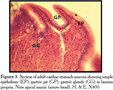
<p style="text-autospace: none; vertical-align: middle" align="justify"> <span lang="EN-US" style="font-family: Verdana; color: black; letter-spacing: -.25pt"> <font size="2">Histologically, the cardiac stomach epithelium covering was of  simple columnar epithelium. Each individual columnar cell had a basal nucleus  and mucin in the apical cytoplasm. The epithelium also contained intraepithelial  leukocytes (<a href="#fig3">Figure 3</a>). Gastric pits were also seen (<a href="#fig3">Figure 3</a>). The lamina propria contained gastric glands; beneath the glands were collagen connective  tissue fibres. The <i>tunica muscularis </i>contained smooth muscle cells in an  inner </font></span> <span lang="EN-US" style="font-family: Verdana; color: black"><font size="2"> circular and o<span style="letter-spacing: -.1pt">uter longitudinal orientation. <i>Myenteric plexus</i> was observed between the two muscle layers. Tunica  serosa was observed. Subserosal blood vessels were also seen. The fundic stomach  was covered by simple columnar mucous epithelium with apical mucin. The gastric  pits were narrower and deeper than those observed in the cardiac stomach  (<a href="#fig4">Figures 4, 5</a>). The lamina propria contained larger gastric glands than the  cardiac stomach (<a href="#fig4">Figures 4, 6</a>). The pyloric stomach was also covered by a simple  mucous epithelium. The gastric pits were very shallow. The lamina propria  contained no gastric glands (<a href="#fig7">Figure 7</a>). Histochemically, the apical mucin in the  simple columnar epithelium was positive to PAS (<a href="#fig9">Figures 9,10</a>), negative to AB  (<a href="#fig11">Figure 11</a>), when subjected to combined AB and PAS procedures, and only </span> neutral mucin was revealed (<a href="#fig12">Figures 12,13</a>).</font></span></p>     <p style="text-autospace: none; vertical-align: middle" align="center"> <a name="fig3"> <img border="0" src="/img/fbpe/rfcv/v55n1/art02fig3.gif" width="383" height="305"></a></p>     
<p style="text-autospace: none; vertical-align: middle" align="center"> <a name="fig4"> <img border="0" src="/img/fbpe/rfcv/v55n1/art02fig4.gif" width="387" height="382"></a></p>     
<p style="text-autospace: none; vertical-align: middle" align="center"> <img border="0" src="/img/fbpe/rfcv/v55n1/art02fig5.gif" width="385" height="331"></p>     
<p style="text-autospace: none; vertical-align: middle" align="center"> <img border="0" src="/img/fbpe/rfcv/v55n1/art02fig6.gif" width="385" height="339"></p>     
<p style="text-autospace: none; vertical-align: middle" align="center"> <a name="fig7"> <img border="0" src="/img/fbpe/rfcv/v55n1/art02fig7.gif" width="387" height="363"></a></p>     
]]></body>
<body><![CDATA[<p style="text-autospace: none; vertical-align: middle" align="center"> <img border="0" src="/img/fbpe/rfcv/v55n1/art02fig8.gif" width="383" height="344"></p>     
<p style="text-autospace: none; vertical-align: middle" align="center"> <a name="fig9"> <img border="0" src="/img/fbpe/rfcv/v55n1/art02fig9.gif" width="385" height="299"></a></p>     
<p style="text-autospace: none; vertical-align: middle" align="center"> <img border="0" src="/img/fbpe/rfcv/v55n1/art02fig10.gif" width="384" height="337"></p>     
<p style="text-autospace: none; vertical-align: middle" align="center"> <a name="fig11"> <img border="0" src="/img/fbpe/rfcv/v55n1/art02fig11.gif" width="383" height="328"></a></p>     
<p style="text-autospace: none; vertical-align: middle" align="center"> <a name="fig12"> <img border="0" src="/img/fbpe/rfcv/v55n1/art02fig12.gif" width="384" height="298"></a></p>     
<p style="text-autospace: none; vertical-align: middle" align="center"> <img border="0" src="/img/fbpe/rfcv/v55n1/art02fig13.gif" width="384" height="345"></p>     
<p style="text-autospace: none; vertical-align: center" align="justify"><b> <span lang="EN-US" style="font-family: Verdana; color: black; text-transform: uppercase"> <font size="2">Discussion</font></span></b></p>     <p style="text-autospace: none; vertical-align: middle" align="justify"> <span lang="EN-US" style="font-family: Verdana; color: black"><font size="2"> Grossly, the stomach was J-shaped. This shape has been reported in <i> Oncorhynchus mykiss</i> [2] and may help extend the time feed stays in the  organ, thereby ensuring greater degree of digestion by gastric enzymes. An  U-shaped stomach has been reported in <i>Salmo</i>, <i>Coregonus</i> and <i> Clupea</i> [1], <i>Glyptosternums maculatum</i> [18]; The Y-shape stomach has  been reported in <i>Oreochromis niloticus</i> [3], <i>Micropogonias furnieri</i>  [5], and the straight stomach in <i>Esox</i> [1]. The cardiac, fundic and  pyloric regions were seen as reported in some teleosts [18]. The general  microanatomy of the regions was similar, except for the absence of gastric  glands in the pylorus, which may be attributed to the need for reduced gastric  acid secretion that may interfere with the needed alkaline medium for digestive  processes in the intestine by pancreatic enzymes [19]. Also, this pyloric  stomach like in most teleosts stomach was characterized by absence of gastric  glands, well developed thick <i>tunica muscularis</i> comprising mainly circular  muscle. The reduced submucosa with collagen fibres is a&nbsp; modification to serve  the gizzard to triturate and mix [5, 20, 21].</font></span></p>     <p style="text-autospace: none; vertical-align: middle" align="justify"> <span lang="EN-US" style="font-family: Verdana; color: black"><font size="2">The  mucosal folds seen in the regions were highest in fundic stomach but shallowest  in the pyloric region. The cardiac stomach was in-between. This has been  reported in the walking catfish <i>Clarias batrachus</i> [6], and may be related  to the development of the gastric glands.</font></span></p>     <p style="text-autospace: none; vertical-align: middle" align="justify"> <span lang="EN-US" style="font-family: Verdana; color: black"><font size="2">The  gastric epithelium was of simple columnar absorptive type with apical mucin. No  goblet cells were observed. This finding is supported by previous studies seen  in literature [2,10, 22-25]. In the bay snook <i>Petenia splendida</i> (Gunther),  the gastric mucosa was lined by simple cuboidal epithelium [26]. The apical  region of the columnar epithelia containing mucin is involved in preventing  auto- digestion and reducing the effect of gastric acid content [2, 24, 26, 27].</font></span></p>     ]]></body>
<body><![CDATA[<p style="text-autospace: none; vertical-align: middle" align="justify"> <span lang="EN-US" style="font-family: Verdana; color: black"><font size="2">The  location of gastric glands is species – specific and has been related to feeding  habits.&nbsp; In this study, the gastric glands were seen only in cardia and fundus.  Absence of gastric gland in pylorus has been reported by Zacarias <i>et al</i>.  [28] in their study of the <i>Paralichtlys&nbsp; californicus </i>larvae. Also, Yang <i>et al.</i> [10] in <i>Pelteobagrus fulvidraco</i> and Xiong <i>et al. </i> [18] in <i>Glyposternum maculatum</i> reported the same findings. In <i>Diplodus  puntazzo</i>, <i>Pleuronectes ferruginea</i>, summer flounder – <i>Paralichthys  dentatus</i>, the fundic region is the only glandular region. In <i>Diplodus  sargus</i>, <i>Umbrina cirrosa</i> and <i>Paudoro pagrus</i> only the cardiac  region was seen to be glandular. On the other hand, in <i>Pandora pagellus</i>  the entire stomach is glandular [25]. The <i>Oreochromis niloticus</i> stomach  is entirety glandular [3]. Only the presence of gastric glands in the cardias  and fundus has also been reported by Chen <i>et al.</i> [29] in their work on  Kingfish <i>Seriola lalandi</i>. They also reported the presence of PAS&nbsp;  positive apical borders of the epithelia as seen in this study. The fundic  region was described by them, to be the largest region of the stomach. This  report supports our findings where the fundus formed the base and two-thirds  (2/3) of the J-shaped stomach; and this may be related to the level of  development of gastric glands in this region and the need for more contact time  between feed and the secreted enzymes since this species is an omnivore. Cao and  Wang [21] reported the absence of gastric glands in the pylorus in the study of  yellow catfish.</font></span></p>     <p style="text-autospace: none; vertical-align: middle" align="justify"> <span lang="EN-US" style="font-family: Verdana; color: black"><font size="2">The  cells of the gastric glands will probably be oxyntopeptic cells [9, 10, 18 30]  producing both pepsinogen and hydrochloric acid, but some researchers have  reported two separate cell types: mucous cell and another one with zymogen  granules [21], but further investigation should be done to ascertain this claim.  The fibrocytes may be producing collagen to surround gastric glands in their  lobules or the cells may be myoepithelial cell producing contractile fibres that  will help elaborate the secretory products of the oxyntopeptic cells, but  further investigation is required to verify the nature and function of these  cells.</font></span></p>     <p align="justify"> <span lang="EN-US" style="font-family: Verdana; color: black"><font size="2">The  collagen bundle in the submucosa and thick <i>tunica muscularis</i> smooth  muscles may be for supporting and strengthening since stratum compactun is  lacking in this species [31]. The dense smooth muscle layer seen in the pyloric  sphincter has been reported and involuntarily controls rate of food passage,  affecting retention time and volumetric capacity of the stomach [4, 32]. These  dense circular smooth muscles in the sphincter may also be involved in  trituration [20, 21]. The presence of skeletal muscle in pyloric sphincter has  been reported [3] and is associated with voluntary control over food entry into  the intestine.</font></span></p>     <p align="justify"> <span lang="EN-US" style="font-family: Verdana; color: black"><font size="2">The  inner circular and outer longitudinal smooth muscle layers of the <i>tunica  muscularis</i> of the stomach observed have been reported and is involved in  involuntary contraction for food digestion [4,5,18].&nbsp; The myenteric plexus seen  is for nerve supply and stimulation [2, 4]. Presence of serosa consisting of  mesothelial cells and loose connective tissue containing small blood vessels as  seen in this study has been reported [33].</font></span></p>     <p align="justify"> <span lang="EN-US" style="font-family: Verdana; color: black"><font size="2"> Mucin histochemistry revealed that only neutral mucin was seen in the stomach.  This is consistent with previous findings in most teleosts [5, 6, 21, 24, 26].  The presence of only neutral mucin on the stomach epithelia has been suggested  to have a role in transport of macromolecules, increase in digestive efficiency,  to defend against bacteria, buffering effect on high acidity of stomach content  this exerting a preventing proteolytic damage to epithelium [25-27, 34, 35].</font></span></p>     <p align="justify"> <span lang="EN-US" style="font-family: Verdana; color: black"><font size="2">The  presence of acid mucin in the stomach has been reported in <i>Umbrina cirrosa</i>  and is associated with prevention of damage to gut epithelium, acting as  lubricant to fibre-rich materials [27]; also, acid mucin has been reported in <i> Oreochromis niloticus </i>[3, 32].</font></span></p>     <p style="text-autospace: none; vertical-align: center" align="justify"><b> <span lang="EN-US" style="font-family: Verdana; color: black; text-transform: uppercase"> <font size="2">Reference </font></span></b></p>     <!-- ref --><p style="text-autospace: none; vertical-align: middle" align="justify"> <span lang="EN-US" style="font-family: Verdana; color: black"><font size="2"> 1.&nbsp;Smith LS. Digestion in Teleost fishes. FAO Corp Doc Repository. 1978; 1-19. </font></span>&nbsp;&nbsp;&nbsp;&nbsp;&nbsp;&nbsp;&nbsp;&nbsp;[&#160;<a href="javascript:void(0);" onclick="javascript: window.open('/scielo.php?script=sci_nlinks&ref=1823068&pid=S0258-6576201400010000200001&lng=','','width=640,height=500,resizable=yes,scrollbars=1,menubar=yes,');">Links</a>&#160;]<!-- end-ref --><!-- ref --><p style="text-autospace: none; vertical-align: middle" align="justify"> <span lang="EN-US" style="font-family: Verdana; color: black"><font size="2">2.&nbsp;Ezeasor  DN. The fine structure of the gastric epithelium of the rainbow trout, <i>Salmo  gairdneri</i>, Richardson. J Fish Biol. 1981; 19:611-27.</font></span>&nbsp;&nbsp;&nbsp;&nbsp;&nbsp;&nbsp;&nbsp;&nbsp;[&#160;<a href="javascript:void(0);" onclick="javascript: window.open('/scielo.php?script=sci_nlinks&ref=1823069&pid=S0258-6576201400010000200002&lng=','','width=640,height=500,resizable=yes,scrollbars=1,menubar=yes,');">Links</a>&#160;]<!-- end-ref --><!-- ref --><p style="text-autospace: none; vertical-align: middle" align="justify"> <span lang="EN-US" style="font-family: Verdana; color: black"><font size="2">3.&nbsp;Caceci  T, El-Habback HA, Smith SA, Smith BJ. The stomach of <i>Oreochromis niloticus</i>  has three regions. J Fish Biol. 1997; 50:939-52.</font></span>&nbsp;&nbsp;&nbsp;&nbsp;&nbsp;&nbsp;&nbsp;&nbsp;[&#160;<a href="javascript:void(0);" onclick="javascript: window.open('/scielo.php?script=sci_nlinks&ref=1823070&pid=S0258-6576201400010000200003&lng=','','width=640,height=500,resizable=yes,scrollbars=1,menubar=yes,');">Links</a>&#160;]<!-- end-ref --><!-- ref --><p style="text-autospace: none; vertical-align: middle" align="justify"> <span lang="EN-US" style="font-family: Verdana; color: black"><font size="2"> 4.&nbsp;Albrecht MP, Ferreira MFN, Caramasch EP. Anatomical features and histology of  the digestive tract of two related neotropical omnivorous fishes (Characiformes;  Anostomidae). </font></span> <span lang="ES-VE" style="font-family: Verdana; color: black"><font size="2">J&nbsp;  Fish Biol. 2001; 58: 419- 30.</font></span>&nbsp;&nbsp;&nbsp;&nbsp;&nbsp;&nbsp;&nbsp;&nbsp;[&#160;<a href="javascript:void(0);" onclick="javascript: window.open('/scielo.php?script=sci_nlinks&ref=1823071&pid=S0258-6576201400010000200004&lng=','','width=640,height=500,resizable=yes,scrollbars=1,menubar=yes,');">Links</a>&#160;]<!-- end-ref --><!-- ref --><p align="justify"><font size="2" face="Verdana">5. Diaz AO, Garcia AM, Figueroa  DE, Goldemberg AL. The mucosa of the digestive tract in <i>Micropogonias  furmieri</i>: A light and Electron microscope approach. Anat Histol Embryol.  2008; 37:251-56.</font>&nbsp;&nbsp;&nbsp;&nbsp;&nbsp;&nbsp;&nbsp;&nbsp;[&#160;<a href="javascript:void(0);" onclick="javascript: window.open('/scielo.php?script=sci_nlinks&ref=1823072&pid=S0258-6576201400010000200005&lng=','','width=640,height=500,resizable=yes,scrollbars=1,menubar=yes,');">Links</a>&#160;]<!-- end-ref --><!-- ref --><p align="justify"><font size="2" face="Verdana">6. Raji AR, Norouzi E.  Histological and histochemical study on the alimentary canal in walking catfish  (<i>Clarias batrachus</i>) and piranha (<i>Serrasalmus nattereri</i>). Iran J  Vet Res Shiraz Univ. 2010; 11:255-61.</font>&nbsp;&nbsp;&nbsp;&nbsp;&nbsp;&nbsp;&nbsp;&nbsp;[&#160;<a href="javascript:void(0);" onclick="javascript: window.open('/scielo.php?script=sci_nlinks&ref=1823073&pid=S0258-6576201400010000200006&lng=','','width=640,height=500,resizable=yes,scrollbars=1,menubar=yes,');">Links</a>&#160;]<!-- end-ref --><!-- ref --><p style="text-autospace: none; vertical-align: middle" align="justify"> <span lang="EN-US" style="font-family: Verdana; color: black"><font size="2">7.&nbsp;Khanna  SS. Histology of the digestive tract of a teleost, <i>Clarias baltrachus</i>  (L). J Zoo Soc of India. 1964; 16:53-8.</font></span>&nbsp;&nbsp;&nbsp;&nbsp;&nbsp;&nbsp;&nbsp;&nbsp;[&#160;<a href="javascript:void(0);" onclick="javascript: window.open('/scielo.php?script=sci_nlinks&ref=1823074&pid=S0258-6576201400010000200007&lng=','','width=640,height=500,resizable=yes,scrollbars=1,menubar=yes,');">Links</a>&#160;]<!-- end-ref --><!-- ref --><p style="text-autospace: none; vertical-align: middle" align="justify"> <span lang="EN-US" style="font-family: Verdana; color: black"><font size="2"> 8.&nbsp;Bishop C, Odense PH. Morphology of the digestive tract of the cod, <i>Gadus  morhua</i>. J Fish Resh Board of Canada. 1966; 23:1607-615.</font></span>&nbsp;&nbsp;&nbsp;&nbsp;&nbsp;&nbsp;&nbsp;&nbsp;[&#160;<a href="javascript:void(0);" onclick="javascript: window.open('/scielo.php?script=sci_nlinks&ref=1823075&pid=S0258-6576201400010000200008&lng=','','width=640,height=500,resizable=yes,scrollbars=1,menubar=yes,');">Links</a>&#160;]<!-- end-ref --><!-- ref --><p style="text-autospace: none; vertical-align: middle" align="justify"> <span lang="EN-US" style="font-family: Verdana; color: black"><font size="2"> 9.&nbsp;Barrington EJW. The alimentary canal and digestion. In “the physiology of  fishes” (M.E. Brown, ed), Vol. 1, Ch.3. 1957; p. 109-61.</font></span>&nbsp;&nbsp;&nbsp;&nbsp;&nbsp;&nbsp;&nbsp;&nbsp;[&#160;<a href="javascript:void(0);" onclick="javascript: window.open('/scielo.php?script=sci_nlinks&ref=1823076&pid=S0258-6576201400010000200009&lng=','','width=640,height=500,resizable=yes,scrollbars=1,menubar=yes,');">Links</a>&#160;]<!-- end-ref --><!-- ref --><p style="text-autospace: none; vertical-align: middle" align="justify"> <font size="2" face="Verdana">10. Yang R, Xie C, Fan Q, Gao C, Fang L. Ontogeny  of the digestive tract in yellow catfish <i>Pelteobagrus fulvidraco</i> larvae.  Aquaculture. 2010; 302:112-23.</font>&nbsp;&nbsp;&nbsp;&nbsp;&nbsp;&nbsp;&nbsp;&nbsp;[&#160;<a href="javascript:void(0);" onclick="javascript: window.open('/scielo.php?script=sci_nlinks&ref=1823077&pid=S0258-6576201400010000200010&lng=','','width=640,height=500,resizable=yes,scrollbars=1,menubar=yes,');">Links</a>&#160;]<!-- end-ref --><!-- ref --><p style="text-autospace: none; vertical-align: middle" align="justify"> <span lang="EN-US" style="font-family: Verdana; color: black"><font size="2">11.  Chan VM. The histology of the alimentary tract of the deep-water gurnard <i> Perisledious longispatha </i>(Goode and Bean). Univ Neb Std Linc. 1941; 41:7-30.</font></span>&nbsp;&nbsp;&nbsp;&nbsp;&nbsp;&nbsp;&nbsp;&nbsp;[&#160;<a href="javascript:void(0);" onclick="javascript: window.open('/scielo.php?script=sci_nlinks&ref=1823078&pid=S0258-6576201400010000200011&lng=','','width=640,height=500,resizable=yes,scrollbars=1,menubar=yes,');">Links</a>&#160;]<!-- end-ref --><!-- ref --><p style="text-autospace: none; vertical-align: middle" align="justify"> <span lang="EN-US" style="font-family: Verdana; color: black"><font size="2">12.  Pillay TVR. Studies on the food, feeding habits and alimentary tract of the grey  mullet, Mugil tade forskal. Proced of the Nat Inst Sci India. 1953; 19:777-827.</font></span>&nbsp;&nbsp;&nbsp;&nbsp;&nbsp;&nbsp;&nbsp;&nbsp;[&#160;<a href="javascript:void(0);" onclick="javascript: window.open('/scielo.php?script=sci_nlinks&ref=1823079&pid=S0258-6576201400010000200012&lng=','','width=640,height=500,resizable=yes,scrollbars=1,menubar=yes,');">Links</a>&#160;]<!-- end-ref --><!-- ref --><p align="justify"> <span lang="EN-US" style="font-family: Verdana; color: black"><font size="2"> 13.&nbsp;Bancroft JD, Stevens A. Theory and practice of histological techniques.  (Churchill Livingstone, New York, USA. 1977; p. 148-155.</font></span>&nbsp;&nbsp;&nbsp;&nbsp;&nbsp;&nbsp;&nbsp;&nbsp;[&#160;<a href="javascript:void(0);" onclick="javascript: window.open('/scielo.php?script=sci_nlinks&ref=1823080&pid=S0258-6576201400010000200013&lng=','','width=640,height=500,resizable=yes,scrollbars=1,menubar=yes,');">Links</a>&#160;]<!-- end-ref --><!-- ref --><p style="text-autospace: none; vertical-align: middle" align="justify"> <font size="2" face="Verdana">14. Steedman HF. Alcian blue 8G: a new stain for  mucin. J Micro Sci. 1950; 91:477-79.</font>&nbsp;&nbsp;&nbsp;&nbsp;&nbsp;&nbsp;&nbsp;&nbsp;[&#160;<a href="javascript:void(0);" onclick="javascript: window.open('/scielo.php?script=sci_nlinks&ref=1823081&pid=S0258-6576201400010000200014&lng=','','width=640,height=500,resizable=yes,scrollbars=1,menubar=yes,');">Links</a>&#160;]<!-- end-ref --><!-- ref --><p style="text-autospace: none; vertical-align: middle" align="justify"> <span lang="EN-US" style="font-family: Verdana; color: black"><font size="2">15.  Lev R, Spicer SS. Specific staining of sulphated groups with alcian blue at low  pH. J Histochem Cytochem. 1964; 12:309-310.&nbsp;&nbsp;&nbsp; </font></span>&nbsp;&nbsp;&nbsp;&nbsp;&nbsp;&nbsp;&nbsp;&nbsp;[&#160;<a href="javascript:void(0);" onclick="javascript: window.open('/scielo.php?script=sci_nlinks&ref=1823082&pid=S0258-6576201400010000200015&lng=','','width=640,height=500,resizable=yes,scrollbars=1,menubar=yes,');">Links</a>&#160;]<!-- end-ref --><!-- ref --><p style="text-autospace: none; vertical-align: middle" align="justify"> <span lang="EN-US" style="font-family: Verdana; color: black"><font size="2">16.  Lillie RD, Greco J. Mact diastase ptyalin in place of saliva in the  identification of glycogen. Stain Tech. 1947; 22:67-70.</font></span>&nbsp;&nbsp;&nbsp;&nbsp;&nbsp;&nbsp;&nbsp;&nbsp;[&#160;<a href="javascript:void(0);" onclick="javascript: window.open('/scielo.php?script=sci_nlinks&ref=1823083&pid=S0258-6576201400010000200016&lng=','','width=640,height=500,resizable=yes,scrollbars=1,menubar=yes,');">Links</a>&#160;]<!-- end-ref --><!-- ref --><p style="text-autospace: none; vertical-align: middle" align="justify"> <span lang="EN-US" style="font-family: Verdana; color: black"><font size="2">17.  Ikpegbu E, Nlebedum UC, Nnadozie O, Agbakwuru I. Fast Green FCF or Ehrlich’s  hematoxylin as counterstain to periodic acid Schiff reaction: A comparative  study. Histologic 2011; 54:29-30.</font></span>&nbsp;&nbsp;&nbsp;&nbsp;&nbsp;&nbsp;&nbsp;&nbsp;[&#160;<a href="javascript:void(0);" onclick="javascript: window.open('/scielo.php?script=sci_nlinks&ref=1823084&pid=S0258-6576201400010000200017&lng=','','width=640,height=500,resizable=yes,scrollbars=1,menubar=yes,');">Links</a>&#160;]<!-- end-ref --><!-- ref --><p style="text-autospace: none; vertical-align: middle" align="justify"> <span lang="EN-US" style="font-family: Verdana; color: black"><font size="2">18.  Xiong&nbsp; D, Zhang L, Yu H, Xie C, Kong Y, Zeng Y, Huo B, Liu Z. A study of  morphological and histology of the alimentary tract of <i>Glyptosternum  maculatum</i> (Sisoridae, silurifores). Acta Zool. 2011; 96:161-69.</font></span>&nbsp;&nbsp;&nbsp;&nbsp;&nbsp;&nbsp;&nbsp;&nbsp;[&#160;<a href="javascript:void(0);" onclick="javascript: window.open('/scielo.php?script=sci_nlinks&ref=1823085&pid=S0258-6576201400010000200018&lng=','','width=640,height=500,resizable=yes,scrollbars=1,menubar=yes,');">Links</a>&#160;]<!-- end-ref --><!-- ref --><p style="text-autospace: none; vertical-align: middle" align="justify"> <span lang="EN-US" style="font-family: Verdana; color: black"><font size="2">19.  Ribeiro L, Sarazquete C, Dinis MT. Histological and histochemical development of  the digestive system of <i>Solea senegalensis</i> (Kaup, 1858) larve. Aquacul.  1999; 171:293-308.</font></span>&nbsp;&nbsp;&nbsp;&nbsp;&nbsp;&nbsp;&nbsp;&nbsp;[&#160;<a href="javascript:void(0);" onclick="javascript: window.open('/scielo.php?script=sci_nlinks&ref=1823086&pid=S0258-6576201400010000200019&lng=','','width=640,height=500,resizable=yes,scrollbars=1,menubar=yes,');">Links</a>&#160;]<!-- end-ref --><!-- ref --><p style="text-autospace: none; vertical-align: middle" align="justify"> <span lang="EN-US" style="font-family: Verdana; color: black"><font size="2">20.  Schmitz EH, Baker CD. Digestive anatomy of the gizzard shad, dorosoma cepedianum  and the threadfin shad, <i>D. petenens.</i> Trans Ameri Mic Soc. 1969;  88:525-46.</font></span>&nbsp;&nbsp;&nbsp;&nbsp;&nbsp;&nbsp;&nbsp;&nbsp;[&#160;<a href="javascript:void(0);" onclick="javascript: window.open('/scielo.php?script=sci_nlinks&ref=1823087&pid=S0258-6576201400010000200020&lng=','','width=640,height=500,resizable=yes,scrollbars=1,menubar=yes,');">Links</a>&#160;]<!-- end-ref --><!-- ref --><p style="text-autospace: none; vertical-align: middle" align="justify"> <font size="2" face="Verdana">21. Cao JX, Wang WM. Histology and mucin  hstochemistry of the digestive tract of Yellow Catfish <i>Petteobagrus  fulvidraco</i>. Anat Histol Embryol. 2009; 38:254-61.</font>&nbsp;&nbsp;&nbsp;&nbsp;&nbsp;&nbsp;&nbsp;&nbsp;[&#160;<a href="javascript:void(0);" onclick="javascript: window.open('/scielo.php?script=sci_nlinks&ref=1823088&pid=S0258-6576201400010000200021&lng=','','width=640,height=500,resizable=yes,scrollbars=1,menubar=yes,');">Links</a>&#160;]<!-- end-ref --><!-- ref --><p style="text-autospace: none; vertical-align: middle" align="justify"> <span lang="EN-US" style="font-family: Verdana; color: black"><font size="2">22.  Sarieyyupoglu M, Girgin A, Koprucu S. Histological study in the digestive tract  on larval development of rainbow trout (<i>Oncorhynchus mykiss</i>, Walbaum,  1792). Truk J Zool. 2000; 24:199-205.</font></span>&nbsp;&nbsp;&nbsp;&nbsp;&nbsp;&nbsp;&nbsp;&nbsp;[&#160;<a href="javascript:void(0);" onclick="javascript: window.open('/scielo.php?script=sci_nlinks&ref=1823089&pid=S0258-6576201400010000200022&lng=','','width=640,height=500,resizable=yes,scrollbars=1,menubar=yes,');">Links</a>&#160;]<!-- end-ref --><!-- ref --><p style="text-autospace: none; vertical-align: middle" align="justify"> <span lang="EN-US" style="font-family: Verdana; color: black"><font size="2">23.  Hamlin HJ, Hunt von Herbing I, Kling&nbsp; LJ. Histological and morphological  evulation of the digestive tract and associated organs of haddock throughout  post – hatching ontogeny. J Fish Biol. 2000; 57:716-32.</font></span>&nbsp;&nbsp;&nbsp;&nbsp;&nbsp;&nbsp;&nbsp;&nbsp;[&#160;<a href="javascript:void(0);" onclick="javascript: window.open('/scielo.php?script=sci_nlinks&ref=1823090&pid=S0258-6576201400010000200023&lng=','','width=640,height=500,resizable=yes,scrollbars=1,menubar=yes,');">Links</a>&#160;]<!-- end-ref --><!-- ref --><p style="text-autospace: none; vertical-align: middle" align="justify"> <span lang="EN-US" style="font-family: Verdana; color: black"><font size="2">24.  Kozaric Z, Kuzir S, Petrinec Z, Gjurevic E, Bozic M. The development of the  digestive tract in larval European Catfish (<i>Silures glanis</i> L.). Anat  Histol Embroyol. 2008; 37:141-46.</font></span>&nbsp;&nbsp;&nbsp;&nbsp;&nbsp;&nbsp;&nbsp;&nbsp;[&#160;<a href="javascript:void(0);" onclick="javascript: window.open('/scielo.php?script=sci_nlinks&ref=1823091&pid=S0258-6576201400010000200024&lng=','','width=640,height=500,resizable=yes,scrollbars=1,menubar=yes,');">Links</a>&#160;]<!-- end-ref --><!-- ref --><p style="text-autospace: none; vertical-align: middle" align="justify"> <span lang="EN-US" style="font-family: Verdana; color: black"><font size="2">25.  Micale V, Mughia U. Comparative ontogeny of the digestive tract in Sharpsnout  sea Bream <i>Diplodus puntazzo</i> C. and common <i>Pandora Pagellus enthrinus</i>  L. Open Mar Biol J. 2011; 5:31-4.</font></span>&nbsp;&nbsp;&nbsp;&nbsp;&nbsp;&nbsp;&nbsp;&nbsp;[&#160;<a href="javascript:void(0);" onclick="javascript: window.open('/scielo.php?script=sci_nlinks&ref=1823092&pid=S0258-6576201400010000200025&lng=','','width=640,height=500,resizable=yes,scrollbars=1,menubar=yes,');">Links</a>&#160;]<!-- end-ref --><!-- ref --><p style="text-autospace: none; vertical-align: middle" align="justify"> <font size="2" face="Verdana">26. Trevino L, Alvarez-Gonzalez CA, Perales-Garcia  N, Avevalo-Galan L, Uscanga-Martinez A, Morquez-Couturier G, Fernandez I,  Gisbert E. A histological study of the organogensis of the digestive system in  baysnook (<i>Peternia splendid</i> Gunther, 1862) from hatching to the juvenile  stage. J Appl Ichthyol. 2011; 27:73-82.</font>&nbsp;&nbsp;&nbsp;&nbsp;&nbsp;&nbsp;&nbsp;&nbsp;[&#160;<a href="javascript:void(0);" onclick="javascript: window.open('/scielo.php?script=sci_nlinks&ref=1823093&pid=S0258-6576201400010000200026&lng=','','width=640,height=500,resizable=yes,scrollbars=1,menubar=yes,');">Links</a>&#160;]<!-- end-ref --><!-- ref --><p style="text-autospace: none; vertical-align: middle" align="justify"> <span lang="EN-US" style="font-family: Verdana; color: black"><font size="2">27.  Pedini V, Scocco P, Radaelhi G, Fagioli O, Ceccarelli P. Carbohydrate  histochemistry of the alimentary canal of the Shi. </font></span> <span lang="ES-VE" style="font-family: Verdana; color: black"><font size="2"> Drun, <i>Umbrina Cirrosa</i>. Anatomia Histologia Embryologia. 2001; 30:345-49.</font></span>&nbsp;&nbsp;&nbsp;&nbsp;&nbsp;&nbsp;&nbsp;&nbsp;[&#160;<a href="javascript:void(0);" onclick="javascript: window.open('/scielo.php?script=sci_nlinks&ref=1823094&pid=S0258-6576201400010000200027&lng=','','width=640,height=500,resizable=yes,scrollbars=1,menubar=yes,');">Links</a>&#160;]<!-- end-ref --><!-- ref --><p style="text-autospace: none; vertical-align: middle" align="justify"> <span lang="ES-VE" style="font-family: Verdana; color: black"><font size="2">28.  Zacarias-Soto M, Muguet JB,&nbsp; Lzao JP. </font></span> <span lang="EN-US" style="font-family: Verdana; color: black"><font size="2"> Proteolytic activity in California Habitat larvae (<i>Paralichthys californiaus</i>)  J World Aquacul Soc. 2006; 37:175-85.</font></span>&nbsp;&nbsp;&nbsp;&nbsp;&nbsp;&nbsp;&nbsp;&nbsp;[&#160;<a href="javascript:void(0);" onclick="javascript: window.open('/scielo.php?script=sci_nlinks&ref=1823095&pid=S0258-6576201400010000200028&lng=','','width=640,height=500,resizable=yes,scrollbars=1,menubar=yes,');">Links</a>&#160;]<!-- end-ref --><!-- ref --><p style="text-autospace: none; vertical-align: middle" align="justify"> <span lang="EN-US" style="font-family: Verdana; color: black"><font size="2">29.  Chen BN, Qin JG, Kumar MS, Wayne H, Clarke S. Ontogenetic development of the  digestive system in yellowtail Kingfish <i>Seriola lalandi</i> Larvae. Aquacul.  2006; 256:489- 501.</font></span>&nbsp;&nbsp;&nbsp;&nbsp;&nbsp;&nbsp;&nbsp;&nbsp;[&#160;<a href="javascript:void(0);" onclick="javascript: window.open('/scielo.php?script=sci_nlinks&ref=1823096&pid=S0258-6576201400010000200029&lng=','','width=640,height=500,resizable=yes,scrollbars=1,menubar=yes,');">Links</a>&#160;]<!-- end-ref --><!-- ref --><p style="text-autospace: none; vertical-align: middle" align="justify"> <span lang="EN-US" style="font-family: Verdana; color: black"><font size="2">30.  Verma SR, Tyagi MP. A comparative histological study of the stomach of a few  teleost fishes. Gegen. Morph Jah. 1974; 120:244-53.</font></span>&nbsp;&nbsp;&nbsp;&nbsp;&nbsp;&nbsp;&nbsp;&nbsp;[&#160;<a href="javascript:void(0);" onclick="javascript: window.open('/scielo.php?script=sci_nlinks&ref=1823097&pid=S0258-6576201400010000200030&lng=','','width=640,height=500,resizable=yes,scrollbars=1,menubar=yes,');">Links</a>&#160;]<!-- end-ref --><!-- ref --><p style="text-autospace: none; vertical-align: middle" align="justify"> <span lang="EN-US" style="font-family: Verdana; color: black"><font size="2">31.  Burnstock G. The morphology of the gut of the brown trout (<i>Salmo trutta</i>).  Quart J Micro Sci. 1959; 100:183-98.</font></span>&nbsp;&nbsp;&nbsp;&nbsp;&nbsp;&nbsp;&nbsp;&nbsp;[&#160;<a href="javascript:void(0);" onclick="javascript: window.open('/scielo.php?script=sci_nlinks&ref=1823098&pid=S0258-6576201400010000200031&lng=','','width=640,height=500,resizable=yes,scrollbars=1,menubar=yes,');">Links</a>&#160;]<!-- end-ref --><!-- ref --><p style="text-autospace: none; vertical-align: middle" align="justify"> <span lang="EN-US" style="font-family: Verdana; color: black"><font size="2">32.  Morrison CM, Wright Jr. A study of the histology of the digestive tract of the  Nile tilapia. J Fish Biol. 1999; 54:597-606.</font></span>&nbsp;&nbsp;&nbsp;&nbsp;&nbsp;&nbsp;&nbsp;&nbsp;[&#160;<a href="javascript:void(0);" onclick="javascript: window.open('/scielo.php?script=sci_nlinks&ref=1823099&pid=S0258-6576201400010000200032&lng=','','width=640,height=500,resizable=yes,scrollbars=1,menubar=yes,');">Links</a>&#160;]<!-- end-ref --><!-- ref --><p style="text-autospace: none; vertical-align: middle" align="justify"> <span lang="EN-US" style="font-family: Verdana; color: black"><font size="2">33.  Arellano JM, Storch V, Sarasquete C. Histological and histochemical observations  in the stomach of the Senegal sole <i>Solea senegalensis</i> Histol Histopathol.  2001; 16:511-21.</font></span>&nbsp;&nbsp;&nbsp;&nbsp;&nbsp;&nbsp;&nbsp;&nbsp;[&#160;<a href="javascript:void(0);" onclick="javascript: window.open('/scielo.php?script=sci_nlinks&ref=1823100&pid=S0258-6576201400010000200033&lng=','','width=640,height=500,resizable=yes,scrollbars=1,menubar=yes,');">Links</a>&#160;]<!-- end-ref --><!-- ref --><p align="justify"> <span lang="EN-US" style="font-family: Verdana; color: black"><font size="2"> 34.&nbsp;Scocco P, Menghi G, Ceccarelli P. Glycohistochemistry of the Tilapian spp.  stomach. J Fish Biol. 1996; 49:584-93.</font></span>&nbsp;&nbsp;&nbsp;&nbsp;&nbsp;&nbsp;&nbsp;&nbsp;[&#160;<a href="javascript:void(0);" onclick="javascript: window.open('/scielo.php?script=sci_nlinks&ref=1823101&pid=S0258-6576201400010000200034&lng=','','width=640,height=500,resizable=yes,scrollbars=1,menubar=yes,');">Links</a>&#160;]<!-- end-ref --><!-- ref --><p align="justify"> <font size="2" face="Verdana">35. Elbal MT, Garcia Hernandez MP, Lozano MT,  Agulleiro B. Development of the digestive tract of gilthead sea bream (<i>Sparus  aurata</i> L.). Light and electron microscopic studies. Aquacul. 2004;  234:215-38.</font>&nbsp;&nbsp;&nbsp;&nbsp;&nbsp;&nbsp;&nbsp;&nbsp;[&#160;<a href="javascript:void(0);" onclick="javascript: window.open('/scielo.php?script=sci_nlinks&ref=1823102&pid=S0258-6576201400010000200035&lng=','','width=640,height=500,resizable=yes,scrollbars=1,menubar=yes,');">Links</a>&#160;]<!-- end-ref --> ]]></body>
<back>
<ref-list>
<ref id="B1">
<label>1</label><nlm-citation citation-type="journal">
<person-group person-group-type="author">
<name>
<surname><![CDATA[Smith]]></surname>
<given-names><![CDATA[LS]]></given-names>
</name>
</person-group>
<article-title xml:lang="en"><![CDATA[Digestion in Teleost fishes]]></article-title>
<source><![CDATA[FAO Corp Doc Repository.]]></source>
<year>1978</year>
<page-range>1-19</page-range></nlm-citation>
</ref>
<ref id="B2">
<label>2</label><nlm-citation citation-type="journal">
<person-group person-group-type="author">
<name>
<surname><![CDATA[Ezeasor]]></surname>
<given-names><![CDATA[DN]]></given-names>
</name>
</person-group>
<article-title xml:lang="en"><![CDATA[The fine structure of the gastric epithelium of the rainbow trout, Salmo gairdneri, Richardson]]></article-title>
<source><![CDATA[J Fish Biol.]]></source>
<year>1981</year>
<volume>19</volume>
<page-range>611-27</page-range></nlm-citation>
</ref>
<ref id="B3">
<label>3</label><nlm-citation citation-type="journal">
<person-group person-group-type="author">
<name>
<surname><![CDATA[Caceci]]></surname>
<given-names><![CDATA[T]]></given-names>
</name>
<name>
<surname><![CDATA[El-Habback]]></surname>
<given-names><![CDATA[HA]]></given-names>
</name>
<name>
<surname><![CDATA[Smith]]></surname>
<given-names><![CDATA[SA]]></given-names>
</name>
<name>
<surname><![CDATA[Smith]]></surname>
<given-names><![CDATA[BJ]]></given-names>
</name>
</person-group>
<article-title xml:lang="en"><![CDATA[The stomach of Oreochromis niloticus has three regions]]></article-title>
<source><![CDATA[J Fish Biol.]]></source>
<year>1997</year>
<volume>50</volume>
<page-range>939-52</page-range></nlm-citation>
</ref>
<ref id="B4">
<label>4</label><nlm-citation citation-type="journal">
<person-group person-group-type="author">
<name>
<surname><![CDATA[Albrecht]]></surname>
<given-names><![CDATA[MP]]></given-names>
</name>
<name>
<surname><![CDATA[Ferreira]]></surname>
<given-names><![CDATA[MFN]]></given-names>
</name>
<name>
<surname><![CDATA[Caramasch]]></surname>
<given-names><![CDATA[EP]]></given-names>
</name>
</person-group>
<article-title xml:lang="en"><![CDATA[Anatomical features and histology of the digestive tract of two related neotropical omnivorous fishes (Characiformes; Anostomidae)]]></article-title>
<source><![CDATA[J Fish Biol.]]></source>
<year>2001</year>
<volume>58</volume>
<page-range>419- 30</page-range></nlm-citation>
</ref>
<ref id="B5">
<label>5</label><nlm-citation citation-type="journal">
<person-group person-group-type="author">
<name>
<surname><![CDATA[Diaz]]></surname>
<given-names><![CDATA[AO]]></given-names>
</name>
<name>
<surname><![CDATA[Garcia]]></surname>
<given-names><![CDATA[AM]]></given-names>
</name>
<name>
<surname><![CDATA[Figueroa]]></surname>
<given-names><![CDATA[DE]]></given-names>
</name>
<name>
<surname><![CDATA[Goldemberg]]></surname>
<given-names><![CDATA[AL]]></given-names>
</name>
</person-group>
<article-title xml:lang="en"><![CDATA[The mucosa of the digestive tract in Micropogonias furmieri: A light and Electron microscope approach]]></article-title>
<source><![CDATA[Anat Histol Embryol.]]></source>
<year>2008</year>
<volume>37</volume>
<page-range>251-56</page-range></nlm-citation>
</ref>
<ref id="B6">
<label>6</label><nlm-citation citation-type="journal">
<person-group person-group-type="author">
<name>
<surname><![CDATA[Raji]]></surname>
<given-names><![CDATA[AR]]></given-names>
</name>
<name>
<surname><![CDATA[Norouzi]]></surname>
<given-names><![CDATA[E]]></given-names>
</name>
</person-group>
<article-title xml:lang="en"><![CDATA[Histological and histochemical study on the alimentary canal in walking catfish (Clarias batrachus) and piranha (Serrasalmus nattereri)]]></article-title>
<source><![CDATA[Iran J Vet Res Shiraz Univ.]]></source>
<year>2010</year>
<volume>11</volume>
<page-range>255-61</page-range></nlm-citation>
</ref>
<ref id="B7">
<label>7</label><nlm-citation citation-type="journal">
<person-group person-group-type="author">
<name>
<surname><![CDATA[Khanna]]></surname>
<given-names><![CDATA[SS]]></given-names>
</name>
</person-group>
<article-title xml:lang="en"><![CDATA[Histology of the digestive tract of a teleost, Clarias baltrachus (L)]]></article-title>
<source><![CDATA[J Zoo Soc of India.]]></source>
<year>1964</year>
<volume>16</volume>
<page-range>53-8</page-range></nlm-citation>
</ref>
<ref id="B8">
<label>8</label><nlm-citation citation-type="journal">
<person-group person-group-type="author">
<name>
<surname><![CDATA[Bishop]]></surname>
<given-names><![CDATA[C]]></given-names>
</name>
<name>
<surname><![CDATA[Odense]]></surname>
<given-names><![CDATA[PH]]></given-names>
</name>
</person-group>
<article-title xml:lang="en"><![CDATA[Morphology of the digestive tract of the cod, Gadus morhua]]></article-title>
<source><![CDATA[J Fish Resh Board of Canada.]]></source>
<year>1966</year>
<volume>23</volume>
<page-range>1607-615</page-range></nlm-citation>
</ref>
<ref id="B9">
<label>9</label><nlm-citation citation-type="">
<person-group person-group-type="author">
<name>
<surname><![CDATA[Barrington]]></surname>
<given-names><![CDATA[EJW]]></given-names>
</name>
</person-group>
<article-title xml:lang="en"><![CDATA[The alimentary canal and digestion]]></article-title>
<person-group person-group-type="editor">
<name>
<surname><![CDATA[Brown]]></surname>
<given-names><![CDATA[M.E]]></given-names>
</name>
</person-group>
<source><![CDATA[“the physiology of fishes”]]></source>
<year>1957</year>
<volume>1</volume>
<page-range>109-61</page-range></nlm-citation>
</ref>
<ref id="B10">
<label>10</label><nlm-citation citation-type="journal">
<person-group person-group-type="author">
<name>
<surname><![CDATA[Yang]]></surname>
<given-names><![CDATA[R]]></given-names>
</name>
<name>
<surname><![CDATA[Xie]]></surname>
<given-names><![CDATA[C]]></given-names>
</name>
<name>
<surname><![CDATA[Fan]]></surname>
<given-names><![CDATA[Q]]></given-names>
</name>
<name>
<surname><![CDATA[Gao]]></surname>
<given-names><![CDATA[C]]></given-names>
</name>
<name>
<surname><![CDATA[Fang]]></surname>
<given-names><![CDATA[L]]></given-names>
</name>
</person-group>
<article-title xml:lang="en"><![CDATA[Ontogeny of the digestive tract in yellow catfish Pelteobagrus fulvidraco larvae]]></article-title>
<source><![CDATA[Aquaculture.]]></source>
<year>2010</year>
<volume>302</volume>
<page-range>112-23</page-range></nlm-citation>
</ref>
<ref id="B11">
<label>11</label><nlm-citation citation-type="journal">
<person-group person-group-type="author">
<name>
<surname><![CDATA[Chan]]></surname>
<given-names><![CDATA[VM]]></given-names>
</name>
</person-group>
<article-title xml:lang="en"><![CDATA[The histology of the alimentary tract of the deep-water gurnard Perisledious longispatha (Goode and Bean)]]></article-title>
<source><![CDATA[Univ Neb Std Linc.]]></source>
<year>1941</year>
<volume>41</volume>
<page-range>7-30</page-range></nlm-citation>
</ref>
<ref id="B12">
<label>12</label><nlm-citation citation-type="journal">
<person-group person-group-type="author">
<name>
<surname><![CDATA[Pillay]]></surname>
<given-names><![CDATA[TVR]]></given-names>
</name>
</person-group>
<article-title xml:lang="en"><![CDATA[Studies on the food, feeding habits and alimentary tract of the grey mullet, Mugil tade forskal]]></article-title>
<source><![CDATA[Proced of the Nat Inst Sci India.]]></source>
<year>1953</year>
<volume>19</volume>
<page-range>777-827</page-range></nlm-citation>
</ref>
<ref id="B13">
<label>13</label><nlm-citation citation-type="book">
<person-group person-group-type="author">
<name>
<surname><![CDATA[Bancroft]]></surname>
<given-names><![CDATA[JD]]></given-names>
</name>
<name>
<surname><![CDATA[Stevens]]></surname>
<given-names><![CDATA[A]]></given-names>
</name>
</person-group>
<source><![CDATA[Theory and practice of histological techniques]]></source>
<year>1977</year>
<page-range>148-155</page-range><publisher-loc><![CDATA[New York ]]></publisher-loc>
<publisher-name><![CDATA[Churchill Livingstone]]></publisher-name>
</nlm-citation>
</ref>
<ref id="B14">
<label>14</label><nlm-citation citation-type="journal">
<person-group person-group-type="author">
<name>
<surname><![CDATA[Steedman]]></surname>
<given-names><![CDATA[HF]]></given-names>
</name>
</person-group>
<article-title xml:lang="en"><![CDATA[Alcian blue 8G: a new stain for mucin]]></article-title>
<source><![CDATA[J Micro Sci.]]></source>
<year>1950</year>
<volume>91</volume>
<page-range>477-79</page-range></nlm-citation>
</ref>
<ref id="B15">
<label>15</label><nlm-citation citation-type="journal">
<person-group person-group-type="author">
<name>
<surname><![CDATA[Lev]]></surname>
<given-names><![CDATA[R]]></given-names>
</name>
<name>
<surname><![CDATA[Spicer]]></surname>
<given-names><![CDATA[SS]]></given-names>
</name>
</person-group>
<article-title xml:lang="en"><![CDATA[Specific staining of sulphated groups with alcian blue at low pH]]></article-title>
<source><![CDATA[J Histochem Cytochem.]]></source>
<year>1964</year>
<volume>12</volume>
<page-range>309-310</page-range></nlm-citation>
</ref>
<ref id="B16">
<label>16</label><nlm-citation citation-type="journal">
<person-group person-group-type="author">
<name>
<surname><![CDATA[Lillie]]></surname>
<given-names><![CDATA[RD]]></given-names>
</name>
<name>
<surname><![CDATA[Greco]]></surname>
<given-names><![CDATA[J]]></given-names>
</name>
</person-group>
<article-title xml:lang="en"><![CDATA[Mact diastase ptyalin in place of saliva in the identification of glycogen]]></article-title>
<source><![CDATA[Stain Tech.]]></source>
<year>1947</year>
<volume>22</volume>
<page-range>67-70</page-range></nlm-citation>
</ref>
<ref id="B17">
<label>17</label><nlm-citation citation-type="journal">
<person-group person-group-type="author">
<name>
<surname><![CDATA[Ikpegbu]]></surname>
<given-names><![CDATA[E]]></given-names>
</name>
<name>
<surname><![CDATA[Nlebedum]]></surname>
<given-names><![CDATA[UC]]></given-names>
</name>
<name>
<surname><![CDATA[Nnadozie]]></surname>
<given-names><![CDATA[O]]></given-names>
</name>
<name>
<surname><![CDATA[Agbakwuru]]></surname>
<given-names><![CDATA[I]]></given-names>
</name>
</person-group>
<article-title xml:lang="en"><![CDATA[Fast Green FCF or Ehrlich’s hematoxylin as counterstain to periodic acid Schiff reaction: A comparative study]]></article-title>
<source><![CDATA[Histologic]]></source>
<year>2011</year>
<volume>54</volume>
<page-range>29-30</page-range></nlm-citation>
</ref>
<ref id="B18">
<label>18</label><nlm-citation citation-type="journal">
<person-group person-group-type="author">
<name>
<surname><![CDATA[Xiong]]></surname>
<given-names><![CDATA[D]]></given-names>
</name>
<name>
<surname><![CDATA[Zhang]]></surname>
<given-names><![CDATA[L]]></given-names>
</name>
<name>
<surname><![CDATA[Yu]]></surname>
<given-names><![CDATA[H]]></given-names>
</name>
<name>
<surname><![CDATA[Xie]]></surname>
<given-names><![CDATA[C]]></given-names>
</name>
<name>
<surname><![CDATA[Kong]]></surname>
<given-names><![CDATA[Y]]></given-names>
</name>
<name>
<surname><![CDATA[Zeng]]></surname>
<given-names><![CDATA[Y]]></given-names>
</name>
<name>
<surname><![CDATA[Huo]]></surname>
<given-names><![CDATA[B]]></given-names>
</name>
<name>
<surname><![CDATA[Liu]]></surname>
<given-names><![CDATA[Z]]></given-names>
</name>
</person-group>
<article-title xml:lang="en"><![CDATA[A study of morphological and histology of the alimentary tract of Glyptosternum maculatum (Sisoridae, silurifores)]]></article-title>
<source><![CDATA[Acta Zool.]]></source>
<year>2011</year>
<volume>96</volume>
<page-range>161-69</page-range></nlm-citation>
</ref>
<ref id="B19">
<label>19</label><nlm-citation citation-type="journal">
<person-group person-group-type="author">
<name>
<surname><![CDATA[Ribeiro]]></surname>
<given-names><![CDATA[L]]></given-names>
</name>
<name>
<surname><![CDATA[Sarazquete]]></surname>
<given-names><![CDATA[C]]></given-names>
</name>
<name>
<surname><![CDATA[Dinis]]></surname>
<given-names><![CDATA[MT]]></given-names>
</name>
</person-group>
<article-title xml:lang="en"><![CDATA[Histological and histochemical development of the digestive system of Solea senegalensis (Kaup, 1858) larve]]></article-title>
<source><![CDATA[Aquacul.]]></source>
<year>1999</year>
<volume>171</volume>
<page-range>293-308</page-range></nlm-citation>
</ref>
<ref id="B20">
<label>20</label><nlm-citation citation-type="journal">
<person-group person-group-type="author">
<name>
<surname><![CDATA[Schmitz]]></surname>
<given-names><![CDATA[EH]]></given-names>
</name>
<name>
<surname><![CDATA[Baker]]></surname>
<given-names><![CDATA[CD]]></given-names>
</name>
</person-group>
<article-title xml:lang="en"><![CDATA[Digestive anatomy of the gizzard shad, dorosoma cepedianum and the threadfin shad, D. petenens]]></article-title>
<source><![CDATA[Trans Ameri Mic Soc.]]></source>
<year>1969</year>
<volume>88</volume>
<page-range>525-46</page-range></nlm-citation>
</ref>
<ref id="B21">
<label>21</label><nlm-citation citation-type="journal">
<person-group person-group-type="author">
<name>
<surname><![CDATA[Cao]]></surname>
<given-names><![CDATA[JX]]></given-names>
</name>
<name>
<surname><![CDATA[Wang]]></surname>
<given-names><![CDATA[WM]]></given-names>
</name>
</person-group>
<article-title xml:lang="en"><![CDATA[Histology and mucin hstochemistry of the digestive tract of Yellow Catfish Petteobagrus fulvidraco]]></article-title>
<source><![CDATA[Anat Histol Embryol.]]></source>
<year>2009</year>
<volume>38</volume>
<page-range>254-61</page-range></nlm-citation>
</ref>
<ref id="B22">
<label>22</label><nlm-citation citation-type="journal">
<person-group person-group-type="author">
<name>
<surname><![CDATA[Sarieyyupoglu]]></surname>
<given-names><![CDATA[M]]></given-names>
</name>
<name>
<surname><![CDATA[Girgin]]></surname>
<given-names><![CDATA[A]]></given-names>
</name>
<name>
<surname><![CDATA[Koprucu]]></surname>
<given-names><![CDATA[S]]></given-names>
</name>
</person-group>
<article-title xml:lang="en"><![CDATA[Histological study in the digestive tract on larval development of rainbow trout (Oncorhynchus mykiss, Walbaum, 1792)]]></article-title>
<source><![CDATA[Truk J Zool.]]></source>
<year>2000</year>
<volume>24</volume>
<page-range>199-205</page-range></nlm-citation>
</ref>
<ref id="B23">
<label>23</label><nlm-citation citation-type="journal">
<person-group person-group-type="author">
<name>
<surname><![CDATA[Hamlin]]></surname>
<given-names><![CDATA[HJ]]></given-names>
</name>
<name>
<surname><![CDATA[Hunt von Herbing]]></surname>
<given-names><![CDATA[I]]></given-names>
</name>
<name>
<surname><![CDATA[Kling]]></surname>
<given-names><![CDATA[LJ]]></given-names>
</name>
</person-group>
<article-title xml:lang="en"><![CDATA[Histological and morphological evulation of the digestive tract and associated organs of haddock throughout post - hatching ontogeny]]></article-title>
<source><![CDATA[J Fish Biol.]]></source>
<year>2000</year>
<volume>57</volume>
<page-range>716-32</page-range></nlm-citation>
</ref>
<ref id="B24">
<label>24</label><nlm-citation citation-type="journal">
<person-group person-group-type="author">
<name>
<surname><![CDATA[Kozaric]]></surname>
<given-names><![CDATA[Z]]></given-names>
</name>
<name>
<surname><![CDATA[Kuzir]]></surname>
<given-names><![CDATA[S]]></given-names>
</name>
<name>
<surname><![CDATA[Petrinec]]></surname>
<given-names><![CDATA[Z]]></given-names>
</name>
<name>
<surname><![CDATA[Gjurevic]]></surname>
<given-names><![CDATA[E]]></given-names>
</name>
<name>
<surname><![CDATA[Bozic]]></surname>
<given-names><![CDATA[M]]></given-names>
</name>
</person-group>
<article-title xml:lang="en"><![CDATA[The development of the digestive tract in larval European Catfish (Silures glanis L.)]]></article-title>
<source><![CDATA[Anat Histol Embroyol.]]></source>
<year>2008</year>
<volume>37</volume>
<page-range>141-46</page-range></nlm-citation>
</ref>
<ref id="B25">
<label>25</label><nlm-citation citation-type="journal">
<person-group person-group-type="author">
<name>
<surname><![CDATA[Micale]]></surname>
<given-names><![CDATA[V]]></given-names>
</name>
<name>
<surname><![CDATA[Mughia]]></surname>
<given-names><![CDATA[U]]></given-names>
</name>
</person-group>
<article-title xml:lang="en"><![CDATA[Comparative ontogeny of the digestive tract in Sharpsnout sea Bream Diplodus puntazzo C. and common Pandora Pagellus enthrinus L]]></article-title>
<source><![CDATA[Open Mar Biol J.]]></source>
<year>2011</year>
<volume>5</volume>
<page-range>31-4</page-range></nlm-citation>
</ref>
<ref id="B26">
<label>26</label><nlm-citation citation-type="journal">
<person-group person-group-type="author">
<name>
<surname><![CDATA[Trevino]]></surname>
<given-names><![CDATA[L]]></given-names>
</name>
<name>
<surname><![CDATA[Alvarez-Gonzalez]]></surname>
<given-names><![CDATA[CA]]></given-names>
</name>
<name>
<surname><![CDATA[Perales-Garcia]]></surname>
<given-names><![CDATA[N]]></given-names>
</name>
<name>
<surname><![CDATA[Avevalo-Galan]]></surname>
<given-names><![CDATA[L]]></given-names>
</name>
<name>
<surname><![CDATA[Uscanga-Martinez]]></surname>
<given-names><![CDATA[A]]></given-names>
</name>
<name>
<surname><![CDATA[Morquez-Couturier]]></surname>
<given-names><![CDATA[G]]></given-names>
</name>
<name>
<surname><![CDATA[Fernandez]]></surname>
<given-names><![CDATA[I]]></given-names>
</name>
<name>
<surname><![CDATA[Gisbert]]></surname>
<given-names><![CDATA[E]]></given-names>
</name>
</person-group>
<article-title xml:lang="en"><![CDATA[A histological study of the organogensis of the digestive system in baysnook (Peternia splendid Gunther, 1862) from hatching to the juvenile stage]]></article-title>
<source><![CDATA[J Appl Ichthyol.]]></source>
<year>2011</year>
<volume>27</volume>
<page-range>73-82</page-range></nlm-citation>
</ref>
<ref id="B27">
<label>27</label><nlm-citation citation-type="journal">
<person-group person-group-type="author">
<name>
<surname><![CDATA[Pedini]]></surname>
<given-names><![CDATA[V]]></given-names>
</name>
<name>
<surname><![CDATA[Scocco]]></surname>
<given-names><![CDATA[P]]></given-names>
</name>
<name>
<surname><![CDATA[Radaelhi]]></surname>
<given-names><![CDATA[G]]></given-names>
</name>
<name>
<surname><![CDATA[Fagioli]]></surname>
<given-names><![CDATA[O]]></given-names>
</name>
<name>
<surname><![CDATA[Ceccarelli]]></surname>
<given-names><![CDATA[P]]></given-names>
</name>
</person-group>
<article-title xml:lang="en"><![CDATA[Carbohydrate histochemistry of the alimentary canal of the Shi. Drun, Umbrina Cirrosa]]></article-title>
<source><![CDATA[Anatomia Histologia Embryologia.]]></source>
<year>2001</year>
<volume>30</volume>
<page-range>345-49</page-range></nlm-citation>
</ref>
<ref id="B28">
<label>28</label><nlm-citation citation-type="journal">
<person-group person-group-type="author">
<name>
<surname><![CDATA[Zacarias-Soto]]></surname>
<given-names><![CDATA[M]]></given-names>
</name>
<name>
<surname><![CDATA[Muguet]]></surname>
<given-names><![CDATA[JB]]></given-names>
</name>
<name>
<surname><![CDATA[Lzao]]></surname>
<given-names><![CDATA[JP]]></given-names>
</name>
</person-group>
<article-title xml:lang="en"><![CDATA[Proteolytic activity in California Habitat larvae (Paralichthys californiaus)]]></article-title>
<source><![CDATA[J World Aquacul Soc.]]></source>
<year>2006</year>
<volume>37</volume>
<page-range>175-85</page-range></nlm-citation>
</ref>
<ref id="B29">
<label>29</label><nlm-citation citation-type="journal">
<person-group person-group-type="author">
<name>
<surname><![CDATA[Chen]]></surname>
<given-names><![CDATA[BN]]></given-names>
</name>
<name>
<surname><![CDATA[Qin]]></surname>
<given-names><![CDATA[JG]]></given-names>
</name>
<name>
<surname><![CDATA[Kumar]]></surname>
<given-names><![CDATA[MS]]></given-names>
</name>
<name>
<surname><![CDATA[Wayne]]></surname>
<given-names><![CDATA[H]]></given-names>
</name>
<name>
<surname><![CDATA[Clarke]]></surname>
<given-names><![CDATA[S]]></given-names>
</name>
</person-group>
<article-title xml:lang="en"><![CDATA[Ontogenetic development of the digestive system in yellowtail Kingfish Seriola lalandi Larvae]]></article-title>
<source><![CDATA[Aquacul.]]></source>
<year>2006</year>
<volume>256</volume>
<page-range>489- 501</page-range></nlm-citation>
</ref>
<ref id="B30">
<label>30</label><nlm-citation citation-type="journal">
<person-group person-group-type="author">
<name>
<surname><![CDATA[Verma]]></surname>
<given-names><![CDATA[SR]]></given-names>
</name>
<name>
<surname><![CDATA[Tyagi]]></surname>
<given-names><![CDATA[MP]]></given-names>
</name>
</person-group>
<article-title xml:lang="en"><![CDATA[A comparative histological study of the stomach of a few teleost fishes]]></article-title>
<source><![CDATA[Gegen. Morph Jah.]]></source>
<year>1974</year>
<volume>120</volume>
<page-range>244-53</page-range></nlm-citation>
</ref>
<ref id="B31">
<label>31</label><nlm-citation citation-type="journal">
<person-group person-group-type="author">
<name>
<surname><![CDATA[Burnstock]]></surname>
<given-names><![CDATA[G]]></given-names>
</name>
</person-group>
<article-title xml:lang="en"><![CDATA[The morphology of the gut of the brown trout (Salmo trutta)]]></article-title>
<source><![CDATA[Quart J Micro Sci.]]></source>
<year>1959</year>
<volume>100</volume>
<page-range>183-98</page-range></nlm-citation>
</ref>
<ref id="B32">
<label>32</label><nlm-citation citation-type="journal">
<person-group person-group-type="author">
<name>
<surname><![CDATA[Morrison]]></surname>
<given-names><![CDATA[CM]]></given-names>
</name>
<name>
<surname><![CDATA[Wright]]></surname>
<given-names><![CDATA[Jr]]></given-names>
</name>
</person-group>
<article-title xml:lang="en"><![CDATA[A study of the histology of the digestive tract of the Nile tilapia]]></article-title>
<source><![CDATA[J Fish Biol.]]></source>
<year>1999</year>
<volume>54</volume>
<page-range>597-606</page-range></nlm-citation>
</ref>
<ref id="B33">
<label>33</label><nlm-citation citation-type="journal">
<person-group person-group-type="author">
<name>
<surname><![CDATA[Arellano]]></surname>
<given-names><![CDATA[JM]]></given-names>
</name>
<name>
<surname><![CDATA[Storch]]></surname>
<given-names><![CDATA[V]]></given-names>
</name>
<name>
<surname><![CDATA[Sarasquete]]></surname>
<given-names><![CDATA[C]]></given-names>
</name>
</person-group>
<article-title xml:lang="en"><![CDATA[Histological and histochemical observations in the stomach of the Senegal sole Solea senegalensis]]></article-title>
<source><![CDATA[Histol Histopathol.]]></source>
<year>2001</year>
<volume>16</volume>
<page-range>511-21</page-range></nlm-citation>
</ref>
<ref id="B34">
<label>34</label><nlm-citation citation-type="journal">
<person-group person-group-type="author">
<name>
<surname><![CDATA[Scocco]]></surname>
<given-names><![CDATA[P]]></given-names>
</name>
<name>
<surname><![CDATA[Menghi]]></surname>
<given-names><![CDATA[G]]></given-names>
</name>
<name>
<surname><![CDATA[Ceccarelli]]></surname>
<given-names><![CDATA[P]]></given-names>
</name>
</person-group>
<article-title xml:lang="en"><![CDATA[Glycohistochemistry of the Tilapian spp. stomach]]></article-title>
<source><![CDATA[J Fish Biol.]]></source>
<year>1996</year>
<volume>49</volume>
<page-range>584-93</page-range></nlm-citation>
</ref>
<ref id="B35">
<label>35</label><nlm-citation citation-type="journal">
<person-group person-group-type="author">
<name>
<surname><![CDATA[Elbal]]></surname>
<given-names><![CDATA[MT]]></given-names>
</name>
<name>
<surname><![CDATA[Garcia Hernandez]]></surname>
<given-names><![CDATA[MP]]></given-names>
</name>
<name>
<surname><![CDATA[Lozano]]></surname>
<given-names><![CDATA[MT]]></given-names>
</name>
<name>
<surname><![CDATA[Agulleiro]]></surname>
<given-names><![CDATA[B]]></given-names>
</name>
</person-group>
<article-title xml:lang="en"><![CDATA[Development of the digestive tract of gilthead sea bream (Sparus aurata L.): Light and electron microscopic studies]]></article-title>
<source><![CDATA[Aquacul.]]></source>
<year>2004</year>
<volume>234</volume>
<page-range>215-38</page-range></nlm-citation>
</ref>
</ref-list>
</back>
</article>