<?xml version="1.0" encoding="ISO-8859-1"?><article xmlns:mml="http://www.w3.org/1998/Math/MathML" xmlns:xlink="http://www.w3.org/1999/xlink" xmlns:xsi="http://www.w3.org/2001/XMLSchema-instance">
<front>
<journal-meta>
<journal-id>0258-6576</journal-id>
<journal-title><![CDATA[Revista de la Facultad de Ciencias Veterinarias]]></journal-title>
<abbrev-journal-title><![CDATA[Rev. Fac. Cienc. Vet.]]></abbrev-journal-title>
<issn>0258-6576</issn>
<publisher>
<publisher-name><![CDATA[Revista de la Facultad de Ciencias Veterinarias. Universidad Central de Venezuela.]]></publisher-name>
</publisher>
</journal-meta>
<article-meta>
<article-id>S0258-65762014000200001</article-id>
<title-group>
<article-title xml:lang="en"><![CDATA[The Kidney and Adrenal Gland of the African Palm Squirrel Epixerus ebii: A Microanatomical Observation]]></article-title>
<article-title xml:lang="es"><![CDATA[El Riñón y la Glándula Adrenal de la Ardilla de la Palma Africana Epixerus ebii: Una Observación Microanatómica]]></article-title>
</title-group>
<contrib-group>
<contrib contrib-type="author">
<name>
<surname><![CDATA[Ikpegbu]]></surname>
<given-names><![CDATA[Ekele]]></given-names>
</name>
<xref ref-type="aff" rid="A01"/>
</contrib>
<contrib contrib-type="author">
<name>
<surname><![CDATA[Nlebedum]]></surname>
<given-names><![CDATA[Uchenna]]></given-names>
</name>
<xref ref-type="aff" rid="A01"/>
</contrib>
<contrib contrib-type="author">
<name>
<surname><![CDATA[Ibe]]></surname>
<given-names><![CDATA[C.S]]></given-names>
</name>
<xref ref-type="aff" rid="A01"/>
</contrib>
</contrib-group>
<aff id="A01">
<institution><![CDATA[,Michael Okpara University of Agriculture Department of Veterinary Anatomy ]]></institution>
<addr-line><![CDATA[Umudike Abia State]]></addr-line>
<country>Nigeria</country>
</aff>
<pub-date pub-type="pub">
<day>00</day>
<month>12</month>
<year>2014</year>
</pub-date>
<pub-date pub-type="epub">
<day>00</day>
<month>12</month>
<year>2014</year>
</pub-date>
<volume>55</volume>
<numero>2</numero>
<fpage>60</fpage>
<lpage>67</lpage>
<copyright-statement/>
<copyright-year/>
<self-uri xlink:href="http://ve.scielo.org/scielo.php?script=sci_arttext&amp;pid=S0258-65762014000200001&amp;lng=en&amp;nrm=iso"></self-uri><self-uri xlink:href="http://ve.scielo.org/scielo.php?script=sci_abstract&amp;pid=S0258-65762014000200001&amp;lng=en&amp;nrm=iso"></self-uri><self-uri xlink:href="http://ve.scielo.org/scielo.php?script=sci_pdf&amp;pid=S0258-65762014000200001&amp;lng=en&amp;nrm=iso"></self-uri><abstract abstract-type="short" xml:lang="en"><p><![CDATA[The kidneys and adrenal glands of the African palm Squirrel (Epixerus ebii), were subjected to histological and mucin histochemical studies. The kidney was covered by a capsule of periodic acid of shiff (PAS) positive connective tissue fibres. The kidney parenchyma was composed of large peripheral cortex and smaller centrally placed medulla. The cortex contained renal corpuscle, proximal convoluted tubules, distal convoluted tubules and juxtaglomerullar complex. At the hilus, the renal artery and vein, ureters, and nerve fibres were surrounded by adipose tissue. The renal medullar was composed of Henles’ loop and collecting ducts. The adrenal gland cortex contained zona glomerulosa, zona fasciculate and zona reticulata. The zona fasciculata was the largest of the zones containing polyhedrally shaped cells with less basophilic nuclei and very foamy cytoplasm. Some cells of zona fasciculate were binucleated. The adrenal medulla contained mostly glandular cells, few ganglion cells and capillaries. This study establishes that the organs investigated are typical of mammals. It will fill the knowledge gap, help wildlife clinicians in disease diagnosis of these organs in this species.]]></p></abstract>
<abstract abstract-type="short" xml:lang="es"><p><![CDATA[Los riñones y las glándulas adrenales de la ardilla de la Palma Africana (Epixerus ebii) fueron objeto de estudios histológicos y de estudios histoquímicos con mucina. El riñón estaba cubierto por una cápsula de fibras de tejido conectivo que resultaron positivas a la coloración con ácido peryódico de Schiff (PAS). El parénquima renal estaba constituido por una gran corteza periférica y una médula más pequeña, ubicada centralmente. La corteza contenía el corpúsculo renal, los túbulos contorneados proximales, los túbulos contorneados distales y un complejo yuxtaglomerular. A nivel del hilio del órgano, la arteria y vena renales, los uréteres y las fibras nerviosas estaban rodeados de tejido adiposo. La médula renal estaba compuesta por el asa de Henle y los túbulos colectores. La corteza de la glándula adrenal contenía las zonas glomerular, fasciculada y reticular. La zona fasciculada era la mayor de las tres y contenía células con forma poliédrica con núcleos menos basofilícos y citoplasma muy esponjoso. Algunas células de la zona fasciculada eran binucleadas. La médula adrenal contenía principalmente células glandulares y pocas células ganglionares y capilares. Los resultados de este estudio establecieron que los órganos investigados son típicos de los mamíferos. Esta investigación llenará la brecha de conocimiento existente y ayudará a los clínicos de fauna silvestre en el diagnostico de enfermedades de estos órganos en esta especies.]]></p></abstract>
<kwd-group>
<kwd lng="en"><![CDATA[Diagnosis]]></kwd>
<kwd lng="en"><![CDATA[adrenal glands]]></kwd>
<kwd lng="en"><![CDATA[kidneys]]></kwd>
<kwd lng="en"><![CDATA[squirrels]]></kwd>
<kwd lng="en"><![CDATA[animal histology]]></kwd>
<kwd lng="en"><![CDATA[animal disease]]></kwd>
<kwd lng="en"><![CDATA[wildlife]]></kwd>
<kwd lng="en"><![CDATA[Nigeria]]></kwd>
<kwd lng="es"><![CDATA[Diagnóstico]]></kwd>
<kwd lng="es"><![CDATA[glándulas suprarrenales]]></kwd>
<kwd lng="es"><![CDATA[riñones]]></kwd>
<kwd lng="es"><![CDATA[ardilla]]></kwd>
<kwd lng="es"><![CDATA[histología animal]]></kwd>
<kwd lng="es"><![CDATA[enfermedades de los animales]]></kwd>
<kwd lng="es"><![CDATA[fauna]]></kwd>
<kwd lng="es"><![CDATA[Nigeria]]></kwd>
</kwd-group>
</article-meta>
</front><body><![CDATA[ <p align="center" style="text-autospace: none; vertical-align: middle"><b> <span lang="EN-US" style="font-family: Verdana; color: black">The Kidney and  Adrenal Gland of the African Palm Squirrel </span><i> <span lang="EN-US" style="font-family: Verdana; color: black">Epixerus ebii</span></i><span lang="EN-US" style="font-family: Verdana; color: black">: </span><span lang="EN-US" style="font-family: Verdana">A Microanatomical  Observation</span></b></p>     <p align="center" style="text-autospace: none; vertical-align: middle"><b><i> <span lang="ES-VE" style="font-family: Verdana; color: black">El Riñón y la  Glándula Adrenal de la Ardilla de la Palma Africana Epixerus ebii: </span></i> <span style="font-family: Verdana">Una Observación Microanatómica</span></b></p>     <p align="center" style="text-autospace: none; vertical-align: middle"> <span lang="EN-US" style="font-family: Verdana; color: black"><font size="2"> Ikpegbu Ekele<sup>*,1</sup>, Nlebedum Uchenna<sup>*</sup>, and Ibe C.S.</font><sup><font size="2">*</font></sup></span></p>     <p align="justify"><sup><span lang="EN-US" style="font-family: Verdana"> <font size="2">*</font></span></sup><span lang="EN-US" style="font-family: Verdana"><font size="2">Department  of Veterinary Anatomy, Michael Okpara University of Agriculture, Umudike, Abia  State, Nigeria</font></span></p>     <p align="justify"><b><span lang="ES-VE" style="font-family: Verdana"> <font size="2">Correo-E: </font><a href="mailto:fikpegbu@yahoo.com"> <font size="2">fikpegbu@yahoo.com</font></a></span></b></p>     <p align="justify"><span lang="es-ve"><font face="Verdana" size="2"><sup>1</sup>  A quien debe dirigirse la correspondencia (To whom correspondence should be  addressed)</font></span></p>     <p style="text-align: justify; text-autospace: none; vertical-align: center"><b> <span lang="EN-US" style="font-family: Verdana; color: black"><font size="2"> Abstract</font></span></b></p>     <p style="text-align: justify; text-autospace: none; vertical-align: middle"> <span lang="EN-US" style="font-family: Verdana; color: black"><font size="2">The  kidneys and adrenal glands of the African palm Squirrel (<i>Epixerus ebii</i>),  were subjected to histological and mucin histochemical studies. The kidney was  covered by a capsule of periodic acid of shiff (PAS) positive connective tissue  fibres. The kidney parenchyma was composed of large peripheral cortex and  smaller centrally placed medulla. The cortex contained renal corpuscle, proximal  convoluted tubules, distal convoluted tubules and juxtaglomerullar complex. At  the hilus, the renal artery and vein, ureters, and nerve fibres were surrounded  by adipose tissue. The renal medullar was composed of Henles’ loop and  collecting ducts. </font></span> <span lang="ES-VE" style="font-family: Verdana; color: black"><font size="2">The  adrenal gland cortex contained zona glomerulosa, zona fasciculate and zona  reticulata. </font></span> <span lang="EN-US" style="font-family: Verdana; color: black"><font size="2">The  zona fasciculata was the largest of the zones containing polyhedrally shaped  cells with less basophilic nuclei and very foamy cytoplasm. Some cells of zona  fasciculate were binucleated. The adrenal medulla contained mostly glandular  cells, few ganglion cells and capillaries. This study establishes that the  organs investigated are typical of mammals. It will fill the knowledge gap, help  wildlife clinicians in disease diagnosis of these organs in this species.</font></span></p>     <p style="line-height: normal" align="justify"> <span lang="EN-US" style="font-family: Verdana"><font size="2">(<b>Key words:</b> <span style="letter-spacing: -.1pt">Diagnosis</span>; adrenal glands; kidneys;  squirrels; animal histology; animal disease; wildlife; Nigeria)</font></span></p>     <p style="text-align: justify; text-autospace: none; vertical-align: center"><b> <span lang="EN-US" style="font-family: Verdana; color: black"><font size="2"> Resumen</font></span></b></p>     ]]></body>
<body><![CDATA[<p align="justify"> <span lang="ES-VE" style="font-family: Verdana; color: black; letter-spacing: -.1pt"> <font size="2">Los riñones y las glándulas adrenales de la ardilla de la Palma  Africana (<i>Epixerus ebii</i>) fueron objeto de estudios histológicos y de  estudios histoquímicos con mucina. El riñón estaba cubierto por una cápsula de  fibras de tejido conectivo que resultaron positivas a la coloración con ácido  peryódico de Schiff (PAS). El parénquima renal estaba constituido por una gran  corteza periférica y una médula más pequeña, ubicada centralmente. La corteza  contenía el corpúsculo renal, los túbulos contorneados proximales, los túbulos  contorneados distales y un complejo yuxtaglomerular. A nivel del hilio del  órgano, la arteria y vena renales, los uréteres y las fibras nerviosas estaban  rodeados de tejido adiposo. La médula renal estaba compuesta por el asa de Henle  y los túbulos colectores. La corteza de la glándula adrenal contenía las zonas  glomerular, fasciculada y reticular. La zona fasciculada era la mayor de las  tres y contenía células con forma poliédrica con núcleos menos basofilícos y  citoplasma muy esponjoso. Algunas células de la zona fasciculada eran  binucleadas. La médula adrenal contenía principalmente células glandulares y  pocas células ganglionares y capilares. Los resultados de este estudio  establecieron que los órganos investigados son típicos de los mamíferos. Esta  investigación llenará la brecha de conocimiento existente y ayudará a los  clínicos de fauna silvestre en el diagnostico de enfermedades de estos órganos </font></span><span lang="ES-VE" style="font-family: Verdana; color: black"> <font size="2">en esta especies.</font></span></p>     <p style="line-height: normal" align="justify"> <span lang="ES-VE" style="font-family: Verdana; letter-spacing: -.1pt"> <font size="2">(<b>Palabras clave</b>: </font></span> <span lang="ES-TRAD" style="font-family: Verdana"><font size="2">Diagnóstico</font></span><span lang="ES-VE" style="font-family: Verdana; letter-spacing: -.1pt"><font size="2">;  glándulas suprarrenales; riñones; ardilla; </font></span> <span lang="ES-TRAD" style="font-family: Verdana"><font size="2">histología  animal; enfermedades de los animales; </font></span> <span lang="ES-VE" style="font-family: Verdana; letter-spacing: -.1pt"> <font size="2">fauna; Nigeria)</font></span></p>     <p style="line-height: normal" align="justify"> <span style="letter-spacing: -0.1pt" lang="es-ve"><font face="Verdana" size="2"> Recibido: 05/09/14 - Aprobado: 20/11/14</font></span></p>     <p style="text-align: justify; text-autospace: none; vertical-align: center"><b> <span lang="EN-US" style="font-family: Verdana; color: black"><font size="2"> Introduction</font></span></b></p>     <p style="text-align: justify; text-autospace: none; vertical-align: middle"> <span lang="EN-US" style="font-family: Verdana; color: black"><font size="2">&nbsp;<span style="vertical-align: center">Rodent</span>  biology has become an area of increasing research interest. This may be  attributed to their use as a ready source of animal protein, experimental  animals, and zoonotic diseases [1-3]. The African Palm Squirrel (<i>Epixerus  ebii</i>), is a popular dark brown coloured rodent native of Eastern Nigeria.  Its activities include pest on fruits and crops, exotic in game reserve; and a  source of meat to locals As part of our continued investigation on its biology,  a research was performed on the micro-morphology of the kidneys and adrenal  glands to establish their &nbsp;normal histology, as there is dearth of information  in available literature. These organs have elicited a lot of research interest  in other animals like the effect of medroxy progesterone acetate on adrenal  glands of Indian Palm Squirrel (<i>Funambulus pennati</i> Wroughton) [4];&nbsp;&nbsp;  volume estimation of the bovine renal parenchyma [5];&nbsp; morphological features of  fetal and adult adrenal glands in kano brown goats (<i>Capra hircus)</i> [6].</font></span></p>     <p style="text-align: justify; text-autospace: none; vertical-align: middle"> <span lang="EN-US" style="font-family: Verdana; color: black"><font size="2"> This study is very significant because the kidneys are involved in regulating  body homeostasis [7-11]; while the adrenal glands play important role in  controlling the hydro-electrolytic balance, and in pH regulation; and also the  flight and fight response of the body [12]. In this paper, we present our  findings on the histology and mucin histochemistry of the kidneys and adrenal  glands of the African palm squirrel. The result will be of interest to  zoologists, histologists, physiologists, and pathologists, especially those  involved in wild animal practice.</font></span></p>     <p style="text-align: justify; text-autospace: none; vertical-align: center"><b> <span lang="EN-US" style="font-family: Verdana; color: black"><font size="2"> Materials and Methods</font></span></b></p>     <p style="text-align: justify; text-autospace: none; vertical-align: middle"> <span lang="EN-US" style="font-family: Verdana; color: black"><font size="2"> Five adult African palm squirrels (<i>Epixerus ebii</i>) of both sexes were  captured in the wild from Olokoro Umuahia in the State of Abia, Nigeria, from  March to November 2012, using metal cage traps for the study. Olokoro Umuahia is  located in the rainforest vegetation of southern Nigeria, which is characterized  by heavy rains and thick well grown mangrove forest trees. The animals were  immediately transferred to the veterinary anatomy laboratory of Michael Okpara  University of Agriculture, Umudike, for acclimatization. During this period, the  animals were fed with grasses, oil palm fruit and water <i>ad libitum</i>. The  squirrels on the day of sacrifice, were euthanized&nbsp; with deep chloroform  sedation . The animals averaged a weight of 163.5g&nbsp; after weighing with a  Mettler balance (Model Ohaus scout PRO-200) with a sensitivity of 0.1g. The  animals were&nbsp; placed on dorsal recumbency and cut open through mid ventral  incision from the inguinal region to the mandibular symphysis. The kidneys and  adrenal glands were dissected out and slices fixed in 10% neutral buffered  formalin. The tissues were passed through graded ethanol, cleared in xylene,  impregnated and embedded in paraffin wax. Sections of 5µm thick were obtained  with a Leitz microtome model 1512. They were stained with haematoxylin and eosin  [13] for light microscopy examination and periodic acid of Schiff (PAS)  procedure with and without prior digestion with diastase [14, 15]. The slides  were examined and photomicrographs taken with a Motican 2001 camera (Motican  U.K.) attached to an Olympus microscope.</font></span></p>     <p style="text-align: justify; text-autospace: none; vertical-align: center"><b> <span lang="EN-US" style="font-family: Verdana; color: black"><font size="2"> Results</font></span></b></p>     <p style="text-align: justify; text-autospace: none; vertical-align: middle"><b> <i><span lang="EN-US" style="font-family: Verdana; color: black"><font size="2"> Kidneys</font></span></i></b><span lang="EN-US" style="font-family: Verdana; color: black"><font size="2">:  The kidneys were coated by two sheaths. An outer single layer of simple squamous  cells and inner layer of thin regular collagen fibres of about four cells layer  thick (<a href="#fig1">Figure 1</a>). The cortex contained prominent renal corpuscles while the  medulla was composed mainly of medullary rays of collecting ducts at low  magnification (<a href="#fig2">Figures 2, 3</a>). At the hilus, the renal vein, renal artery,  ureters, and nerve fibres were surrounded by a dense layer of unilocular adipose  tissue and scanty loose irregular connective tissue fibres (<a href="#fig2">Figure 2</a>).</font></span></p>     ]]></body>
<body><![CDATA[<p style="text-align: center; text-autospace: none; vertical-align: middle"> <a name="fig1"> <img border="0" src="/img/fbpe/rfcv/v55n2/art01fig1.gif" width="367" height="402"></a></p>     
<p style="text-align: center; text-autospace: none; vertical-align: middle"> <a name="fig2"> <img border="0" src="/img/fbpe/rfcv/v55n2/art01fig2.gif" width="367" height="397"></a></p>     
<p style="text-align: center; text-autospace: none; vertical-align: middle"> <a name="fig3"> <img border="0" src="/img/fbpe/rfcv/v55n2/art01fig3.gif" width="367" height="434"></a></p>     
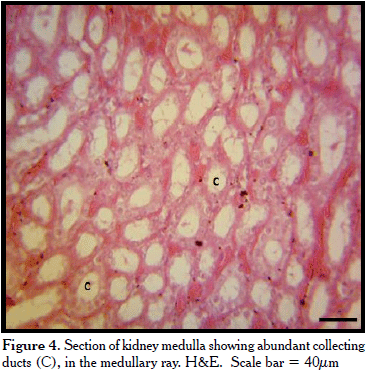
<p style="text-align: justify; text-autospace: none; vertical-align: middle"> <span lang="EN-US" style="font-family: Verdana; color: black"><font size="2">In  the cortex, the renal corpuscle contained a tuft of glomerular vessels inside  the Bowmans’ cup. The urinary space was prominent and clear (<a href="#fig3">Figure 3</a>). The  proximal convoluted tubules (PCT) were lined by dark staining simple cuboidal  cells with prominent brush border that reduced the luminal diameter or even  partial occlusion in some PCT (<a href="#fig3">Figure 3</a>). The distal convoluted tubule (DCT) was  lined by light staining simple cuboidal cells with no brush border (<a href="#fig1">Figure 1</a>).  The DCT simple cuboidal cells, which were very close to the renal corpuscle,  changed to simple columnar cells, thus forming the macula densa of the  juxtaglomerular complex or apparatus (JGC) (<a href="#fig3">Figure 3</a>). In most microscopic view  in the cortex, more PCTs were observed than renal corpuscles and DCTs.</font></span></p>     <p style="text-align: justify; text-autospace: none; vertical-align: middle"> <span lang="EN-US" style="font-family: Verdana; color: black"><font size="2">The  medulla contained mostly collecting ducts lined by simple cuboidal epithelium  and few loops of Henle lined by simple squamous cells (<a href="#fig4">Figure 4</a>).</font></span></p>     <p style="text-align: center; text-autospace: none; vertical-align: middle"> <a name="fig4"> <img border="0" src="/img/fbpe/rfcv/v55n2/art01fig4.gif" width="366" height="373"></a></p>     
<p style="text-align: justify; text-autospace: none; vertical-align: middle"> <span lang="EN-US" style="font-family: Verdana; color: black"><font size="2"> Mucin histochemistry reaction revealed that the epithelial basement membrane of  the kidney tubules, ureters, and blood vessels was PAS positive (<a href="#fig5">Figure 5</a>). The  tunica media of the renal artery was PAS positive (<a href="#fig6">Figure 6</a>), while the poorly  developed tunica media; and well developed tunica adventitia were PAS negative.</font></span></p>     <p style="text-align: center; text-autospace: none; vertical-align: middle"> <a name="fig5"> <img border="0" src="/img/fbpe/rfcv/v55n2/art01fig5.gif" width="368" height="394"></a></p>     
<p style="text-align: center; text-autospace: none; vertical-align: middle"> <a name="fig6"> <img border="0" src="/img/fbpe/rfcv/v55n2/art01fig6.gif" width="369" height="375"></a></p>     
<p style="text-align: justify; text-autospace: none; vertical-align: middle"> <span lang="EN-US" style="font-family: Verdana; color: black"><font size="2"> Adrenal Glands: At low magnification, the adrenal glands were coated by a thin  capsule. The gland parenchyma contained a large cortex and a relatively small  medulla (<a href="#fig7">Figure 7</a>). The cortex contained three clearly demarcated zones- an  outer subcapsular basophilic area of about 1/3&nbsp; of the cortex; a middle lighter  staining area with abundant vacuoles about ½ of the cortex; and an inner  basophilic peri-medulla zone about 1/6 of the cortex (<a href="#fig7">Figure 7</a>). The medulla was  light stained with blood vessels (<a href="#fig7">Figure 7</a>).</font></span></p>     ]]></body>
<body><![CDATA[<p style="text-align: center; text-autospace: none; vertical-align: middle"> <a name="fig7"> <img border="0" src="/img/fbpe/rfcv/v55n2/art01fig7.gif" width="369" height="411"></a></p>     
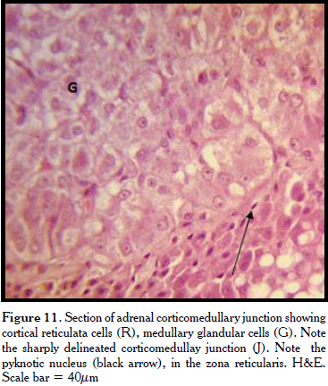
<p style="text-align: justify; text-autospace: none; vertical-align: middle"> <span lang="EN-US" style="font-family: Verdana; color: black"><font size="2">At  higher magnification, the capsule contained thin layer of PAS positive  connective tissue fibres whose trabeculae penetrated the cortical parenchyma  (<a href="#fig7">Figs. 7</a>, <a href="#fig8">8</a>, <a href="#fig9">9</a>). The cortical subcapsular zone (zona glomerulosa) contained  cords of cells. These polyhedral cells arranged in acini had centrally placed  basophilic nuclei and evenly acidophilic cytoplasm (<a href="#fig8">Figure 8</a>). Few vacuolated  structures and blood vessels were also observed. In the middle cortical zone  (the zona fasciculata), the cords contained polyhedral- shaped cells with less  basophilic vesicular nuclei with foamy cytoplasm (<a href="#fig10">Figure 10</a>). Some cells were binucleated. The blood vessels were more abundant while the vacuoles were larger  and more numerous. The inner third zone (the zona reticulata) contained freely  anastomosing cells. The cell nucleus was very basophilic to pyknotic while the  cytoplasm was less foamy than the fasciculata cells (<a href="#fig10">Figure 10</a>). Some reticulata  cells contained brown pigments. These adrenal cortical zones were not clearly  demarcated. Abundant blood vessels were observed, especially near the  corticomedullary junction which was well delineated (<a href="#fig11">Figure 11</a>).</font></span></p>     <p style="text-align: center; text-autospace: none; vertical-align: middle"> <a name="fig8"> <img border="0" src="/img/fbpe/rfcv/v55n2/art01fig8.gif" width="367" height="425"></a></p>     
<p style="text-align: center; text-autospace: none; vertical-align: middle"> <a name="fig9"> <img border="0" src="/img/fbpe/rfcv/v55n2/art01fig9.gif" width="365" height="413"></a></p>     
<p style="text-align: center; text-autospace: none; vertical-align: middle"> <a name="fig10"> <img border="0" src="/img/fbpe/rfcv/v55n2/art01fig10.gif" width="365" height="387"></a></p>     
<p style="text-align: center; text-autospace: none; vertical-align: middle"> <a name="fig11"> <img border="0" src="/img/fbpe/rfcv/v55n2/art01fig11.gif" width="367" height="432"></a></p>     
<p style="text-align: justify; text-autospace: none; vertical-align: middle"> <span lang="EN-US" style="font-family: Verdana; color: black"><font size="2">The  medulla contained glandular cells, ganglion cells, and capillaries, including  some sinusoids (</font><span style="letter-spacing: -.1pt"><font size="2"><a href="#fig11">Figure  11</a>). The basement membrane of the capillary endothelium was PAS positive. The  glandular cells were large polyhedral to columnar cells with vesicular nuclei  and slightly basophilic granular cytoplasm.</font></span></span></p>     <p style="text-align: justify; text-autospace: none; vertical-align: center"><b> <span lang="EN-US" style="font-family: Verdana; color: black; letter-spacing: -.15pt"> <font size="2">Discussion</font></span></b></p>     <p style="text-align: justify; text-autospace: none; vertical-align: middle"> <span lang="EN-US" style="font-family: Verdana; color: black; letter-spacing: -.25pt"> <font size="2">The renal capsule of regular collagen tissue maintains kidney  shape and protects the underlying tissue. A thin fibro-muscular capsule has been  seen in the <i>Camelus dromedaries</i> [11]; but a capsule containing collagen  and reticular fibres has been reported in the rabbit [16]. The differences in  capsular composition may be related to functional adaptation or species  variation.&nbsp; The simple squamous cells of the mesothelium are part of peritoneal  coat on the kidney. The adipose tissue observed is part of the peri-renal fat  that helps hold the kidneys in place in the abdomen [17]. This well developed  unilocular renal adipose tissue suggests that the animal is actively feeding and  not hibernating, as adipose tissue physiology of hibernating mammals has been  described in literature [18]. The absence of brown fat also supports the fact  that tree squirrels like the species under study do not hibernate. The presence  of adipose tissue around the kidney hilus, surrounding its borders has been  reported in the <i>Camelus dromedaries </i>[11]; and albino rats [19]. The  presence of the renal artery, renal vein, ureters and nerve fibres at the hilus  is typical of mammalian kidneys [20].</font></span></p>     <p style="text-align: justify; text-autospace: none; vertical-align: middle"> <span lang="EN-US" style="font-family: Verdana; color: black; letter-spacing: -.1pt"> <font size="2">The PCT epithelium of simple cuboidal cells agrees with the  findings recorded in albino rats [19]. The more abundant PCT than DCT in this  species has been reported in the hedgehog [21]. This relative difference can be  related to the increased need for selective re-absorption of essential  substances like water, glucose, amino acids and various ions, which the animal  has to adequately conserve in the wild. The same reason can be adduced for the  well developed collecting ducts that will enable the Palm Squirrel conserve  water in the wild since access to water bodies is limited, hence the most  probable source of water will be re-absorbed from ingested food through the  large intestine. Species differences in rodent renal histology have revealed  that structure is related to function especially with variation in habitat and  the need for resources conservation [22]. The presence of PAS positive basement  m</font></span><span lang="EN-US" style="font-family: Verdana; color: black"><font size="2">embrane  and renal artery tunica media fibres indicates glycogen storage and this has  been reported [21].&nbsp; The absence of PAS positive entities especially in the  luminal surface of the tubules has been reported in hedgehog [21], while the  positive PAS reaction of kidney tubules in birds have been associated with  aiding the elimination of uric acid from urine [23]. The presence of macula  densa is very significant as its complex with JG cells, the juxtaglomerular  complex produces renin, which regulates the secretion of aldosterone by cells  the zona glomerulosa of the adrenal glands. The presence of macula densa and  renin in JG cells has been reported in the hedgehog like other mammals [21].</font></span></p>     ]]></body>
<body><![CDATA[<p style="text-align: justify; text-autospace: none; vertical-align: middle"> <span lang="EN-US" style="font-family: Verdana; color: black"><font size="2">The  relatively larger adrenal cortex and smaller medulla is typical of mammalian  adrenal cortex [24]. The observation of the zona fasiculata as the most abundant  portion of the cortex has been reported in the porcupine <i>Hystrix cristata</i>  [24]; kano brown goats <i>Capra hircus</i> [6]. The presence of vesicular zona  fasciulata cells nuclei indicates an active cell involved in protein synthesis  most probably the glucocorticoids cortisone and cortisol. The mostly foamy  cytoplasm of the zona fasciculata cells and vacuoles is due to the presence of  fat droplets [24]. Also the pyknotic nuclei seen in the reticularis cells have  been reported in goats and rabbits, where they are related to apoptosis, as  cells originating from outermost layers of zona glomerulosa migrate to the  reticulate for their final death [26, 27]. This migration theory can also  explain the presence of brown pigments of the zona reticulata, which are  intracellular deposits and debris due to age long cell activity.&nbsp; The abundant  vascularization reflects the need to effectively transport hormones produced by  the adrenal glands to the target organ, being a ductless gland.</font></span></p>     <p style="text-align: justify; text-autospace: none; vertical-align: middle"> <span lang="EN-US" style="font-family: Verdana; color: black"><font size="2">The  absence of clear demarcation of the adrenal cortical zones has been reported in  the goat, where the zona fasciculata and zona reticularis lacked clear  distinction [28], this contrasts with the histology in the rabbit [27] and the  buffalo [29], where the zones were clearly delineated.</font></span></p>     <p style="text-align: justify; text-autospace: none; vertical-align: middle"> <span lang="EN-US" style="font-family: Verdana; color: black"><font size="2">The  clearly delineated corticomedullary junction as observed in this study has also  been seen in rabbits [27] and day old foal [30]. But an intermingled cortical  and medullary junction has been observed in birds [31]; a festooned  corticomedullary junction has been reported in the whale and it was related to  its pseudolobulated appearance [32].&nbsp; The presence of well developed abundant  medullary glandular cells can be related to the need to produce adequate  epinephrine and norepinephrine for flight and fight response. This is important  for the survival of the Palm Squirrel in the wild, especially escape from  predators. The neurotransmitter norepinephrine, released at the sympathetic end  of adrenergic fibres is of immense physiologic importance [33].</font></span></p>     <p align="justify"> <span lang="EN-US" style="font-family: Verdana; color: black"><font size="2">In  conclusion, the results of the findings of the micro-morphology study of the  kidneys and adrenal glands of the Palm Squirrel, will help physiologists and  zoologists in understanding its functional adaptation in the wild. It will also  facilitate clinicians in disease diagnosis in the species, especially the wild  life Veterinarians.</font></span></p>     <p style="text-align: justify; text-autospace: none; vertical-align: center"><b> <span lang="EN-US" style="font-family: Verdana; color: black"><font size="2"> Reference</font></span></b></p>     <!-- ref --><p style="text-align: justify; text-autospace: none; vertical-align: middle"> <span lang="EN-US" style="font-family: Verdana; color: black"><font size="2">1.&nbsp;Asibey  EOA, Addo PG.. The grasscutter, a promising animal for meat production. In:  Turnham D (ed). African perspective practices and policies supporting  sustainable development (Scandinavian seminar college Denmark, in association  with Weaver Press, Harare, Zimbabwe). Available in: <a href="http://www.cdr.dk/sscafrica/as&adgh.htm"> http://www.cdr.dk/sscafrica/as &amp; adgh.htm</a>. [access date: 19 abril 2013]. 2000.</font></span>&nbsp;&nbsp;&nbsp;&nbsp;&nbsp;&nbsp;&nbsp;&nbsp;[&#160;<a href="javascript:void(0);" onclick="javascript: window.open('/scielo.php?script=sci_nlinks&ref=1824096&pid=S0258-6576201400020000100001&lng=','','width=640,height=500,resizable=yes,scrollbars=1,menubar=yes,');">Links</a>&#160;]<!-- end-ref --><!-- ref --><p style="text-align: justify; text-autospace: none; vertical-align: middle"> <span lang="EN-US" style="font-family: Verdana; color: black"><font size="2">2.&nbsp;Ogunsanmi  AO, Ozegbe PC, Ogunjobi O, Taiwo VO, Adu JO. Haematological plasma biochemistry  and whole blood minerals of the captive adult African grasscutter (<i>Thryonomis  swinderianus</i> Temminck). Trop Vet. 2002; 20: 27.</font></span>&nbsp;&nbsp;&nbsp;&nbsp;&nbsp;&nbsp;&nbsp;&nbsp;[&#160;<a href="javascript:void(0);" onclick="javascript: window.open('/scielo.php?script=sci_nlinks&ref=1824097&pid=S0258-6576201400020000100002&lng=','','width=640,height=500,resizable=yes,scrollbars=1,menubar=yes,');">Links</a>&#160;]<!-- end-ref --><!-- ref --><p style="text-align: justify; text-autospace: none; vertical-align: middle"> <span lang="EN-US" style="font-family: Verdana; color: black"><font size="2">3.&nbsp;Opara  MN, Ike KA, Okoli IC. Haematology and plasma biochemistry of the wild adult  African grasscutter (<i>Thrynomys swinderianus</i>). J Amer Sci. 2006; 2: 17-22.</font></span>&nbsp;&nbsp;&nbsp;&nbsp;&nbsp;&nbsp;&nbsp;&nbsp;[&#160;<a href="javascript:void(0);" onclick="javascript: window.open('/scielo.php?script=sci_nlinks&ref=1824098&pid=S0258-6576201400020000100003&lng=','','width=640,height=500,resizable=yes,scrollbars=1,menubar=yes,');">Links</a>&#160;]<!-- end-ref --><!-- ref --><p style="text-align: justify; text-autospace: none; vertical-align: middle"> <font size="2" face="Verdana">4. Pradhan VP. The effect of medroxy progesterone  acetate on adrenal gland of Indian Palm Squirrel <i>Funambulus pennati</i> (Wroughton).  Internat J of Life Sci. 2013; 1(3):198-201.</font>&nbsp;&nbsp;&nbsp;&nbsp;&nbsp;&nbsp;&nbsp;&nbsp;[&#160;<a href="javascript:void(0);" onclick="javascript: window.open('/scielo.php?script=sci_nlinks&ref=1824099&pid=S0258-6576201400020000100004&lng=','','width=640,height=500,resizable=yes,scrollbars=1,menubar=yes,');">Links</a>&#160;]<!-- end-ref --><!-- ref --><p style="text-align: justify; text-autospace: none; vertical-align: middle"> <span lang="EN-US" style="font-family: Verdana; color: black"><font size="2">5.&nbsp;Lenger  OF, Akosman MS. An urological model: volume estimation of the bovine renal  parenchyma. Bulgarian J Agric Sci. 2013; 19(4): 848-853.</font></span>&nbsp;&nbsp;&nbsp;&nbsp;&nbsp;&nbsp;&nbsp;&nbsp;[&#160;<a href="javascript:void(0);" onclick="javascript: window.open('/scielo.php?script=sci_nlinks&ref=1824100&pid=S0258-6576201400020000100005&lng=','','width=640,height=500,resizable=yes,scrollbars=1,menubar=yes,');">Links</a>&#160;]<!-- end-ref --><!-- ref --><p style="text-align: justify; text-autospace: none; vertical-align: middle"> <span lang="EN-US" style="font-family: Verdana; color: black"><font size="2">6.&nbsp;Nwaogu  IC, Francis B. 2009. Morphological features of fetal and adult adrenal glands in  kano brown goats (<i>Capra hircus</i>). Anim Res Internat. 2009; 6(1):953-957.</font></span>&nbsp;&nbsp;&nbsp;&nbsp;&nbsp;&nbsp;&nbsp;&nbsp;[&#160;<a href="javascript:void(0);" onclick="javascript: window.open('/scielo.php?script=sci_nlinks&ref=1824101&pid=S0258-6576201400020000100006&lng=','','width=640,height=500,resizable=yes,scrollbars=1,menubar=yes,');">Links</a>&#160;]<!-- end-ref --><!-- ref --><p style="text-align: justify; text-autospace: none; vertical-align: middle"> <span lang="EN-US" style="font-family: Verdana; color: black"><font size="2"> 7.&nbsp;Moffat DB. The Mammalian Kidney. Cambridge University Press, London, 1975; p.  1-211.</font></span>&nbsp;&nbsp;&nbsp;&nbsp;&nbsp;&nbsp;&nbsp;&nbsp;[&#160;<a href="javascript:void(0);" onclick="javascript: window.open('/scielo.php?script=sci_nlinks&ref=1824102&pid=S0258-6576201400020000100007&lng=','','width=640,height=500,resizable=yes,scrollbars=1,menubar=yes,');">Links</a>&#160;]<!-- end-ref --><!-- ref --><p style="text-align: justify; text-autospace: none; vertical-align: middle"> <span lang="EN-US" style="font-family: Verdana; color: black"><font size="2">8.&nbsp;Holz  PH, Raidal SR. Comparative renal anatomy of exotic species. Vet. Clin. North Am  Exot Anim Pract. 2006; 9:1-11.</font></span>&nbsp;&nbsp;&nbsp;&nbsp;&nbsp;&nbsp;&nbsp;&nbsp;[&#160;<a href="javascript:void(0);" onclick="javascript: window.open('/scielo.php?script=sci_nlinks&ref=1824103&pid=S0258-6576201400020000100008&lng=','','width=640,height=500,resizable=yes,scrollbars=1,menubar=yes,');">Links</a>&#160;]<!-- end-ref --><!-- ref --><p style="text-align: justify; text-autospace: none; vertical-align: middle"> <span lang="EN-US" style="font-family: Verdana; color: black"><font size="2">9.&nbsp;Oyeanusi  BI, Adeniyi AA, Ayo JO, Ibe CS, Onyeanusi CG. A comparative study of the urinary  system of the African giant rat (<i>Cricetomys gambianus </i>Waterhouse) and the  Wistar rat. Pak J Nutri. 2009; 8: 1043-1047.</font></span>&nbsp;&nbsp;&nbsp;&nbsp;&nbsp;&nbsp;&nbsp;&nbsp;[&#160;<a href="javascript:void(0);" onclick="javascript: window.open('/scielo.php?script=sci_nlinks&ref=1824104&pid=S0258-6576201400020000100009&lng=','','width=640,height=500,resizable=yes,scrollbars=1,menubar=yes,');">Links</a>&#160;]<!-- end-ref --><!-- ref --><p style="text-align: justify; text-autospace: none; vertical-align: middle"> <span lang="EN-US" style="font-family: Verdana; color: black"><font size="2">10.  Olukole SG. Morphometric analysis of the kidneys of the adult domesticated  African great cane rat (<i>Thryonomys swinderianus</i>). Eur J Anat. 2009;  13(3):117-120.</font></span>&nbsp;&nbsp;&nbsp;&nbsp;&nbsp;&nbsp;&nbsp;&nbsp;[&#160;<a href="javascript:void(0);" onclick="javascript: window.open('/scielo.php?script=sci_nlinks&ref=1824105&pid=S0258-6576201400020000100010&lng=','','width=640,height=500,resizable=yes,scrollbars=1,menubar=yes,');">Links</a>&#160;]<!-- end-ref --><!-- ref --><p style="text-align: justify; text-autospace: none; vertical-align: middle"> <font face="Verdana" size="2">11. Salehi E, Sharifabad MM. 2012. Kidney  morphogenesis during development in Camelus dromedaries embr yos. J Anim Vet  Adv. 2012; 11(6):822-825.</font>&nbsp;&nbsp;&nbsp;&nbsp;&nbsp;&nbsp;&nbsp;&nbsp;[&#160;<a href="javascript:void(0);" onclick="javascript: window.open('/scielo.php?script=sci_nlinks&ref=1824106&pid=S0258-6576201400020000100011&lng=','','width=640,height=500,resizable=yes,scrollbars=1,menubar=yes,');">Links</a>&#160;]<!-- end-ref --><!-- ref --><p style="text-align: justify; text-autospace: none; vertical-align: middle"> <font face="Verdana" size="2">12. Malatesta M, Fakan S, Zancanaro C. Cell and  tissue structural modifications in hibernating dormice. Hystrix It. J. Mamm.  2005; 16(1):41-52.</font>&nbsp;&nbsp;&nbsp;&nbsp;&nbsp;&nbsp;&nbsp;&nbsp;[&#160;<a href="javascript:void(0);" onclick="javascript: window.open('/scielo.php?script=sci_nlinks&ref=1824107&pid=S0258-6576201400020000100012&lng=','','width=640,height=500,resizable=yes,scrollbars=1,menubar=yes,');">Links</a>&#160;]<!-- end-ref --><!-- ref --><p style="text-align: justify; text-autospace: none; vertical-align: middle"> <span lang="EN-US" style="font-family: Verdana; color: black"><font size="2">13.  Bancroft J.D and Stevens A. 1990. Theory and <span style="text-transform: uppercase">p</span>ractice of <span style="text-transform: uppercase">h</span>istological <span style="text-transform: uppercase">t</span>echniques. Third Edition.  Churchill Livingstone, London. p. 88-89.</font></span>&nbsp;&nbsp;&nbsp;&nbsp;&nbsp;&nbsp;&nbsp;&nbsp;[&#160;<a href="javascript:void(0);" onclick="javascript: window.open('/scielo.php?script=sci_nlinks&ref=1824108&pid=S0258-6576201400020000100013&lng=','','width=640,height=500,resizable=yes,scrollbars=1,menubar=yes,');">Links</a>&#160;]<!-- end-ref --><!-- ref --><p style="text-align: justify; text-autospace: none; vertical-align: middle"> <span lang="EN-US" style="font-family: Verdana; color: black"><font size="2">14.  Lillie RD, Greco J. Mact diastase ptyalin in place of saliva in the  identification of glycogen. Stain Tech. 1947; 22:67-70.</font></span>&nbsp;&nbsp;&nbsp;&nbsp;&nbsp;&nbsp;&nbsp;&nbsp;[&#160;<a href="javascript:void(0);" onclick="javascript: window.open('/scielo.php?script=sci_nlinks&ref=1824109&pid=S0258-6576201400020000100014&lng=','','width=640,height=500,resizable=yes,scrollbars=1,menubar=yes,');">Links</a>&#160;]<!-- end-ref --><!-- ref --><p style="text-align: justify; text-autospace: none; vertical-align: middle"> <font size="2" face="Verdana">15. Ikpegbu E, Nlebedum UC, Nnadozie O, Agbakwuru  I. Fast Green FCF or Ehrlich’s hematoxylin as counterstain to periodic acid  Schiff reaction: A comparative study. Histologic. 2011; 54:29-30.</font>&nbsp;&nbsp;&nbsp;&nbsp;&nbsp;&nbsp;&nbsp;&nbsp;[&#160;<a href="javascript:void(0);" onclick="javascript: window.open('/scielo.php?script=sci_nlinks&ref=1824110&pid=S0258-6576201400020000100015&lng=','','width=640,height=500,resizable=yes,scrollbars=1,menubar=yes,');">Links</a>&#160;]<!-- end-ref --><!-- ref --><p style="text-align: justify; text-autospace: none; vertical-align: middle"> <span lang="EN-US" style="font-family: Verdana; color: black"><font size="2">16.  Al-Jeboril JGA, Al-Badri AMS, Jassim BA. 2014. Study of the anatomical and  histomorphological description of the kidney in adult white rabbits female “New  Zealand strain”. Wld J Pharm Pharmaceut Sci.&nbsp; 2014; 3(6):40-51.</font></span>&nbsp;&nbsp;&nbsp;&nbsp;&nbsp;&nbsp;&nbsp;&nbsp;[&#160;<a href="javascript:void(0);" onclick="javascript: window.open('/scielo.php?script=sci_nlinks&ref=1824111&pid=S0258-6576201400020000100016&lng=','','width=640,height=500,resizable=yes,scrollbars=1,menubar=yes,');">Links</a>&#160;]<!-- end-ref --><!-- ref --><p style="text-align: justify; text-autospace: none; vertical-align: middle"> <span lang="EN-US" style="font-family: Verdana; color: black"><font size="2">17.  Singh J. Textbook of Human Histology. Jaypee Brothers, India. 2006; p. 70-72.</font></span>&nbsp;&nbsp;&nbsp;&nbsp;&nbsp;&nbsp;&nbsp;&nbsp;[&#160;<a href="javascript:void(0);" onclick="javascript: window.open('/scielo.php?script=sci_nlinks&ref=1824112&pid=S0258-6576201400020000100017&lng=','','width=640,height=500,resizable=yes,scrollbars=1,menubar=yes,');">Links</a>&#160;]<!-- end-ref --><!-- ref --><p style="text-align: justify; text-autospace: none; vertical-align: middle"> <span lang="EN-US" style="font-family: Verdana; color: black"><font size="2">18.  Himms-Hagen J. Brown <span style="text-transform: uppercase">a</span>dipose  tissue and <span style="text-transform: uppercase">c</span>old <span style="text-transform: uppercase">a</span>cclimatation. In: Trayhurn, P.,  and Nicholls, D.G. (eds). Brown Adipose Tissue. Edward Arnold, London: 1986;  214-268.</font></span>&nbsp;&nbsp;&nbsp;&nbsp;&nbsp;&nbsp;&nbsp;&nbsp;[&#160;<a href="javascript:void(0);" onclick="javascript: window.open('/scielo.php?script=sci_nlinks&ref=1824113&pid=S0258-6576201400020000100018&lng=','','width=640,height=500,resizable=yes,scrollbars=1,menubar=yes,');">Links</a>&#160;]<!-- end-ref --><!-- ref --><p style="text-align: justify; text-autospace: none; vertical-align: middle"> <span lang="EN-US" style="font-family: Verdana; color: black"><font size="2">19.  Al-Samawy ERM. Morphological and histological study of the kidneys on the Albino  rats. Al-Anbar J Vet Sci. 2012; 5(1):115-119. </font></span>&nbsp;&nbsp;&nbsp;&nbsp;&nbsp;&nbsp;&nbsp;&nbsp;[&#160;<a href="javascript:void(0);" onclick="javascript: window.open('/scielo.php?script=sci_nlinks&ref=1824114&pid=S0258-6576201400020000100019&lng=','','width=640,height=500,resizable=yes,scrollbars=1,menubar=yes,');">Links</a>&#160;]<!-- end-ref --><!-- ref --><p style="text-align: justify; text-autospace: none; vertical-align: middle"> <span lang="EN-US" style="font-family: Verdana; color: black"><font size="2">20.  Sandeep GJ, Deepak AY, Gorahnath RT. Case report: Varicosity of the  communicating vein between the left renal vein and the left ascending lumbar  vein mimicking a renal artery aneurysm: Report of an unusual site of varicose  veins and a novel hypothesis to explain its association with abdominal pain.  Indian J Radiol Imaging. 2011; 21(1): 24-27.</font></span>&nbsp;&nbsp;&nbsp;&nbsp;&nbsp;&nbsp;&nbsp;&nbsp;[&#160;<a href="javascript:void(0);" onclick="javascript: window.open('/scielo.php?script=sci_nlinks&ref=1824115&pid=S0258-6576201400020000100020&lng=','','width=640,height=500,resizable=yes,scrollbars=1,menubar=yes,');">Links</a>&#160;]<!-- end-ref --><!-- ref --><p style="text-align: justify; text-autospace: none; vertical-align: middle"> <font size="2" face="Verdana">21. Nabipour A, Dehghani H. Light and electron  microscopic features of the kidney in hedgehog (<i>Hemiechinus auritus</i>). J  Vet Anat. 2012; 5(1): 91-106.</font>&nbsp;&nbsp;&nbsp;&nbsp;&nbsp;&nbsp;&nbsp;&nbsp;[&#160;<a href="javascript:void(0);" onclick="javascript: window.open('/scielo.php?script=sci_nlinks&ref=1824116&pid=S0258-6576201400020000100021&lng=','','width=640,height=500,resizable=yes,scrollbars=1,menubar=yes,');">Links</a>&#160;]<!-- end-ref --><!-- ref --><p style="text-align: justify; text-autospace: none; vertical-align: middle"> <span lang="EN-US" style="font-family: Verdana; color: black"><font size="2">22.  Samuelson, D. A 2007: Textbook of Veterinary Histology. Saunders, St. Louis. USA.  2007; p. 372-390.</font></span>&nbsp;&nbsp;&nbsp;&nbsp;&nbsp;&nbsp;&nbsp;&nbsp;[&#160;<a href="javascript:void(0);" onclick="javascript: window.open('/scielo.php?script=sci_nlinks&ref=1824117&pid=S0258-6576201400020000100022&lng=','','width=640,height=500,resizable=yes,scrollbars=1,menubar=yes,');">Links</a>&#160;]<!-- end-ref --><!-- ref --><p style="text-align: justify; text-autospace: none; vertical-align: middle"> <span lang="EN-US" style="font-family: Verdana; color: black"><font size="2">23.  Casotti G. Effects of season on kidney morphology in house sparrows. J.  Experimental Biol. 2001; 204: 1201-1206.</font></span>&nbsp;&nbsp;&nbsp;&nbsp;&nbsp;&nbsp;&nbsp;&nbsp;[&#160;<a href="javascript:void(0);" onclick="javascript: window.open('/scielo.php?script=sci_nlinks&ref=1824118&pid=S0258-6576201400020000100023&lng=','','width=640,height=500,resizable=yes,scrollbars=1,menubar=yes,');">Links</a>&#160;]<!-- end-ref --><!-- ref --><p style="text-align: justify; text-autospace: none; vertical-align: middle"> <font face="Verdana" size="2">24. Yilmazl S, Girgin A. 2005. Light and electron  microscopic observations on the structure of the porcupine (Hystrix cristata)  adrenal gland. Vet Arhiv. 2005; 75 (3): 265-272.</font>&nbsp;&nbsp;&nbsp;&nbsp;&nbsp;&nbsp;&nbsp;&nbsp;[&#160;<a href="javascript:void(0);" onclick="javascript: window.open('/scielo.php?script=sci_nlinks&ref=1824119&pid=S0258-6576201400020000100024&lng=','','width=640,height=500,resizable=yes,scrollbars=1,menubar=yes,');">Links</a>&#160;]<!-- end-ref --><!-- ref --><p style="text-align: justify; text-autospace: none; vertical-align: middle"> <font size="2" face="Verdana">25. Peng KM, Song H, Liu HZ, Zhang JB, Lu ZQ, Liu  ZW, Liu YX. Histological study of the adrenal gland of african white rhinoceros.  Pak Vet J. 2012; 32(3): 394-397.</font>&nbsp;&nbsp;&nbsp;&nbsp;&nbsp;&nbsp;&nbsp;&nbsp;[&#160;<a href="javascript:void(0);" onclick="javascript: window.open('/scielo.php?script=sci_nlinks&ref=1824120&pid=S0258-6576201400020000100025&lng=','','width=640,height=500,resizable=yes,scrollbars=1,menubar=yes,');">Links</a>&#160;]<!-- end-ref --><!-- ref --><p style="text-align: justify; text-autospace: none; vertical-align: middle"> <span lang="EN-US" style="font-family: Verdana; color: black"><font size="2">26.  Roy MK, Sinha RD, Prasad J. 1987. Certain micrometric observation on the adrenal  cortex of normal and vasectomized goats. Ind J Anim Sci. 1987; 57(11):  1163-1165.</font></span>&nbsp;&nbsp;&nbsp;&nbsp;&nbsp;&nbsp;&nbsp;&nbsp;[&#160;<a href="javascript:void(0);" onclick="javascript: window.open('/scielo.php?script=sci_nlinks&ref=1824121&pid=S0258-6576201400020000100026&lng=','','width=640,height=500,resizable=yes,scrollbars=1,menubar=yes,');">Links</a>&#160;]<!-- end-ref --><!-- ref --><p style="text-align: justify; text-autospace: none; vertical-align: middle"> <span lang="EN-US" style="font-family: Verdana; color: black"><font size="2">27.  Farhan OR. 2013. Morphological study of adrenal gland in prenatal and postnatal  periods of domestic Rabbit. Bas J Vet Res. 2013; 12(1): 253-264.</font></span>&nbsp;&nbsp;&nbsp;&nbsp;&nbsp;&nbsp;&nbsp;&nbsp;[&#160;<a href="javascript:void(0);" onclick="javascript: window.open('/scielo.php?script=sci_nlinks&ref=1824122&pid=S0258-6576201400020000100027&lng=','','width=640,height=500,resizable=yes,scrollbars=1,menubar=yes,');">Links</a>&#160;]<!-- end-ref --><!-- ref --><p style="text-align: justify; text-autospace: none; vertical-align: middle"> <span lang="EN-US" style="font-family: Verdana; color: black"><font size="2">28.  Eurell JA. Veterinary Histology. Tenton new Media. Library of congress. 2004;  34-36.</font></span>&nbsp;&nbsp;&nbsp;&nbsp;&nbsp;&nbsp;&nbsp;&nbsp;[&#160;<a href="javascript:void(0);" onclick="javascript: window.open('/scielo.php?script=sci_nlinks&ref=1824123&pid=S0258-6576201400020000100028&lng=','','width=640,height=500,resizable=yes,scrollbars=1,menubar=yes,');">Links</a>&#160;]<!-- end-ref --><!-- ref --><p style="text-align: justify; text-autospace: none; vertical-align: middle"> <span lang="EN-US" style="font-family: Verdana; color: black"><font size="2">29.  Mohammed NI. Anatomical and Histological study of Adrenal Gland in Iraqi Buffalo <i>Bubalus bubalis </i>with reference to the Seasonal Changes. MSc.<span style="text-transform: uppercase">t</span>hesis,  Veterinary Medicine College, Baghdad University. 2007; p.190.</font></span>&nbsp;&nbsp;&nbsp;&nbsp;&nbsp;&nbsp;&nbsp;&nbsp;[&#160;<a href="javascript:void(0);" onclick="javascript: window.open('/scielo.php?script=sci_nlinks&ref=1824124&pid=S0258-6576201400020000100029&lng=','','width=640,height=500,resizable=yes,scrollbars=1,menubar=yes,');">Links</a>&#160;]<!-- end-ref --><!-- ref --><p style="text-align: justify; text-autospace: none; vertical-align: middle"> <span lang="EN-US" style="font-family: Verdana; color: black"><font size="2">30.  Sharma A. Histomorphological observation on the adrenal gland of day old foal.  Ind J Vet Sci. 2013; 1(2): 31-38.</font></span>&nbsp;&nbsp;&nbsp;&nbsp;&nbsp;&nbsp;&nbsp;&nbsp;[&#160;<a href="javascript:void(0);" onclick="javascript: window.open('/scielo.php?script=sci_nlinks&ref=1824125&pid=S0258-6576201400020000100030&lng=','','width=640,height=500,resizable=yes,scrollbars=1,menubar=yes,');">Links</a>&#160;]<!-- end-ref --><!-- ref --><p style="text-align: justify; text-autospace: none; vertical-align: middle"> <span lang="EN-US" style="font-family: Verdana; color: black"><font size="2">31.  Basha SH, Kannan TA, Geetha RC. Age related of the adrenal gland in Japans quail  (<i>Coturnix coturnix </i>japonica). Tamilnadu J Vet Anim Sci. 2009; 5(5):  198-202.</font></span>&nbsp;&nbsp;&nbsp;&nbsp;&nbsp;&nbsp;&nbsp;&nbsp;[&#160;<a href="javascript:void(0);" onclick="javascript: window.open('/scielo.php?script=sci_nlinks&ref=1824126&pid=S0258-6576201400020000100031&lng=','','width=640,height=500,resizable=yes,scrollbars=1,menubar=yes,');">Links</a>&#160;]<!-- end-ref --><!-- ref --><p style="text-align: justify; text-autospace: none; vertical-align: middle"> <font face="Verdana" size="2">32. Carballeria A, Brown JW., Fishman LM, Trujillo  D, Odell DK. The adrenal gland of stranded Whales (<i>Kogia breviceps</i> and <i> Mesoplodon europaeus</i>): Morphology, hormonal contents and biosynthsis of  corticosteroids. Gen Comp Endocrinol. 1987; 68(2): 293-303.</font>&nbsp;&nbsp;&nbsp;&nbsp;&nbsp;&nbsp;&nbsp;&nbsp;[&#160;<a href="javascript:void(0);" onclick="javascript: window.open('/scielo.php?script=sci_nlinks&ref=1824127&pid=S0258-6576201400020000100032&lng=','','width=640,height=500,resizable=yes,scrollbars=1,menubar=yes,');">Links</a>&#160;]<!-- end-ref --><!-- ref --><p align="justify"> <span lang="EN-US" style="font-family: Verdana; color: black"><font size="2"> 33.&nbsp;Engelhardt WV, Breves G. Nebennierenhormone. In Physiologie der Haustiere,  Stuttgart: Enke im Hippokrates Verlag. 2000; p. 345-354.</font></span>&nbsp;&nbsp;&nbsp;&nbsp;&nbsp;&nbsp;&nbsp;&nbsp;[&#160;<a href="javascript:void(0);" onclick="javascript: window.open('/scielo.php?script=sci_nlinks&ref=1824128&pid=S0258-6576201400020000100033&lng=','','width=640,height=500,resizable=yes,scrollbars=1,menubar=yes,');">Links</a>&#160;]<!-- end-ref --> ]]></body>
<back>
<ref-list>
<ref id="B1">
<label>1</label><nlm-citation citation-type="book">
<person-group person-group-type="author">
<name>
<surname><![CDATA[Asibey]]></surname>
<given-names><![CDATA[EOA]]></given-names>
</name>
<name>
<surname><![CDATA[Addo]]></surname>
<given-names><![CDATA[PG]]></given-names>
</name>
</person-group>
<article-title xml:lang="en"><![CDATA[The grasscutter, a promising animal for meat production]]></article-title>
<person-group person-group-type="editor">
<name>
<surname><![CDATA[Turnham]]></surname>
<given-names><![CDATA[D]]></given-names>
</name>
</person-group>
<source><![CDATA[African perspective practices and policies supporting sustainable development]]></source>
<year></year>
<publisher-loc><![CDATA[Harare ]]></publisher-loc>
<publisher-name><![CDATA[Scandinavian seminar college Denmark, in association with Weaver Press]]></publisher-name>
</nlm-citation>
</ref>
<ref id="B2">
<label>2</label><nlm-citation citation-type="journal">
<person-group person-group-type="author">
<name>
<surname><![CDATA[Ogunsanmi]]></surname>
<given-names><![CDATA[AO]]></given-names>
</name>
<name>
<surname><![CDATA[Ozegbe]]></surname>
<given-names><![CDATA[PC]]></given-names>
</name>
<name>
<surname><![CDATA[Ogunjobi]]></surname>
<given-names><![CDATA[O]]></given-names>
</name>
<name>
<surname><![CDATA[Taiwo]]></surname>
<given-names><![CDATA[VO]]></given-names>
</name>
<name>
<surname><![CDATA[Adu]]></surname>
<given-names><![CDATA[JO]]></given-names>
</name>
</person-group>
<article-title xml:lang="en"><![CDATA[Haematological plasma biochemistry and whole blood minerals of the captive adult African grasscutter (Thryonomis swinderianus Temminck)]]></article-title>
<source><![CDATA[Trop Vet.]]></source>
<year>2002</year>
<volume>20</volume>
<page-range>27</page-range></nlm-citation>
</ref>
<ref id="B3">
<label>3</label><nlm-citation citation-type="journal">
<person-group person-group-type="author">
<name>
<surname><![CDATA[Opara]]></surname>
<given-names><![CDATA[MN]]></given-names>
</name>
<name>
<surname><![CDATA[Ike]]></surname>
<given-names><![CDATA[KA]]></given-names>
</name>
<name>
<surname><![CDATA[Okoli]]></surname>
<given-names><![CDATA[IC]]></given-names>
</name>
</person-group>
<article-title xml:lang="en"><![CDATA[Haematology and plasma biochemistry of the wild adult African grasscutter (Thrynomys swinderianus)]]></article-title>
<source><![CDATA[J Amer Sci.]]></source>
<year>2006</year>
<volume>2</volume>
<page-range>17-22</page-range></nlm-citation>
</ref>
<ref id="B4">
<label>4</label><nlm-citation citation-type="journal">
<person-group person-group-type="author">
<name>
<surname><![CDATA[Pradhan]]></surname>
<given-names><![CDATA[VP]]></given-names>
</name>
</person-group>
<article-title xml:lang="en"><![CDATA[The effect of medroxy progesterone acetate on adrenal gland of Indian Palm Squirrel Funambulus pennati (Wroughton)]]></article-title>
<source><![CDATA[Internat J of Life Sci.]]></source>
<year>2013</year>
<volume>1</volume>
<numero>3</numero>
<issue>3</issue>
<page-range>198-201</page-range></nlm-citation>
</ref>
<ref id="B5">
<label>5</label><nlm-citation citation-type="journal">
<person-group person-group-type="author">
<name>
<surname><![CDATA[Lenger]]></surname>
<given-names><![CDATA[OF]]></given-names>
</name>
<name>
<surname><![CDATA[Akosman]]></surname>
<given-names><![CDATA[MS]]></given-names>
</name>
</person-group>
<article-title xml:lang="en"><![CDATA[An urological model: volume estimation of the bovine renal parenchyma]]></article-title>
<source><![CDATA[Bulgarian J Agric Sci.]]></source>
<year>2013</year>
<volume>19</volume>
<numero>4</numero>
<issue>4</issue>
<page-range>848-853</page-range></nlm-citation>
</ref>
<ref id="B6">
<label>6</label><nlm-citation citation-type="journal">
<person-group person-group-type="author">
<name>
<surname><![CDATA[Nwaogu]]></surname>
<given-names><![CDATA[IC]]></given-names>
</name>
<name>
<surname><![CDATA[Francis]]></surname>
<given-names><![CDATA[B]]></given-names>
</name>
</person-group>
<article-title xml:lang="en"><![CDATA[Morphological features of fetal and adult adrenal glands in kano brown goats (Capra hircus)]]></article-title>
<source><![CDATA[Anim Res Internat.]]></source>
<year>2009</year>
<month>20</month>
<day>09</day>
<volume>6</volume>
<numero>1</numero>
<issue>1</issue>
<page-range>953-957</page-range></nlm-citation>
</ref>
<ref id="B7">
<label>7</label><nlm-citation citation-type="book">
<person-group person-group-type="author">
<name>
<surname><![CDATA[Moffat]]></surname>
<given-names><![CDATA[DB]]></given-names>
</name>
</person-group>
<source><![CDATA[The Mammalian Kidney]]></source>
<year>1975</year>
<page-range>1-211</page-range><publisher-loc><![CDATA[London ]]></publisher-loc>
<publisher-name><![CDATA[Cambridge University Press]]></publisher-name>
</nlm-citation>
</ref>
<ref id="B8">
<label>8</label><nlm-citation citation-type="journal">
<person-group person-group-type="author">
<name>
<surname><![CDATA[Holz]]></surname>
<given-names><![CDATA[PH]]></given-names>
</name>
<name>
<surname><![CDATA[Raidal]]></surname>
<given-names><![CDATA[SR]]></given-names>
</name>
</person-group>
<article-title xml:lang="en"><![CDATA[Comparative renal anatomy of exotic species]]></article-title>
<source><![CDATA[Vet. Clin. North Am Exot Anim Pract.]]></source>
<year>2006</year>
<volume>9</volume>
<page-range>1-11</page-range></nlm-citation>
</ref>
<ref id="B9">
<label>9</label><nlm-citation citation-type="journal">
<person-group person-group-type="author">
<name>
<surname><![CDATA[Oyeanusi]]></surname>
<given-names><![CDATA[BI]]></given-names>
</name>
<name>
<surname><![CDATA[Adeniyi]]></surname>
<given-names><![CDATA[AA]]></given-names>
</name>
<name>
<surname><![CDATA[Ayo]]></surname>
<given-names><![CDATA[JO]]></given-names>
</name>
<name>
<surname><![CDATA[Ibe]]></surname>
<given-names><![CDATA[CS]]></given-names>
</name>
<name>
<surname><![CDATA[Onyeanusi]]></surname>
<given-names><![CDATA[CG]]></given-names>
</name>
</person-group>
<article-title xml:lang="en"><![CDATA[A comparative study of the urinary system of the African giant rat (Cricetomys gambianus Waterhouse) and the Wistar rat]]></article-title>
<source><![CDATA[Pak J Nutri.]]></source>
<year>2009</year>
<volume>8</volume>
<page-range>1043-1047</page-range></nlm-citation>
</ref>
<ref id="B10">
<label>10</label><nlm-citation citation-type="journal">
<person-group person-group-type="author">
<name>
<surname><![CDATA[Olukole]]></surname>
<given-names><![CDATA[SG]]></given-names>
</name>
</person-group>
<article-title xml:lang="en"><![CDATA[Morphometric analysis of the kidneys of the adult domesticated African great cane rat (Thryonomys swinderianus)]]></article-title>
<source><![CDATA[Eur J Anat.]]></source>
<year>2009</year>
<volume>13</volume>
<numero>3</numero>
<issue>3</issue>
<page-range>117-120</page-range></nlm-citation>
</ref>
<ref id="B11">
<label>11</label><nlm-citation citation-type="journal">
<person-group person-group-type="author">
<name>
<surname><![CDATA[Salehi]]></surname>
<given-names><![CDATA[E]]></given-names>
</name>
<name>
<surname><![CDATA[Sharifabad]]></surname>
<given-names><![CDATA[MM]]></given-names>
</name>
</person-group>
<article-title xml:lang="en"><![CDATA[Kidney morphogenesis during development in Camelus dromedaries embr yos]]></article-title>
<source><![CDATA[J Anim Vet Adv.]]></source>
<year>2012</year>
<month>20</month>
<day>12</day>
<volume>11</volume>
<numero>6</numero>
<issue>6</issue>
<page-range>822-825</page-range></nlm-citation>
</ref>
<ref id="B12">
<label>12</label><nlm-citation citation-type="journal">
<person-group person-group-type="author">
<name>
<surname><![CDATA[Malatesta]]></surname>
<given-names><![CDATA[M]]></given-names>
</name>
<name>
<surname><![CDATA[Fakan]]></surname>
<given-names><![CDATA[S]]></given-names>
</name>
<name>
<surname><![CDATA[Zancanaro]]></surname>
<given-names><![CDATA[C]]></given-names>
</name>
</person-group>
<article-title xml:lang="en"><![CDATA[Cell and tissue structural modifications in hibernating dormice]]></article-title>
<source><![CDATA[Hystrix It. J. Mamm.]]></source>
<year>2005</year>
<volume>16</volume>
<numero>1</numero>
<issue>1</issue>
<page-range>41-52</page-range></nlm-citation>
</ref>
<ref id="B13">
<label>13</label><nlm-citation citation-type="book">
<person-group person-group-type="author">
<name>
<surname><![CDATA[Bancroft]]></surname>
<given-names><![CDATA[J.D]]></given-names>
</name>
<name>
<surname><![CDATA[Stevens]]></surname>
<given-names><![CDATA[A]]></given-names>
</name>
</person-group>
<source><![CDATA[Theory and practice of histological techniques]]></source>
<year>1990</year>
<edition>Third</edition>
<page-range>88-89</page-range><publisher-loc><![CDATA[London ]]></publisher-loc>
<publisher-name><![CDATA[Churchill Livingstone]]></publisher-name>
</nlm-citation>
</ref>
<ref id="B14">
<label>14</label><nlm-citation citation-type="journal">
<person-group person-group-type="author">
<name>
<surname><![CDATA[Lillie]]></surname>
<given-names><![CDATA[RD]]></given-names>
</name>
<name>
<surname><![CDATA[Greco]]></surname>
<given-names><![CDATA[J]]></given-names>
</name>
</person-group>
<article-title xml:lang="en"><![CDATA[Mact diastase ptyalin in place of saliva in the identification of glycogen]]></article-title>
<source><![CDATA[Stain Tech.]]></source>
<year>1947</year>
<volume>22</volume>
<page-range>67-70</page-range></nlm-citation>
</ref>
<ref id="B15">
<label>15</label><nlm-citation citation-type="journal">
<person-group person-group-type="author">
<name>
<surname><![CDATA[Ikpegbu]]></surname>
<given-names><![CDATA[E]]></given-names>
</name>
<name>
<surname><![CDATA[Nlebedum]]></surname>
<given-names><![CDATA[UC]]></given-names>
</name>
<name>
<surname><![CDATA[Nnadozie]]></surname>
<given-names><![CDATA[O]]></given-names>
</name>
<name>
<surname><![CDATA[Agbakwuru]]></surname>
<given-names><![CDATA[I]]></given-names>
</name>
</person-group>
<article-title xml:lang="en"><![CDATA[Fast Green FCF or Ehrlich’s hematoxylin as counterstain to periodic acid Schiff reaction: A comparative study]]></article-title>
<source><![CDATA[Histologic.]]></source>
<year>2011</year>
<volume>54</volume>
<page-range>29-30</page-range></nlm-citation>
</ref>
<ref id="B16">
<label>16</label><nlm-citation citation-type="journal">
<person-group person-group-type="author">
<name>
<surname><![CDATA[Al-Jeboril]]></surname>
<given-names><![CDATA[JGA]]></given-names>
</name>
<name>
<surname><![CDATA[Al-Badri]]></surname>
<given-names><![CDATA[AMS]]></given-names>
</name>
<name>
<surname><![CDATA[Jassim]]></surname>
<given-names><![CDATA[BA]]></given-names>
</name>
</person-group>
<article-title xml:lang="en"><![CDATA[Study of the anatomical and histomorphological description of the kidney in adult white rabbits female “New Zealand strain”]]></article-title>
<source><![CDATA[Wld J Pharm Pharmaceut Sci.]]></source>
<year>2014</year>
<month>20</month>
<day>14</day>
<volume>3</volume>
<numero>6</numero>
<issue>6</issue>
<page-range>40-51</page-range></nlm-citation>
</ref>
<ref id="B17">
<label>17</label><nlm-citation citation-type="book">
<person-group person-group-type="author">
<name>
<surname><![CDATA[Singh]]></surname>
<given-names><![CDATA[J]]></given-names>
</name>
</person-group>
<source><![CDATA[Textbook of Human Histology]]></source>
<year>2006</year>
<page-range>70-72</page-range><publisher-name><![CDATA[Jaypee Brothers]]></publisher-name>
</nlm-citation>
</ref>
<ref id="B18">
<label>18</label><nlm-citation citation-type="book">
<person-group person-group-type="author">
<name>
<surname><![CDATA[Himms-Hagen]]></surname>
<given-names><![CDATA[J]]></given-names>
</name>
</person-group>
<article-title xml:lang="en"><![CDATA[Brown adipose tissue and cold acclimatation]]></article-title>
<person-group person-group-type="editor">
<name>
<surname><![CDATA[Trayhurn]]></surname>
<given-names><![CDATA[P]]></given-names>
</name>
<name>
<surname><![CDATA[Nicholls]]></surname>
<given-names><![CDATA[D.G]]></given-names>
</name>
</person-group>
<source><![CDATA[Brown Adipose Tissue]]></source>
<year>1986</year>
<page-range>214-268</page-range><publisher-loc><![CDATA[London ]]></publisher-loc>
<publisher-name><![CDATA[Edward Arnold]]></publisher-name>
</nlm-citation>
</ref>
<ref id="B19">
<label>19</label><nlm-citation citation-type="journal">
<person-group person-group-type="author">
<name>
<surname><![CDATA[Al-Samawy]]></surname>
<given-names><![CDATA[ERM]]></given-names>
</name>
</person-group>
<article-title xml:lang="en"><![CDATA[Morphological and histological study of the kidneys on the Albino rats]]></article-title>
<source><![CDATA[Al-Anbar J Vet Sci.]]></source>
<year>2012</year>
<volume>5</volume>
<numero>1</numero>
<issue>1</issue>
<page-range>115-119</page-range></nlm-citation>
</ref>
<ref id="B20">
<label>20</label><nlm-citation citation-type="journal">
<person-group person-group-type="author">
<name>
<surname><![CDATA[Sandeep]]></surname>
<given-names><![CDATA[GJ]]></given-names>
</name>
<name>
<surname><![CDATA[Deepak]]></surname>
<given-names><![CDATA[AY]]></given-names>
</name>
<name>
<surname><![CDATA[Gorahnath]]></surname>
<given-names><![CDATA[RT]]></given-names>
</name>
</person-group>
<article-title xml:lang="en"><![CDATA[Case report: Varicosity of the communicating vein between the left renal vein and the left ascending lumbar vein mimicking a renal artery aneurysm: Report of an unusual site of varicose veins and a novel hypothesis to explain its association with abdominal pain]]></article-title>
<source><![CDATA[Indian J Radiol Imaging.]]></source>
<year>2011</year>
<volume>21</volume>
<numero>1</numero>
<issue>1</issue>
<page-range>24-27</page-range></nlm-citation>
</ref>
<ref id="B21">
<label>21</label><nlm-citation citation-type="journal">
<person-group person-group-type="author">
<name>
<surname><![CDATA[Nabipour]]></surname>
<given-names><![CDATA[A]]></given-names>
</name>
<name>
<surname><![CDATA[Dehghani]]></surname>
<given-names><![CDATA[H]]></given-names>
</name>
</person-group>
<article-title xml:lang="en"><![CDATA[Light and electron microscopic features of the kidney in hedgehog (Hemiechinus auritus)]]></article-title>
<source><![CDATA[J Vet Anat.]]></source>
<year>2012</year>
<volume>5</volume>
<numero>1</numero>
<issue>1</issue>
<page-range>91-106</page-range></nlm-citation>
</ref>
<ref id="B22">
<label>22</label><nlm-citation citation-type="book">
<person-group person-group-type="author">
<name>
<surname><![CDATA[Samuelson]]></surname>
<given-names><![CDATA[D]]></given-names>
</name>
</person-group>
<source><![CDATA[A 2007: Textbook of Veterinary Histology]]></source>
<year>2007</year>
<page-range>372-390</page-range><publisher-loc><![CDATA[St. Louis ]]></publisher-loc>
<publisher-name><![CDATA[Saunders]]></publisher-name>
</nlm-citation>
</ref>
<ref id="B23">
<label>23</label><nlm-citation citation-type="journal">
<person-group person-group-type="author">
<name>
<surname><![CDATA[Casotti]]></surname>
<given-names><![CDATA[G]]></given-names>
</name>
</person-group>
<article-title xml:lang="en"><![CDATA[Effects of season on kidney morphology in house sparrows]]></article-title>
<source><![CDATA[J. Experimental Biol.]]></source>
<year>2001</year>
<volume>204</volume>
<page-range>1201-1206</page-range></nlm-citation>
</ref>
<ref id="B24">
<label>24</label><nlm-citation citation-type="journal">
<person-group person-group-type="author">
<name>
<surname><![CDATA[Yilmazl]]></surname>
<given-names><![CDATA[S]]></given-names>
</name>
<name>
<surname><![CDATA[Girgin]]></surname>
<given-names><![CDATA[A]]></given-names>
</name>
</person-group>
<article-title xml:lang="en"><![CDATA[Light and electron microscopic observations on the structure of the porcupine (Hystrix cristata) adrenal gland]]></article-title>
<source><![CDATA[Vet Arhiv.]]></source>
<year>2005</year>
<month>20</month>
<day>05</day>
<volume>75</volume>
<numero>3</numero>
<issue>3</issue>
<page-range>265-272</page-range></nlm-citation>
</ref>
<ref id="B25">
<label>25</label><nlm-citation citation-type="journal">
<person-group person-group-type="author">
<name>
<surname><![CDATA[Peng]]></surname>
<given-names><![CDATA[KM]]></given-names>
</name>
<name>
<surname><![CDATA[Song]]></surname>
<given-names><![CDATA[H]]></given-names>
</name>
<name>
<surname><![CDATA[Liu]]></surname>
<given-names><![CDATA[HZ]]></given-names>
</name>
<name>
<surname><![CDATA[Zhang]]></surname>
<given-names><![CDATA[JB]]></given-names>
</name>
<name>
<surname><![CDATA[Lu]]></surname>
<given-names><![CDATA[ZQ]]></given-names>
</name>
<name>
<surname><![CDATA[Liu]]></surname>
<given-names><![CDATA[ZW]]></given-names>
</name>
<name>
<surname><![CDATA[Liu]]></surname>
<given-names><![CDATA[YX]]></given-names>
</name>
</person-group>
<article-title xml:lang="en"><![CDATA[Histological study of the adrenal gland of african white rhinoceros]]></article-title>
<source><![CDATA[Pak Vet J.]]></source>
<year>2012</year>
<volume>32</volume>
<numero>3</numero>
<issue>3</issue>
<page-range>394-397</page-range></nlm-citation>
</ref>
<ref id="B26">
<label>26</label><nlm-citation citation-type="journal">
<person-group person-group-type="author">
<name>
<surname><![CDATA[Roy]]></surname>
<given-names><![CDATA[MK]]></given-names>
</name>
<name>
<surname><![CDATA[Sinha]]></surname>
<given-names><![CDATA[RD]]></given-names>
</name>
<name>
<surname><![CDATA[Prasad]]></surname>
<given-names><![CDATA[J]]></given-names>
</name>
</person-group>
<article-title xml:lang="en"><![CDATA[Certain micrometric observation on the adrenal cortex of normal and vasectomized goats]]></article-title>
<source><![CDATA[Ind J Anim Sci.]]></source>
<year>1987</year>
<month>19</month>
<day>87</day>
<volume>57</volume>
<numero>11</numero>
<issue>11</issue>
<page-range>1163-1165</page-range></nlm-citation>
</ref>
<ref id="B27">
<label>27</label><nlm-citation citation-type="journal">
<person-group person-group-type="author">
<name>
<surname><![CDATA[Farhan]]></surname>
<given-names><![CDATA[OR]]></given-names>
</name>
</person-group>
<article-title xml:lang="en"><![CDATA[Morphological study of adrenal gland in prenatal and postnatal periods of domestic Rabbit]]></article-title>
<source><![CDATA[Bas J Vet Res.]]></source>
<year>2013</year>
<month>20</month>
<day>13</day>
<volume>12</volume>
<numero>1</numero>
<issue>1</issue>
<page-range>253-264</page-range></nlm-citation>
</ref>
<ref id="B28">
<label>28</label><nlm-citation citation-type="book">
<person-group person-group-type="author">
<name>
<surname><![CDATA[Eurell]]></surname>
<given-names><![CDATA[JA]]></given-names>
</name>
</person-group>
<source><![CDATA[Veterinary Histology]]></source>
<year>2004</year>
<page-range>34-36</page-range><publisher-name><![CDATA[Tenton new Media. Library of congress]]></publisher-name>
</nlm-citation>
</ref>
<ref id="B29">
<label>29</label><nlm-citation citation-type="">
<person-group person-group-type="author">
<name>
<surname><![CDATA[Mohammed]]></surname>
<given-names><![CDATA[NI]]></given-names>
</name>
</person-group>
<source><![CDATA[Anatomical and Histological study of Adrenal Gland in Iraqi Buffalo Bubalus bubalis with reference to the Seasonal Changes]]></source>
<year></year>
<page-range>190</page-range></nlm-citation>
</ref>
<ref id="B30">
<label>30</label><nlm-citation citation-type="journal">
<person-group person-group-type="author">
<name>
<surname><![CDATA[Sharma]]></surname>
<given-names><![CDATA[A]]></given-names>
</name>
</person-group>
<article-title xml:lang="en"><![CDATA[Histomorphological observation on the adrenal gland of day old foal]]></article-title>
<source><![CDATA[Ind J Vet Sci.]]></source>
<year>2013</year>
<volume>1</volume>
<numero>2</numero>
<issue>2</issue>
<page-range>31-38</page-range></nlm-citation>
</ref>
<ref id="B31">
<label>31</label><nlm-citation citation-type="journal">
<person-group person-group-type="author">
<name>
<surname><![CDATA[Basha]]></surname>
<given-names><![CDATA[SH]]></given-names>
</name>
<name>
<surname><![CDATA[Kannan]]></surname>
<given-names><![CDATA[TA]]></given-names>
</name>
<name>
<surname><![CDATA[Geetha]]></surname>
<given-names><![CDATA[RC]]></given-names>
</name>
</person-group>
<article-title xml:lang="en"><![CDATA[Age related of the adrenal gland in Japans quail (Coturnix coturnix japonica)]]></article-title>
<source><![CDATA[Tamilnadu J Vet Anim Sci.]]></source>
<year>2009</year>
<volume>5</volume>
<numero>5</numero>
<issue>5</issue>
<page-range>198-202</page-range></nlm-citation>
</ref>
<ref id="B32">
<label>32</label><nlm-citation citation-type="journal">
<person-group person-group-type="author">
<name>
<surname><![CDATA[Carballeria]]></surname>
<given-names><![CDATA[A]]></given-names>
</name>
<name>
<surname><![CDATA[Brown]]></surname>
<given-names><![CDATA[JW.]]></given-names>
</name>
<name>
<surname><![CDATA[Fishman]]></surname>
<given-names><![CDATA[LM]]></given-names>
</name>
<name>
<surname><![CDATA[Trujillo]]></surname>
<given-names><![CDATA[D]]></given-names>
</name>
<name>
<surname><![CDATA[Odell]]></surname>
<given-names><![CDATA[DK]]></given-names>
</name>
</person-group>
<article-title xml:lang="en"><![CDATA[The adrenal gland of stranded Whales (Kogia breviceps and Mesoplodon europaeus): Morphology, hormonal contents and biosynthsis of corticosteroids]]></article-title>
<source><![CDATA[Gen Comp Endocrinol.]]></source>
<year>1987</year>
<volume>68</volume>
<numero>2</numero>
<issue>2</issue>
<page-range>293-303</page-range></nlm-citation>
</ref>
<ref id="B33">
<label>33</label><nlm-citation citation-type="book">
<person-group person-group-type="author">
<name>
<surname><![CDATA[Engelhardt]]></surname>
<given-names><![CDATA[WV]]></given-names>
</name>
<name>
<surname><![CDATA[Breves]]></surname>
<given-names><![CDATA[G]]></given-names>
</name>
</person-group>
<article-title xml:lang="de"><![CDATA[Nebennierenhormone]]></article-title>
<source><![CDATA[Physiologie der Haustiere]]></source>
<year>2000</year>
<page-range>345-354</page-range><publisher-loc><![CDATA[Stuttgart ]]></publisher-loc>
<publisher-name><![CDATA[Enke im Hippokrates Verlag]]></publisher-name>
</nlm-citation>
</ref>
</ref-list>
</back>
</article>