<?xml version="1.0" encoding="ISO-8859-1"?><article xmlns:mml="http://www.w3.org/1998/Math/MathML" xmlns:xlink="http://www.w3.org/1999/xlink" xmlns:xsi="http://www.w3.org/2001/XMLSchema-instance">
<front>
<journal-meta>
<journal-id>0535-5133</journal-id>
<journal-title><![CDATA[Investigación Clínica]]></journal-title>
<abbrev-journal-title><![CDATA[Invest. clín]]></abbrev-journal-title>
<issn>0535-5133</issn>
<publisher>
<publisher-name><![CDATA[Instituto de Investigaciones Clínicas "Dr. Américo Negrette", Facultad de Medicina, Universidad del Zulia]]></publisher-name>
</publisher>
</journal-meta>
<article-meta>
<article-id>S0535-51332010000400005</article-id>
<title-group>
<article-title xml:lang="en"><![CDATA[Human hepatoma cell line (Hep G2) cellular response to hypothermic stress with recovery: Induction of Hsp70, Hsp60 and Hsf1 expression]]></article-title>
<article-title xml:lang="es"><![CDATA[Respuesta celular de línea de hepatoma humano (HepG2) al estrés hipotérmico con recuperación: Inducción de la expresión de Hsp60, Hsp70, y Hsf1]]></article-title>
</title-group>
<contrib-group>
<contrib contrib-type="author">
<name>
<surname><![CDATA[Rada]]></surname>
<given-names><![CDATA[Alegna]]></given-names>
</name>
<xref ref-type="aff" rid="A01"/>
</contrib>
<contrib contrib-type="author">
<name>
<surname><![CDATA[Merentes]]></surname>
<given-names><![CDATA[Elizabeth]]></given-names>
</name>
<xref ref-type="aff" rid="A02"/>
</contrib>
<contrib contrib-type="author">
<name>
<surname><![CDATA[Rodríguez]]></surname>
<given-names><![CDATA[Marianela]]></given-names>
</name>
<xref ref-type="aff" rid="A01"/>
</contrib>
<contrib contrib-type="author">
<name>
<surname><![CDATA[Anselmi]]></surname>
<given-names><![CDATA[Guillermo]]></given-names>
</name>
<xref ref-type="aff" rid="A01"/>
</contrib>
<contrib contrib-type="author">
<name>
<surname><![CDATA[Strauss]]></surname>
<given-names><![CDATA[Mirian]]></given-names>
</name>
<xref ref-type="aff" rid="A01"/>
</contrib>
</contrib-group>
<aff id="A01">
<institution><![CDATA[,Universidad Central de Venezuela Facultad de Medicina Instituto de Medicina Tropical]]></institution>
<addr-line><![CDATA[ ]]></addr-line>
</aff>
<aff id="A02">
<institution><![CDATA[,Universidad Central de Venezuela Facultad de Ciencias Laboratorio de Cultivo de Tejidos y Biología de Tumores]]></institution>
<addr-line><![CDATA[Caracas ]]></addr-line>
<country>Venezuela</country>
</aff>
<pub-date pub-type="pub">
<day>00</day>
<month>12</month>
<year>2010</year>
</pub-date>
<pub-date pub-type="epub">
<day>00</day>
<month>12</month>
<year>2010</year>
</pub-date>
<volume>51</volume>
<numero>4</numero>
<fpage>479</fpage>
<lpage>488</lpage>
<copyright-statement/>
<copyright-year/>
<self-uri xlink:href="http://ve.scielo.org/scielo.php?script=sci_arttext&amp;pid=S0535-51332010000400005&amp;lng=en&amp;nrm=iso"></self-uri><self-uri xlink:href="http://ve.scielo.org/scielo.php?script=sci_abstract&amp;pid=S0535-51332010000400005&amp;lng=en&amp;nrm=iso"></self-uri><self-uri xlink:href="http://ve.scielo.org/scielo.php?script=sci_pdf&amp;pid=S0535-51332010000400005&amp;lng=en&amp;nrm=iso"></self-uri><abstract abstract-type="short" xml:lang="en"><p><![CDATA[The cell response of human HepG2 cells exposed to hypothermia with rewarming was analyzed. Ultrastructural findings in hypothermic stressed cells showed swollen mitochondria, dispersed chromatin, vacuoles and ring-shape nucleolar reorganization. These changes were coupled with significative differences in the induction of Hsp60, inducible Hsp70 and monomeric Hsf1 in all treated samples, but not in Hsc 70 expression. Cellular response to hypothermia could be associated with the synergistic induction of Hsp expression]]></p></abstract>
<abstract abstract-type="short" xml:lang="es"><p><![CDATA[En este trabajo se analizó la respuesta celular de células HepG2 expuestas a hipotermia con posterior recuperación. Los hallazgos ultraestructurales en células sometidas a estrés hipotérmico incluyeron mitocondrias edematizadas, núcleos picnóticos, vacuolas y reorganización nucleolar en forma de anillo. Tales cambios están relacionados con diferencias significativas en la inducción de la expresión de Hsp60, Hsp70 inducible y Hsf 1 monomérico en todas las muestras tratadas, pero no de Hsc70. La respuesta celular a la hipotermia puede ser relacionada con la inducción sinergística de las Hsp]]></p></abstract>
<kwd-group>
<kwd lng="en"><![CDATA[heat shock proteins]]></kwd>
<kwd lng="en"><![CDATA[ultrastructural damage]]></kwd>
<kwd lng="en"><![CDATA[hypothermia with recovery]]></kwd>
<kwd lng="en"><![CDATA[HepG2 cells]]></kwd>
<kwd lng="es"><![CDATA[proteínas de shock térmico]]></kwd>
<kwd lng="es"><![CDATA[daño estructural]]></kwd>
<kwd lng="es"><![CDATA[hipotermia con recuperación]]></kwd>
<kwd lng="es"><![CDATA[células HepG2]]></kwd>
</kwd-group>
</article-meta>
</front><body><![CDATA[  <BASEFONT SIZE="3"> <MULTICOL GUTTER="31" COLS="2">     <P align="center" style="word-spacing: 0; line-height: 100%"> <B><font color="#1f1a17" face="Verdana" size="3">Human hepatoma cell line (Hep G2) cellular response to hypothermic stress with recovery. Induction of Hsp70, Hsp60 and Hsf1 expression.</font></B></P>     <P align="center" style="word-spacing: 0; line-height: 100%"><font face="Verdana" size="2"><b>Alegna Rada <sup>1</sup>, Elizabeth Merentes <sup>2</sup>, Marianela Rodr&#237;guez <sup>1</sup> Guillermo Anselmi <sup>1</sup> and Mirian Strauss <sup>1</sup>&nbsp;</b></font></P>     <P align="justify" style="word-spacing: 0; line-height: 100%"><font face="Verdana" size="2"><sup>1</sup> Secci&#243;n de Biolog&#237;a Celular, Instituto de Medicina Tropical, Facultad  de Medicina y</font></P>     <P align="justify" style="word-spacing: 0; line-height: 100%"><font face="Verdana" size="2"><sup>2</sup>&nbsp; Laboratorio de Cultivo de Tejidos y Biolog&#237;a de Tumores,  Facultad de Ciencias. Universidad Central de Venezuela. Caracas, Venezuela.</font></P>     <P align="justify" style="word-spacing: 0; line-height: 100%"><font color="#1f1a17" size="2" face="Verdana">Corresponding author: Mirian Strauss. Sección de Biología Celular, Instituto de Medicina Tropical, Facultad de Medicina, Universidad Central de Venezuela. Apdo 47019. Caracas 1041A, Venezuela. Phone: 00-58-212- 6053650, Fax: 00-58-212-2434685. E-mail: <a href="mailto:mstraussve@gmail.com">mstraussve@gmail.com</a>.&nbsp;&nbsp;</font></P>     <P align="justify" style="word-spacing: 0; line-height: 100%"><font size="2"> <B><FONT COLOR="#1f1a17" face="Verdana"> Abstract.</FONT></B></font></P>     <P align="justify" style="word-spacing: 0; line-height: 100%"><FONT COLOR="#1f1a17" size="2" face="Verdana">The cell response of human HepG2 cells exposed to hypothermia  with rewarming was analyzed. Ultrastructural findings in hypothermic stressed  cells showed swollen mitochondria, dispersed chromatin, vacuoles and ring-shape  nucleolar reorganization. These changes were coupled with significative  differences in the induction of Hsp60, inducible Hsp70 and monomeric Hsf1  in all treated samples, but not in Hsc 70 expression. Cellular response  to hypothermia could be associated with the synergistic induction of Hsp  expression.&nbsp; </FONT></P>     <P align="justify" style="word-spacing: 0; line-height: 100%"> <B><FONT COLOR="#1f1a17" size="2" face="Verdana"> Key words:&nbsp;</FONT></B><FONT COLOR="#1f1a17" size="2" face="Verdana">heat shock proteins, ultrastructural damage, hypothermia with recovery,  HepG2 cells.</FONT></P>     <P align="center" style="word-spacing: 0; line-height: 100%"><font size="2"><B><FONT COLOR="#1f1a17" face="Verdana">Respuesta celular de l&#237;nea de hepatoma humano (HepG2) al estr&#233;s hipot&#233;rmico  con recuperaci&#243;n. Inducci&#243;n de la expresi&#243;n de Hsp60, Hsp70, y Hsf1.</FONT></B></font></P>     ]]></body>
<body><![CDATA[<P style="word-spacing: 0; line-height: 100%"><font size="2"><B><FONT COLOR="#1f1a17" face="Verdana">Resumen.</FONT></B></font></P>     <P align="justify" style="word-spacing: 0; line-height: 100%"><FONT COLOR="#1f1a17" size="2" face="Verdana">En este trabajo se analiz&#243; la respuesta celular de c&#233;lulas HepG2  expuestas a hipotermia con posterior recuperaci&#243;n. Los hallazgos ultraestructurales  en c&#233;lulas sometidas a estr&#233;s hipot&#233;rmico incluyeron mitocondrias edematizadas,  n&#250;cleos picn&#243;ticos, vacuolas y reorganizaci&#243;n nucleolar en forma de anillo.  Tales cambios est&#225;n relacionados con diferencias significativas en la inducci&#243;n  de la expresi&#243;n de Hsp60, Hsp70 inducible y Hsf 1 monom&#233;rico en todas las  muestras tratadas, pero no de Hsc70. La respuesta celular a la hipotermia  puede ser relacionada con la inducci&#243;n sinerg&#237;stica de las Hsp.</FONT></P>     <P align="justify" style="word-spacing: 0; line-height: 100%"> <B><FONT COLOR="#1f1a17" size="2" face="Verdana"> Palabras clave:&nbsp;</FONT></B><FONT COLOR="#1f1a17" size="2" face="Verdana"> prote&#237;nas de shock t&#233;rmico, da&#241;o estructural, hipotermia con recuperaci&#243;n,  c&#233;lulas HepG2.</FONT></P>     <P align="justify" style="word-spacing: 0; line-height: 100%"><font SIZE="2" COLOR="#231f20" face="Verdana"><b>Received:</b> 30-11-2009. <b>Accepted: </b>06-05-2010.</font></P> </MULTICOL>     <P align="justify" style="word-spacing: 0; line-height: 100%"> <B><FONT COLOR="#1f1a17" size="2" face="Verdana"> INTRODUCTION&nbsp; </FONT></B> </P>     <P align="justify" style="word-spacing: 0; line-height: 100%"><FONT COLOR="#1f1a17" size="2" face="Verdana"> Thermal stress stimulates a sort of complex responses which are fundamentals  in the preservation of cell survival (1). Particularly in mammals, exposure  to hypothermia or hyperthermia has been related to morphological and physiological  modifications. While tissue exposure to hypothermia has been considered  a strategy of preservation and protection (3, 4) a number of evidences  of cell damage have been described in mammals exposed to hypothermic stress  as a result of the alteration of homeostasis. Such physiological modifications  (2) include losing the integrity of plasma membrane (3), cell breakage,  edema, presence of dispersed chromatin, as well as chromatin condensation  (4, 5), decrease of enzymatic rate, losing calcium and coagulation homeostasis,  free radical provoked-damage, cytoskeleton and plasma membrane alterations,  cell growth delay and cell death (6). Besides the modifications previously  described as a result of thermal stress, cells respond by inducing the  transient expression of heat shock proteins (Hsps) or stress proteins.  These proteins participate in numerous functions including folding of newly  synthesized proteins, transport of proteins into cell compartments, disaggregation  of protein complexes and others functions (7).&nbsp; </FONT></P>     <P align="justify" style="word-spacing: 0; line-height: 100%"><FONT COLOR="#1f1a17" size="2" face="Verdana"> The induction of the Hsps expression is mediated by the induction of the  heat shock transcription factors (HsFs) through binding to the heat shock  element (HSE) in the promoter region of the Hsp genes. The member of the  Hsf family, Hsf1 is involved in the Hsps induction owed to heat shock and  other kinds of stress (8-10). The mechanism of Hsf1 is regulated by phosphorylation  at the post-translational level or interaction with other proteins. Upon  a stressful condition the Hsf monomer is translocated into the nucleus  and trimerizes. The trimer is able to bind the HSE in the promoter region  of Hsp genes (11).&nbsp; </FONT></P>     <P align="justify" style="word-spacing: 0; line-height: 100%"><FONT COLOR="#1f1a17" size="2" face="Verdana"> In human HepG2 cells, sublethal hypothermia induces an increased Hsp70  expression and accumulation. In addition its localization follows a stress  dependent pattern in these cells by an unknown mechanism (12). Although  little is known about the molecular mechanism implied in the induction  of Hsps during hypothermia, the expression and accumulation of different  Hsps could be linked with a synergistic network of mechanisms related to  the stabilization of protein homeostasis. Indeed, the inductions of Hsp60  and Hsp90 in mouse adipose tissue (13) and Hsp90 in human keratinocytes  have been described (4). This work studied the expression of Hsps in a  human hepatoma cell line (HepG2) treated under hypothermic conditions,  followed by rewarming together with the possible subcellular damage associated.&nbsp; </FONT></P>     <P align="justify" style="word-spacing: 0; line-height: 100%"> <B><FONT COLOR="#1f1a17" size="2" face="Verdana"> MATERIALS AND METHODS&nbsp; </FONT></B> </P>     <P align="justify" style="word-spacing: 0; line-height: 100%"><font size="2"> <B><FONT COLOR="#1f1a17" face="Verdana"> Cell culture</FONT></B><FONT COLOR="#1f1a17" face="Verdana">&nbsp;</FONT></font></P>     ]]></body>
<body><![CDATA[<P align="justify" style="word-spacing: 0; line-height: 100%"><FONT COLOR="#1f1a17" size="2" face="Verdana"> Human hepatoma HepG2 cell line kindly provided by Dr. Antonio De Maio from  the University of California, San Diego, USA, was used in all experiments.  The cells were maintained in Dulbecco&#146;s Modified Eagle Medium (Invitrogen),  supplemented with heat inactivated 10% fetal bovine serum (Invitrogen),  in a humidified atmosphere of 5% CO<FONT COLOR="#1f1a17" FACE="Caslon224 Bk BT" SIZE="1"><SUB>2</SUB> at 37&#176;C.&nbsp;</FONT> </FONT></P>     <P align="justify" style="word-spacing: 0; line-height: 100%"><font size="2"> <B><FONT COLOR="#1f1a17" face="Verdana"> Thermal treatment</FONT></B><FONT COLOR="#1f1a17" face="Verdana">&nbsp;</FONT></font></P>     <P align="justify" style="word-spacing: 0; line-height: 100%"><FONT COLOR="#1f1a17" size="2" face="Verdana"> The cells were incubated at 4&#176;C during 40 min, 1 or 2 hours to assess the  hypothermic stress condition. Then cells were transferred back to a 37&#176;C  incubator during 1 for rewarming while control cells were maintained at  37&#176;C.&nbsp;</FONT></P>     <P align="justify" style="word-spacing: 0; line-height: 100%"><font size="2"><B><FONT COLOR="#1f1a17" face="Verdana">Ultrastructure methods</FONT></B><FONT COLOR="#1f1a17" face="Verdana">&nbsp;</FONT></font></P>     <P align="justify" style="word-spacing: 0; line-height: 100%"><FONT COLOR="#1f1a17" size="2" face="Verdana"> &nbsp;Cell samples exposed to 4&#176;C during 1h and recovered at 37&#176;C 1h, where  the highest induction of Hsp70 and Hsp60 was observed, were analyzed by  transmission electron microscopy. The cells were washed with Millonig buffer  and centrifuged at 1200 rpm during 5 minutes, fixed in Karnovsky (320&nbsp;mosmol,  pH 7.4, 2&nbsp;h, 4&#176;C) and post-fixed in presence of osmium tetroxide (2% osmium  tetroxide in Milloning buffer 0.12&nbsp;M, 320&nbsp;mosmol, pH 7.4, 2&nbsp;h, 4&#176;C). The samples  were subsequently dehydrated by incubation in increasing concentrations  of acetone (50%, 70%+uranyl, 80%, 95%, 100%) during 20 minutes and embedded  in Araldyte epoxic resin and, before polymerization, at 60&#176; during 48h.  Thin sections of 100 mm were cut in a Reichert Om U3 ultramicrotome and  counterstained with uranyl acetate (45&nbsp;min, 60&#176;C) and lead citrate (3&nbsp;min,  25&#176;C). The sections were analyzed in a transmission electron microscope  Hitachi H-300, 75&nbsp;kV.&nbsp;</FONT></P>     <P align="justify" style="word-spacing: 0; line-height: 100%"><font size="2"><B><FONT COLOR="#1f1a17" face="Verdana">Western blot analysis</FONT></B><FONT COLOR="#1f1a17" face="Verdana">&nbsp;</FONT></font></P>     <P align="justify" style="word-spacing: 0; line-height: 100%"><FONT COLOR="#1f1a17" size="2" face="Verdana"> Control and thermally stressed cells were lysed in a buffer containing  10 mM Tris-HCl pH 7.4, 0.1M EDTA, 2 mM PMSF, 10mM NaCl and 0.5% Triton  X-100 at 4&#176;C. The lysate was clarified by centrifugation at 14000 r.p.m.  for 10 min at 4&#176;C. Equal amounts of proteins (8.3 &#181;g), determined by Bradford  microassay using albumin as a standard, were boiled in SDS sample buffer  (10mM Tris-HCl, pH 6.8, 2% SDS, 10% 2-<FONT COLOR="#1f1a17">bmercaptoethanol, 10% glycerol and  bromephenol blue) during 5 min. Samples of equal protein loading were separated  by SDS-PAGE (Laemmli, 1970) in duplicated gels using a Bio-Rad mini-gel  system, one of them was stained with Coomassie Brilliant Blue G-250 (Bio-Rad  Laboratories) while the second gel was blotted onto a nitrocellulose membrane  (0.45&#181;m, Bio-Rad Laboratories) and stained with Ponceau Red before inmunoblotting.  Nonspecific protein binding sites were blocked by incubation in PBS pH  7.4; 0.1% Tween 20 and 5% skimmed milk. To examine Hsps expression the  samples were incubated with the following antibodies (1:5000) Anti-Actin  (rabbit polyclonal, Sigma, St. Louis, USA), anti-Hsp70 (rabbit polyclonal,  Stressgen Bioreagents, Ann Arbor, USA), anti-Hsc 70 (rabbit polyclonal,  Stressgen Bioreagents, Ann Arbor, USA) anti-Hsp60 (mouse monoclonal, Sigma,  St. Louis, USA) and anti-monomeric Hsf1 (rabbit polyclonal anti-mouse,  Laboratorio de Bioqu&#237;mica de Par&#225;sitos-IMT-UCV, Caracas, Venezuela). Actin  was used as a loading control. After washing several times with PBS-0.1%  Tween 20, the membrane was incubated with 1:5000 horseradish peroxidase-conjugated  rabbit anti-mouse IgG Peroxidase (Sigma, St. Louis, USA) or goat Anti-rabbit  IgG (Pierce, Rockford, USA). Immunoreactive bands were detected by an enhanced  chemiluminescence detection kit (Pierce) and exposed to Kodak X-ray film  for 10 s. Protein immunoblots were scanned by GS-800 Bio-Rad Densitometer  using the Multi-Analyst program (Bio-Rad).</FONT></FONT></P>     <P align="justify" style="word-spacing: 0; line-height: 100%"><font size="2"><B><FONT COLOR="#1f1a17" face="Verdana">Statistical analysis</FONT></B><FONT COLOR="#1f1a17" face="Verdana">&nbsp;</FONT></font></P>     <P align="justify" style="word-spacing: 0; line-height: 100%"><FONT COLOR="#1f1a17" size="2" face="Verdana"> Relative optical density values of each protein compared to actin from  Western blot analysis are expressed as mean &#177; SEM of three independent  experiments. The statistical significance of the differences between values  was assessed by one-way Analysis of Variance (ANOVA) followed by Duncan  multiple comparison post hoc test. A P&lt;0.05 was considered to be statistically  significant. Statistical analysis was performed using Statistica V.6.0  (StatSoft, Tulsa, USA).&nbsp; </FONT></P>     <P align="justify" style="word-spacing: 0; line-height: 100%"> <B><FONT COLOR="#1f1a17" size="2" face="Verdana"> RESULTS&nbsp; </FONT></B> </P>     ]]></body>
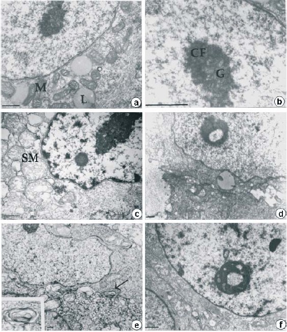
<body><![CDATA[<P align="justify" style="word-spacing: 0; line-height: 100%"><FONT COLOR="#1f1a17" size="2" face="Verdana"> A variety of morphological changes are provoked by hypothermia.&nbsp; </FONT></P>     <P align="justify" style="word-spacing: 0; line-height: 100%"><FONT COLOR="#1f1a17" size="2" face="Verdana"> The ultrastructural findings included controls resembling normal appearance (<a href="#fig1">Fig. 1a,b</a>). In thermally treated samples (4&#176;C,1h and 37&#176;C, 1h; <a href="#fig1"> Fig. 1 c-f</a>) lipid drops were not observed. In contrast, generalized edema (c-f)  dispersed chromatin (c,f), swollen mitochondria (c-f) and autophagic vacuoles  (e) were distinguished giving the appearance of necrotic cells, particularly  in d and e. Ring-shape reorganization was observed (d,f) in the nucleolus  of the thermal treated samples.</FONT></P>     <P align="center" style="word-spacing: 0; line-height: 100%"><a name="fig1"><img border="0" src="/img/fbpe/ic/v51n4/art05fig1.jpg" align="center" width="564" height="654"></a></P>     
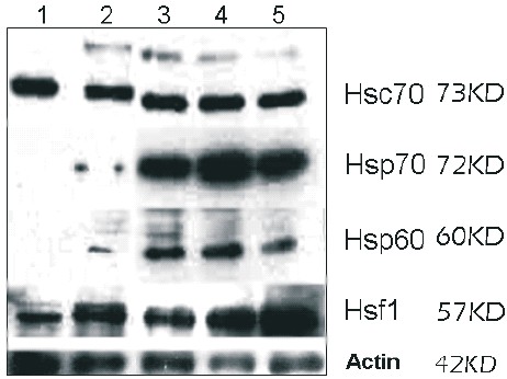
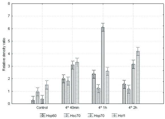
<P align="center" style="word-spacing: 0; line-height: 100%"><font face="Verdana" size="2"><b>Fig. 1.</b> Transmission electron micrographs of human HepG2 cells. Control (a, b) and hypothermically stressed cells with recovery (c-f). There are evident lipids drops (L) and abundant mitochondria in the perinuclear region (M). The typical nucleolus structure with the fibrillar dense (CF) and the granular (G) components is easily distinguished in b. In the cells under hypothermia 4°C during 1h and recovered at 37°C 1h some changes are evident: generalized edema (c-f), picnotic), swollen mitochondria (SM, c-f), as well as autophagic vacuoles (e, arrow and insert) are distinguished. Picnotic nucleus (c,f) were present and in the nucleolus of the thermal treated samples the ring-shape reorganization was observed (d, f). Bar=0, 5ìm.</font></P>     <P align="justify" style="word-spacing: 0; line-height: 100%"><FONT COLOR="#1f1a17" size="2" face="Verdana"> Hypothermia induces Hsp60, the inducible Hsp70 and monomeric Hsf1 but not  Hsc 70 expression. Western blot analysis (<a href="#fig2">Fig. 2</a>) and semiquantitative  densitometry (<a href="#fig3">Fig. 3</a>) showed that Hsp60, Hsp70 and Hsf1 expression was  increased in all treated samples, in contrast to control group. The cells  treated hypothermically during 1h with 1h of recovery, showed the higher  increase of Hsp60 and Hsp70. In the case of Hsf1, the denser band belonged  to the group treated during 2h with 1h of recovery. The lower levels of  expression were observed in Hsc70, while Hsp70 and Hsf1 were the most induced.  As ANOVA showed, no significative changes in Hsc70 were observed between  treatments. In the case of Hsp60, Hsp70 and Hsf1 the relative D.O. difference  was significative to each treated sample compared to the control group  (P&lt;0.05).</FONT></P>     <P align="center" style="word-spacing: 0; line-height: 100%"><a name="fig2"><img border="0" src="/img/fbpe/ic/v51n4/art05fig2.jpg" align="center" width="462" height="346"></a></P>     
<P align="center" style="word-spacing: 0; line-height: 100%"><font face="Verdana" size="2"><b>Fig. 2. </b>Hsc70, Hsp70, Hsp60, Hsf1 and actin expression of hypothermically stressed and recovered human HepG2 cells. Western blot analysis of cells maintained at 37°C (1,2); hypothermically treated 4°C, 40 min (3), 1h (4), 2h (5). Thermal stressed cells were exposed to 37°C, 1h after treatment to rewarming. Relative molecular mass is indicated on the right. Actin was used as a loading control.</font></P>     <P align="center" style="word-spacing: 0; line-height: 100%"><a name="fig3"><img border="0" src="/img/fbpe/ic/v51n4/art05fig3.jpg" align="center" width="580" height="421"></a></P>     
<P align="center" style="word-spacing: 0; line-height: 100%"><font face="Verdana" size="2"><b>Fig. 3.</b> Densitometric analysis of the bands of Hsp60, Hsc70, Hsp70 and Hsf1 in human HepG2 cells hypothermically treated at 4°C during different times as indicated, with one hour of recovery at 37°C. Relative optical density values of each protein compared to actin from Western blot analysis are expressed as mean ± SEM of three independent experiments. The statistical significance of the differences between values was assessed by (ANOVA) followed by Duncan multiple comparison post hoc test. A P&lt;0.05 was considered to be statistically significant. The O.D. showed that Hsp60, Hsp70 and Hsf1 expression was increased in all treated samples, in contrast to control group. The higher increase of Hsp60, Hsp70 was observed in cells hypothermically treated during 1h. To Hsf1 the denser band belonged to the group treated during 2h. The lower levels of expression were observed in Hsc70, while Hsp70 and Hsf1 were the most induced. ANOVA showed no significative changes in Hsc70 between treatments. In the case of Hsp60, Hsp70 and Hsf1 the relative O.D. difference was significative to each treated sample compared to the control group (P&lt;0.05).</font></P>     <P align="justify" style="word-spacing: 0; line-height: 100%"> <B><FONT COLOR="#1f1a17" size="2" face="Verdana"> DISCUSSION&nbsp; </FONT></B> </P>     ]]></body>
<body><![CDATA[<P align="justify" style="word-spacing: 0; line-height: 100%"><FONT COLOR="#1f1a17" size="2" face="Verdana"> This paper reports for the first time monomeric Hsf1 and Hsp60 induction  under hypothermic stress in HepG2 cells, and corroborates the accumulation  of monomeric Hsf1, as has been reported in mice (14). Moreover, Hsp60 has  also been reported in rat brown adipose tissue under whole body hypothermia  (13)&nbsp; </FONT></P>     <P align="justify" style="word-spacing: 0; line-height: 100%"><FONT COLOR="#1f1a17" size="2" face="Verdana"> The increased expression of the heat shock proteins is possibly a natural  protective and homeostatic response of the cell to the deleterious effects  of the hypothermic stress. This response has been associated with the decrease  in the rates of enzymatic processes, enhancement of free radical production  and protein unfolding and misfolding promoted by hypothermia (15). The  differential accumulation of Hsp60 and the inducible form of Hsp70 in all  the stress conditions studied could be related to the assistant in the  appropriate folding of denatured or newly synthesized peptides (16).&nbsp; </FONT></P>     <P align="justify" style="word-spacing: 0; line-height: 100%"><FONT COLOR="#1f1a17" size="2" face="Verdana"> Likewise, it has been suggested that Hsps could be involved in tumorigenesis  and to be essential in the survival of different cancer cells, possibly  by the modulation of the activity of proteins involved in cell cycle and  apoptosis (17, 18), as these proteins are frequently up-regulated in that  kind of cells, as in hepatocellular carcinoma, (19). In this regard, Hsf1  itself is involved in overriding cell-cycle checkpoints and enhanced metastasis  not dependent on Hsps (20, 21), as well downstream products of Hsf1, including  Hsp70 and 27, are related to the increase of the invasive and/or metastatic  capacity of tumors (22). Hsp90 has been involved in maintaining the quality  of proteins in breast and prostate cancer (23). Cooperative promotion as  a coordinated response of Hsp expression might represent one of the mechanisms  involved in the protective effect of hypothermia widely used in medical  strategies and preservation of biological samples (24), possibly due to  thermotolerance.&nbsp; </FONT></P>     <P align="justify" style="word-spacing: 0; line-height: 100%"><FONT COLOR="#1f1a17" size="2" face="Verdana"> Even though cold exposure without rewarming is related with decreased expression  of several Hsps (25), the cytoprotective function of Hsp70 is associated  with its anti-apoptotic effect at the rewarming, instead of during stress  (26). Indeed, it is also understood that a general mechanism still unknown  is involved in the regulation of Hsf1 activity during rewarming in response  to cold stress in various species (11, 14). This mechanism could take place  towards the activation of several transcription factors without its phosphorylation,  while in hyperthermia occurs by the unique activation of Hsf1 preceded  by its hyperphosphorylation (14). Additionally, in response to heat or  cold stress an alternative splicing of the Hsf pre-mRNA has been identified  (11). These observations suggest that the differences in Hsps expression  in hypothermia or hyperthermia conditions are possibly related with the  induction mechanisms.&nbsp; </FONT></P>     <P align="justify" style="word-spacing: 0; line-height: 100%"><FONT COLOR="#1f1a17" size="2" face="Verdana"> The response to hypothermic stress has also been studied in HepG2 cells  by Ohsaka et al. &nbsp;(26, 27). These have identified the induction of mitogen  and stress activated protein kinases, which are closely related with the  mechanisms of signaling implicated in cell repair and survival. It has  been also suggested that the increasing of activity of antioxidants mechanisms  such as the synthesis of catalase, superoxide dismutase and hemo-oxigenase  1 are involved in the response to hypothermia (28), together with the synthesis  of other glycine-rich polypeptides of unknown function (27).&nbsp; </FONT></P>     <P align="justify" style="word-spacing: 0; line-height: 100%"><FONT COLOR="#1f1a17" size="2" face="Verdana"> The possible explanation of the morphological changes has been related  with the effect of the temperature in the molecules and changes in the  rates of metabolic processes. The distribution of cell material is consistent  with the changes in the cytoskeleton provoked by the hypothermia (3) revealed  in the retraction of the material of the nucleus and nucleolar reorganization.  The proteins of the nuclear matrix has been recognized as the most thermal  labile proteins in the cell (29).&nbsp; </FONT></P> <MULTICOL GUTTER="31" COLS="2">     <P align="justify" style="word-spacing: 0; line-height: 100%"><FONT COLOR="#1f1a17" size="2" face="Verdana"> The aggregation of the material around the nucleus in hepatocytes exposed  to hypothermia has been described in non tumoral hepatic cells (5). The  retraction of material to the nucleus and the ring-shape nucleolar reorganization,  and associated severe cell damage has been recognized in different pathologies  in a variety of biological systems (30). In hypothermia, it has been also  possible the recognition of nucleolar components segregation in the ring-shape  structure. In depth this has been interpreted as a consequence of the disengagement  between transcription and protein processing. Hsp70 is associated with  transcription stabilization (31), its detection in the nucleolus could  be related with those processes (30).&nbsp; </FONT></P>     <P align="justify" style="word-spacing: 0; line-height: 100%"><FONT COLOR="#1f1a17" size="2" face="Verdana"> The production of oxygen radicals, ATP depletion and the increased Ca<FONT COLOR="#1f1a17" FACE="Caslon224 Bk BT" SIZE="1"><SUP>2+</SUP></FONT></FONT><font face="Verdana" size="2">concentration  are among the mechanisms associated with the hypothermia-induced morphological  changes in the mitochondria after rewarming (32, 33). The swollen and roundshaped  appearance of the mitochondria showed here, has been also described in  proximal tubular cells (33), together with the generalized cell swelling  in glial cells (34).&nbsp;</font></P>     <P align="justify" style="word-spacing: 0; line-height: 100%"><FONT COLOR="#1f1a17" size="2" face="Verdana"> Synergistic promotion as a coordinated response of Hsp expression might  represent one of the mechanisms involved in the protective effect of hypothermia  widely used in medical strategies and preservation of biological samples.  Cold stress could be an alternative to thermal stress, capable of inducting  Hsp expression, by an unknown mechanism that needs to be determined.&nbsp; </FONT></P>     <P align="justify" style="word-spacing: 0; line-height: 100%"> <B><FONT COLOR="#1f1a17" size="2" face="Verdana"> ACKNOWLEDGEMENTS&nbsp; </FONT></B> </P>     ]]></body>
<body><![CDATA[<P align="justify" style="word-spacing: 0; line-height: 100%"><FONT COLOR="#1f1a17" size="2" face="Verdana"> This work has been supported by the Consejo de Desarrollo Cient&#237;fico y  Human&#237;stico (CDCH) from the Universidad Central de Venezuela and Laboratorios  Elmor S.A. We thank Dr. Antonio De Maio who kindly provided HepG2 cell  line, Dr. Tom&#225;s Hermoso and Lic. Mar&#237;a Narv&#225;ez for the anti-Hsf1 antibody  and Mr. Ra&#250;l Colina for his excellent technical assistance.&nbsp; </FONT></P>     <P align="justify" style="word-spacing: 0; line-height: 100%"> <B><FONT COLOR="#1f1a17" size="2" face="Verdana"> REFERENCES&nbsp; </FONT></B> </P>     <!-- ref --><P align="justify" style="word-spacing: 0; line-height: 100%"><FONT COLOR="#1f1a17" size="2" face="Verdana"> 1.&nbsp;</FONT><font size="2"><FONT COLOR="#1f1a17" face="Verdana">Sonna L, Fujita J, Gaffin S, Lilly C. Effects of heat and cold stress on  mammalian gene expression. J Appl Physiol 2002; 92:1725-1742. &nbsp;</FONT></font>&nbsp;&nbsp;&nbsp;&nbsp;&nbsp;&nbsp;&nbsp;&nbsp;[&#160;<a href="javascript:void(0);" onclick="javascript: window.open('/scielo.php?script=sci_nlinks&ref=1187844&pid=S0535-5133201000040000500001&lng=','','width=640,height=500,resizable=yes,scrollbars=1,menubar=yes,');">Links</a>&#160;]<!-- end-ref --><!-- ref --><P align="justify" style="word-spacing: 0; line-height: 100%"><FONT COLOR="#1f1a17" size="2" face="Verdana"> 2.&nbsp;</FONT><font size="2"><FONT COLOR="#1f1a17" face="Verdana">Lepock J. How do cells respond to their thermal environment?. Int J Hyperthermia  2005; 21: 681-687.&nbsp; </FONT></font>&nbsp;&nbsp;&nbsp;&nbsp;&nbsp;&nbsp;&nbsp;&nbsp;[&#160;<a href="javascript:void(0);" onclick="javascript: window.open('/scielo.php?script=sci_nlinks&ref=1187845&pid=S0535-5133201000040000500002&lng=','','width=640,height=500,resizable=yes,scrollbars=1,menubar=yes,');">Links</a>&#160;]<!-- end-ref --><!-- ref --><P align="justify" style="word-spacing: 0; line-height: 100%"><FONT COLOR="#1f1a17" size="2" face="Verdana"> 3.&nbsp;</FONT><font size="2"><FONT COLOR="#1f1a17" face="Verdana">Welch W. Mammalian stress response: cell physiology, structure/function  of stress proteins, and implications for medicine and disease. Physiol  Rev 1992; 72:1063-1081.&nbsp; </FONT></font>&nbsp;&nbsp;&nbsp;&nbsp;&nbsp;&nbsp;&nbsp;&nbsp;[&#160;<a href="javascript:void(0);" onclick="javascript: window.open('/scielo.php?script=sci_nlinks&ref=1187846&pid=S0535-5133201000040000500003&lng=','','width=640,height=500,resizable=yes,scrollbars=1,menubar=yes,');">Links</a>&#160;]<!-- end-ref --><!-- ref --><P align="justify" style="word-spacing: 0; line-height: 100%"><FONT COLOR="#1f1a17" size="2" face="Verdana"> 4.&nbsp;</FONT><font size="2"><FONT COLOR="#1f1a17" face="Verdana">Holland D, Roberts S, Wood E, Cunliffe W. Cold shock induces the synthesis  of stress proteins in human keratinocytes. J Inv Dermatol 1993; 101:196-199.&nbsp; </FONT></font>&nbsp;&nbsp;&nbsp;&nbsp;&nbsp;&nbsp;&nbsp;&nbsp;[&#160;<a href="javascript:void(0);" onclick="javascript: window.open('/scielo.php?script=sci_nlinks&ref=1187847&pid=S0535-5133201000040000500004&lng=','','width=640,height=500,resizable=yes,scrollbars=1,menubar=yes,');">Links</a>&#160;]<!-- end-ref --><!-- ref --><P align="justify" style="word-spacing: 0; line-height: 100%"><FONT COLOR="#1f1a17" size="2" face="Verdana"> 5.&nbsp;</FONT><font size="2"><FONT COLOR="#1f1a17" face="Verdana">Griffiths N, Evans P. Ultraestructural changes in hypothermically preserved  hepatocytes. Cryobiology 2000; 40:176-181.&nbsp; </FONT></font>&nbsp;&nbsp;&nbsp;&nbsp;&nbsp;&nbsp;&nbsp;&nbsp;[&#160;<a href="javascript:void(0);" onclick="javascript: window.open('/scielo.php?script=sci_nlinks&ref=1187848&pid=S0535-5133201000040000500005&lng=','','width=640,height=500,resizable=yes,scrollbars=1,menubar=yes,');">Links</a>&#160;]<!-- end-ref --><!-- ref --><P align="justify" style="word-spacing: 0; line-height: 100%"><FONT COLOR="#1f1a17" size="2" face="Verdana"> 6.&nbsp;</FONT><font size="2"><FONT COLOR="#1f1a17" face="Verdana">Stefanovich P, Ezzel M, Sheehan S, Tompkins R, Yarmush M, Toner M. Effects  of hypothermia on the function, membrane integrity and cytoeskeletal structure  of hepatocytes. Cryobiology 1995; 32:389-403.&nbsp; </FONT></font>&nbsp;&nbsp;&nbsp;&nbsp;&nbsp;&nbsp;&nbsp;&nbsp;[&#160;<a href="javascript:void(0);" onclick="javascript: window.open('/scielo.php?script=sci_nlinks&ref=1187849&pid=S0535-5133201000040000500006&lng=','','width=640,height=500,resizable=yes,scrollbars=1,menubar=yes,');">Links</a>&#160;]<!-- end-ref --><!-- ref --><P align="justify" style="word-spacing: 0; line-height: 100%"><FONT COLOR="#1f1a17" size="2" face="Verdana"> 7.&nbsp;</FONT><font size="2"><FONT COLOR="#1f1a17" face="Verdana">Zeng X, Bhasin S, Wu X, Lee J, Maffi S, Nichols C, Lee K, Taylor J, Greene  L, Eisenberg E. Hsp70 dynamics in vivo: effect of heat shock and protein  aggregation. J Cell Sci 2004; 117: 4991-5000.&nbsp; </FONT></font>&nbsp;&nbsp;&nbsp;&nbsp;&nbsp;&nbsp;&nbsp;&nbsp;[&#160;<a href="javascript:void(0);" onclick="javascript: window.open('/scielo.php?script=sci_nlinks&ref=1187850&pid=S0535-5133201000040000500007&lng=','','width=640,height=500,resizable=yes,scrollbars=1,menubar=yes,');">Links</a>&#160;]<!-- end-ref --><!-- ref --><P align="justify" style="word-spacing: 0; line-height: 100%"><FONT COLOR="#1f1a17" size="2" face="Verdana"> 8.&nbsp;</FONT><font size="2"><FONT COLOR="#1f1a17" face="Verdana">Baler R, Dahl G, Voellmy R. Activation of human heat shock genes is accompanied  by oligomerization, modification and rapid translocation of heat shock  transcription factor HSF1. Mol Cell Biol 1993; 13: 2486-2496.</FONT></font>&nbsp;&nbsp;&nbsp;&nbsp;&nbsp;&nbsp;&nbsp;&nbsp;[&#160;<a href="javascript:void(0);" onclick="javascript: window.open('/scielo.php?script=sci_nlinks&ref=1187851&pid=S0535-5133201000040000500008&lng=','','width=640,height=500,resizable=yes,scrollbars=1,menubar=yes,');">Links</a>&#160;]<!-- end-ref --><!-- ref --><P align="justify" style="word-spacing: 0; line-height: 100%"><FONT COLOR="#1f1a17" size="2" face="Verdana"> 9.&nbsp;</FONT><font size="2"><FONT COLOR="#1f1a17" face="Verdana">Sarge K, Murphy S, Morimoto R. Activation of heat shock gene transcription  by heat shock factor 1 involves oligomerization, acquisition of DNA-binding  activity, and nuclear localization and can occur in the absence of stress.  Mol Cell Biol 1993; 13:1392-1407.&nbsp; </FONT></font>&nbsp;&nbsp;&nbsp;&nbsp;&nbsp;&nbsp;&nbsp;&nbsp;[&#160;<a href="javascript:void(0);" onclick="javascript: window.open('/scielo.php?script=sci_nlinks&ref=1187852&pid=S0535-5133201000040000500009&lng=','','width=640,height=500,resizable=yes,scrollbars=1,menubar=yes,');">Links</a>&#160;]<!-- end-ref --><!-- ref --><P align="justify" style="word-spacing: 0; line-height: 100%"><FONT COLOR="#1f1a17" size="2" face="Verdana"> 10.&nbsp;</FONT><font size="2"><FONT COLOR="#1f1a17" face="Verdana">Ruell P, Thompson M, Hoffman K. Heat shock proteins as an aid in the treatment  and diagnosis of heat stroke. J Thermal Biol 2009; 34:1-7.&nbsp; </FONT></font>&nbsp;&nbsp;&nbsp;&nbsp;&nbsp;&nbsp;&nbsp;&nbsp;[&#160;<a href="javascript:void(0);" onclick="javascript: window.open('/scielo.php?script=sci_nlinks&ref=1187853&pid=S0535-5133201000040000500010&lng=','','width=640,height=500,resizable=yes,scrollbars=1,menubar=yes,');">Links</a>&#160;]<!-- end-ref --><!-- ref --><P align="justify" style="word-spacing: 0; line-height: 100%"><FONT COLOR="#1f1a17" size="2" face="Verdana"> 11.&nbsp;</FONT><font size="2"><FONT COLOR="#1f1a17" face="Verdana">Fujikake N, Nagai Y, Popiel H, Kano H, Yamaguchi M, Toda T. Alternative  splicing regulates the transcriptional activity of Drosophila heat shock  transcription factor in response to heat/cold stress. FEBS Lett 2005; 579:3842-3848.&nbsp; </FONT></font>&nbsp;&nbsp;&nbsp;&nbsp;&nbsp;&nbsp;&nbsp;&nbsp;[&#160;<a href="javascript:void(0);" onclick="javascript: window.open('/scielo.php?script=sci_nlinks&ref=1187854&pid=S0535-5133201000040000500011&lng=','','width=640,height=500,resizable=yes,scrollbars=1,menubar=yes,');">Links</a>&#160;]<!-- end-ref --><!-- ref --><P align="justify" style="word-spacing: 0; line-height: 100%"><FONT COLOR="#1f1a17" size="2" face="Verdana"> 12.&nbsp;</FONT><font size="2"><FONT COLOR="#1f1a17" face="Verdana">Rada A, Tonino P, Anselmi G, Strauss M. Is hypothermia a stress condition  in HepG2 cells? Expression and localization of Hsp70 in human hepatoma  cell line. Tissue Cell 2005; 37: 59-65.&nbsp; </FONT></font>&nbsp;&nbsp;&nbsp;&nbsp;&nbsp;&nbsp;&nbsp;&nbsp;[&#160;<a href="javascript:void(0);" onclick="javascript: window.open('/scielo.php?script=sci_nlinks&ref=1187855&pid=S0535-5133201000040000500012&lng=','','width=640,height=500,resizable=yes,scrollbars=1,menubar=yes,');">Links</a>&#160;]<!-- end-ref --><!-- ref --><P align="justify" style="word-spacing: 0; line-height: 100%"><FONT COLOR="#1f1a17" size="2" face="Verdana"> 13.&nbsp;</FONT><font size="2"><FONT COLOR="#1f1a17" face="Verdana">Matz J, Blake M, Tatelman H, Lavoi K, Holbrook N. Characterization and  regulation of cold-induced heat shock protein expression in mouse brown  adipose tissue. Am J Physiol 1995; 269:R38-R47. </FONT></font>&nbsp;&nbsp;&nbsp;&nbsp;&nbsp;&nbsp;&nbsp;&nbsp;[&#160;<a href="javascript:void(0);" onclick="javascript: window.open('/scielo.php?script=sci_nlinks&ref=1187856&pid=S0535-5133201000040000500013&lng=','','width=640,height=500,resizable=yes,scrollbars=1,menubar=yes,');">Links</a>&#160;]<!-- end-ref --><!-- ref --><P align="justify" style="word-spacing: 0; line-height: 100%"><FONT COLOR="#1f1a17" size="2" face="Verdana">14.&nbsp;</FONT><font size="2"><FONT COLOR="#1f1a17" face="Verdana">Cullen K, Sarge N. Characterization of hypothermia-induced cellular stress  response in mouse tissues. J Biol Chem 1997; 272: 1742-1746.&nbsp; </FONT></font>&nbsp;&nbsp;&nbsp;&nbsp;&nbsp;&nbsp;&nbsp;&nbsp;[&#160;<a href="javascript:void(0);" onclick="javascript: window.open('/scielo.php?script=sci_nlinks&ref=1187857&pid=S0535-5133201000040000500014&lng=','','width=640,height=500,resizable=yes,scrollbars=1,menubar=yes,');">Links</a>&#160;]<!-- end-ref --><!-- ref --><P align="justify" style="word-spacing: 0; line-height: 100%"><FONT COLOR="#1f1a17" size="2" face="Verdana"> 15.&nbsp;</FONT><font size="2"><FONT COLOR="#1f1a17" face="Verdana">Mestril R. Stress protein involvement in cardioprotection induced by hypothermia.  J Mol Cell Cardiol 2001; 33:2075-2078.&nbsp; </FONT></font>&nbsp;&nbsp;&nbsp;&nbsp;&nbsp;&nbsp;&nbsp;&nbsp;[&#160;<a href="javascript:void(0);" onclick="javascript: window.open('/scielo.php?script=sci_nlinks&ref=1187858&pid=S0535-5133201000040000500015&lng=','','width=640,height=500,resizable=yes,scrollbars=1,menubar=yes,');">Links</a>&#160;]<!-- end-ref --><!-- ref --><P align="justify" style="word-spacing: 0; line-height: 100%"><FONT COLOR="#1f1a17" size="2" face="Verdana"> 16.&nbsp;</FONT><font size="2"><FONT COLOR="#1f1a17" face="Verdana">Young J, Agashe V, Siegers K, Hartl F. Pathways of chaperone-mediated protein  folding in the cytosol. Nat Rev Mol Cell Biol 2004; 5:781-791.&nbsp; </FONT></font>&nbsp;&nbsp;&nbsp;&nbsp;&nbsp;&nbsp;&nbsp;&nbsp;[&#160;<a href="javascript:void(0);" onclick="javascript: window.open('/scielo.php?script=sci_nlinks&ref=1187859&pid=S0535-5133201000040000500016&lng=','','width=640,height=500,resizable=yes,scrollbars=1,menubar=yes,');">Links</a>&#160;]<!-- end-ref --><!-- ref --><P align="justify" style="word-spacing: 0; line-height: 100%"><FONT COLOR="#1f1a17" size="2" face="Verdana"> 17.&nbsp;</FONT><font size="2"><FONT COLOR="#1f1a17" face="Verdana">Garrido C, Gurbuxani S, Ravagnan L, Kroemer G. Heat shock proteins: endogenous  modulators of apoptotic cell&nbsp;death. Biochem Biophys Res Commun 2001; 286:433-442.&nbsp; </FONT></font>&nbsp;&nbsp;&nbsp;&nbsp;&nbsp;&nbsp;&nbsp;&nbsp;[&#160;<a href="javascript:void(0);" onclick="javascript: window.open('/scielo.php?script=sci_nlinks&ref=1187860&pid=S0535-5133201000040000500017&lng=','','width=640,height=500,resizable=yes,scrollbars=1,menubar=yes,');">Links</a>&#160;]<!-- end-ref --><!-- ref --><P align="justify" style="word-spacing: 0; line-height: 100%"><FONT COLOR="#1f1a17" size="2" face="Verdana"> 18.&nbsp;</FONT><font size="2"><FONT COLOR="#1f1a17" face="Verdana">Calderwood SK, Khaleque A, Sawyer D, Ciocca Daniel R. Heat shock proteins  in cancer: chaperones of tumorigenesis. Trends Biochem Sci 2006; 31:164-172.&nbsp; </FONT></font>&nbsp;&nbsp;&nbsp;&nbsp;&nbsp;&nbsp;&nbsp;&nbsp;[&#160;<a href="javascript:void(0);" onclick="javascript: window.open('/scielo.php?script=sci_nlinks&ref=1187861&pid=S0535-5133201000040000500018&lng=','','width=640,height=500,resizable=yes,scrollbars=1,menubar=yes,');">Links</a>&#160;]<!-- end-ref --><!-- ref --><P align="justify" style="word-spacing: 0; line-height: 100%"><FONT COLOR="#1f1a17" size="2" face="Verdana"> 19.&nbsp;</FONT><font size="2"><FONT COLOR="#1f1a17" face="Verdana">Lim S, Park S, Yoo J, Park Y, Kim H, Jang K, Cho J, Yoo B, Jung G, Park  C. Expression of heat shock proteins (HSP27, HSP60, HSP70, HSP90, GRP78,  GRP94) in hepatitis B virus-related hepatocellular carcinomas and dysplastic  nodules. World J Gastroenterol 2005; 11:2072-2079.&nbsp; </FONT></font>&nbsp;&nbsp;&nbsp;&nbsp;&nbsp;&nbsp;&nbsp;&nbsp;[&#160;<a href="javascript:void(0);" onclick="javascript: window.open('/scielo.php?script=sci_nlinks&ref=1187862&pid=S0535-5133201000040000500019&lng=','','width=640,height=500,resizable=yes,scrollbars=1,menubar=yes,');">Links</a>&#160;]<!-- end-ref --><!-- ref --><P align="justify" style="word-spacing: 0; line-height: 100%"><FONT COLOR="#1f1a17" size="2" face="Verdana"> 20.&nbsp;</FONT><font size="2"><FONT COLOR="#1f1a17" face="Verdana">Hoang AT, Huang J, Rudra-Ganguly N, Zheng J, Powell WC, Rabindran SK, Wu  C, Roy-Burma P. A novel association between the human heat shock transcription  factor 1 (HSF1) and prostate adenocarcinoma. Am J Pathol 2000; 156:857-864.&nbsp; </FONT></font>&nbsp;&nbsp;&nbsp;&nbsp;&nbsp;&nbsp;&nbsp;&nbsp;[&#160;<a href="javascript:void(0);" onclick="javascript: window.open('/scielo.php?script=sci_nlinks&ref=1187863&pid=S0535-5133201000040000500020&lng=','','width=640,height=500,resizable=yes,scrollbars=1,menubar=yes,');">Links</a>&#160;]<!-- end-ref --><!-- ref --><P align="justify" style="word-spacing: 0; line-height: 100%"><FONT COLOR="#1f1a17" size="2" face="Verdana"> 21.&nbsp;</FONT><font size="2"><FONT COLOR="#1f1a17" face="Verdana">Wang Y, Theriault J, He H, Gong J, Calderwood S. Expression of a dominant  negative heat shock factor-1 construct inhibits aneuploidy in prostate  carcinoma cells. J Biol Chem 2004; 279: 32651-32659.&nbsp;</FONT></font>&nbsp;&nbsp;&nbsp;&nbsp;&nbsp;&nbsp;&nbsp;&nbsp;[&#160;<a href="javascript:void(0);" onclick="javascript: window.open('/scielo.php?script=sci_nlinks&ref=1187864&pid=S0535-5133201000040000500021&lng=','','width=640,height=500,resizable=yes,scrollbars=1,menubar=yes,');">Links</a>&#160;]<!-- end-ref --><!-- ref --><P align="justify" style="word-spacing: 0; line-height: 100%"><FONT COLOR="#1f1a17" size="2" face="Verdana"> 22.&nbsp;</FONT><font size="2"><FONT COLOR="#1f1a17" face="Verdana">Ciocca D, Calderwood S. Heat shock proteins in cancer: diagnostic, prognostic,  predictive, and treatment implications. Cell Stress Chaperones 2005; 10:86-103.&nbsp; </FONT></font>&nbsp;&nbsp;&nbsp;&nbsp;&nbsp;&nbsp;&nbsp;&nbsp;[&#160;<a href="javascript:void(0);" onclick="javascript: window.open('/scielo.php?script=sci_nlinks&ref=1187865&pid=S0535-5133201000040000500022&lng=','','width=640,height=500,resizable=yes,scrollbars=1,menubar=yes,');">Links</a>&#160;]<!-- end-ref --><!-- ref --><P align="justify" style="word-spacing: 0; line-height: 100%"><FONT COLOR="#1f1a17" size="2" face="Verdana"> 23.&nbsp;</FONT><font size="2"><FONT COLOR="#1f1a17" face="Verdana">Isaacs JS, Xu W, Neckers L. Heat shock protein 90 as a molecular target  for cancer therapeutics. Cancer Cell 2003;&nbsp;3:213-217.&nbsp; </FONT></font>&nbsp;&nbsp;&nbsp;&nbsp;&nbsp;&nbsp;&nbsp;&nbsp;[&#160;<a href="javascript:void(0);" onclick="javascript: window.open('/scielo.php?script=sci_nlinks&ref=1187866&pid=S0535-5133201000040000500023&lng=','','width=640,height=500,resizable=yes,scrollbars=1,menubar=yes,');">Links</a>&#160;]<!-- end-ref --><!-- ref --><p align="justify" style="word-spacing: 0; line-height: 100%"><span lang="EN-US" style="font-size:10.0pt;font-family:Verdana;color:#1F1A17; mso-ansi-language:EN-US">24.&nbsp;Nagao S, Irie K, Kawai N, Nakamura T, Kunishio K, Matsumoto Y. The use of mild hypothermia for patients with severe vasospasm: a preliminary report. </span><span lang="FR" style="font-size:10.0pt;font-family: Verdana;color:#1F1A17;mso-ansi-language:FR">J Clin Neurosci 2003; 10: 208-212.&nbsp; </span><span lang="FR" style="mso-ansi-language:FR"><o:p> </o:p> </span>&nbsp;&nbsp;&nbsp;&nbsp;&nbsp;&nbsp;&nbsp;&nbsp;[&#160;<a href="javascript:void(0);" onclick="javascript: window.open('/scielo.php?script=sci_nlinks&ref=1187867&pid=S0535-5133201000040000500024&lng=','','width=640,height=500,resizable=yes,scrollbars=1,menubar=yes,');">Links</a>&#160;]<!-- end-ref --><!-- ref --><P align="justify" style="word-spacing: 0; line-height: 100%"><font face="Verdana" size="2"> 25.&nbsp;Sonna L, Kuhlmeier M, Carter H, Hasday J, Lilly C, Fairchild K. Effect  of moderate hypothermia on gene expression by THP-1 cells: a DNA microarray  study. Physiol Genomics 2006; 16:91-98. &nbsp;</font>&nbsp;&nbsp;&nbsp;&nbsp;&nbsp;&nbsp;&nbsp;&nbsp;[&#160;<a href="javascript:void(0);" onclick="javascript: window.open('/scielo.php?script=sci_nlinks&ref=1187868&pid=S0535-5133201000040000500025&lng=','','width=640,height=500,resizable=yes,scrollbars=1,menubar=yes,');">Links</a>&#160;]<!-- end-ref --><!-- ref --><P align="justify" style="word-spacing: 0; line-height: 100%"><FONT COLOR="#1f1a17" size="2" face="Verdana"> 26.&nbsp;</FONT><font size="2"><FONT COLOR="#1f1a17" face="Verdana">Kao Y, Goto S, Jawan B, Nakano T, Hsu L, Lin Y, Pan M, Lai C, Sun Ch, Cheng  Y, Tai M, Huang H, Chen Ch. Heat preconditioning ameliorates hepatocyte  viability after cold preservation and rewarming, and modulates its immunoactivity.  Transplant Immunol 2008; 18: 220-231.&nbsp;</FONT></font>&nbsp;&nbsp;&nbsp;&nbsp;&nbsp;&nbsp;&nbsp;&nbsp;[&#160;<a href="javascript:void(0);" onclick="javascript: window.open('/scielo.php?script=sci_nlinks&ref=1187869&pid=S0535-5133201000040000500026&lng=','','width=640,height=500,resizable=yes,scrollbars=1,menubar=yes,');">Links</a>&#160;]<!-- end-ref --><!-- ref --><P align="justify" style="word-spacing: 0; line-height: 100%"><FONT COLOR="#1f1a17" size="2" face="Verdana"> 27.&nbsp;</FONT><font size="2"><FONT COLOR="#1f1a17" face="Verdana">Ohsaka Y, Ohgiya S, Hoshino T, Ishizaki K. Mitochondrial genome-encoded  ATPase subunit 6+8 mRNA increases in human hepatoblastoma cells in response  to nonfatal cold stress. Cryobiology 2000; 40: 92-10.&nbsp; </FONT></font>&nbsp;&nbsp;&nbsp;&nbsp;&nbsp;&nbsp;&nbsp;&nbsp;[&#160;<a href="javascript:void(0);" onclick="javascript: window.open('/scielo.php?script=sci_nlinks&ref=1187870&pid=S0535-5133201000040000500027&lng=','','width=640,height=500,resizable=yes,scrollbars=1,menubar=yes,');">Links</a>&#160;]<!-- end-ref --><!-- ref --><P align="justify" style="word-spacing: 0; line-height: 100%"><FONT COLOR="#1f1a17" size="2" face="Verdana"> 28.&nbsp;</FONT><font size="2"><FONT COLOR="#1f1a17" face="Verdana">Matsumoto K, Honda K, Kobayashi N. Protective effect of heat preconditioning  of rat liver graft resulting in improved transplant survival. Transplantation  2001; 71:862-868.&nbsp; </FONT></font>&nbsp;&nbsp;&nbsp;&nbsp;&nbsp;&nbsp;&nbsp;&nbsp;[&#160;<a href="javascript:void(0);" onclick="javascript: window.open('/scielo.php?script=sci_nlinks&ref=1187871&pid=S0535-5133201000040000500028&lng=','','width=640,height=500,resizable=yes,scrollbars=1,menubar=yes,');">Links</a>&#160;]<!-- end-ref --><!-- ref --><P align="justify" style="word-spacing: 0; line-height: 100%"><FONT COLOR="#1f1a17" size="2" face="Verdana"> 29.&nbsp;</FONT><font size="2"><FONT COLOR="#1f1a17" face="Verdana">Roti Roti J, Kampinga H, Malyapa R, Wright W, Van der Waal R, Xu M. Nuclear  matrix as a target for hyperthermic killing of cancer cells. Cell Stress  Chaperones. 1998; 3:245-255.&nbsp; </FONT></font>&nbsp;&nbsp;&nbsp;&nbsp;&nbsp;&nbsp;&nbsp;&nbsp;[&#160;<a href="javascript:void(0);" onclick="javascript: window.open('/scielo.php?script=sci_nlinks&ref=1187872&pid=S0535-5133201000040000500029&lng=','','width=640,height=500,resizable=yes,scrollbars=1,menubar=yes,');">Links</a>&#160;]<!-- end-ref --><!-- ref --><P align="justify" style="word-spacing: 0; line-height: 100%"><FONT COLOR="#1f1a17" size="2" face="Verdana"> 30.&nbsp;</FONT><font size="2"><FONT COLOR="#1f1a17" face="Verdana">Morcillo G, Gorab E, Tanguay R, Diez J. Specific intranucleolar distribution  of Hsp70 during heat shock in polytene cells. Exp Cell Res 1997; 1:361-370.&nbsp; </FONT></font>&nbsp;&nbsp;&nbsp;&nbsp;&nbsp;&nbsp;&nbsp;&nbsp;[&#160;<a href="javascript:void(0);" onclick="javascript: window.open('/scielo.php?script=sci_nlinks&ref=1187873&pid=S0535-5133201000040000500030&lng=','','width=640,height=500,resizable=yes,scrollbars=1,menubar=yes,');">Links</a>&#160;]<!-- end-ref --><!-- ref --><P align="justify" style="word-spacing: 0; line-height: 100%"><FONT COLOR="#1f1a17" size="2" face="Verdana"> 31.&nbsp;</FONT><font size="2"><FONT COLOR="#1f1a17" face="Verdana">Beck S, De Maio A. Stabilization of protein synthesis in thermotolerant  cells during heat shock. Association of heat shock protein -72 with ribosomal  subunits of polysomes. J Biol Chem 1994; 269:21803-21811.&nbsp; </FONT></font>&nbsp;&nbsp;&nbsp;&nbsp;&nbsp;&nbsp;&nbsp;&nbsp;[&#160;<a href="javascript:void(0);" onclick="javascript: window.open('/scielo.php?script=sci_nlinks&ref=1187874&pid=S0535-5133201000040000500031&lng=','','width=640,height=500,resizable=yes,scrollbars=1,menubar=yes,');">Links</a>&#160;]<!-- end-ref --><!-- ref --><P align="justify" style="word-spacing: 0; line-height: 100%"><FONT COLOR="#1f1a17" size="2" face="Verdana"> 32.&nbsp;</FONT><font size="2"><FONT COLOR="#1f1a17" face="Verdana">Brinkkoetter P, Song H, L&#246;sel R, Schnetzke U, Gottmann U, Feng Y, Hanusch  C, Beck G, Schnuelle P, Wehling M, van der Woude F, Yard B. Hypothermic  injury: the mitochondrial calcium, ATP and ROS love-hate triangle out of  balance. Cell Physiol Biochem 2008; 22:195-204.&nbsp; </FONT></font>&nbsp;&nbsp;&nbsp;&nbsp;&nbsp;&nbsp;&nbsp;&nbsp;[&#160;<a href="javascript:void(0);" onclick="javascript: window.open('/scielo.php?script=sci_nlinks&ref=1187875&pid=S0535-5133201000040000500032&lng=','','width=640,height=500,resizable=yes,scrollbars=1,menubar=yes,');">Links</a>&#160;]<!-- end-ref --><!-- ref --><P align="justify" style="word-spacing: 0; line-height: 100%"><FONT COLOR="#1f1a17" size="2" face="Verdana"> 33.&nbsp;</FONT><font size="2"><FONT COLOR="#1f1a17" face="Verdana">Bartels-Stringer M, Kramers C, Wetzels J, Russel F, Groot H, Rauen U. Hypothermia  causes a marked injury to rat proximal tubular cells that is aggravated  by all currently used preservation solutions. Cryobiology 2003; 47:82-91.&nbsp; </FONT></font>&nbsp;&nbsp;&nbsp;&nbsp;&nbsp;&nbsp;&nbsp;&nbsp;[&#160;<a href="javascript:void(0);" onclick="javascript: window.open('/scielo.php?script=sci_nlinks&ref=1187876&pid=S0535-5133201000040000500033&lng=','','width=640,height=500,resizable=yes,scrollbars=1,menubar=yes,');">Links</a>&#160;]<!-- end-ref --><!-- ref --><P align="justify" style="word-spacing: 0; line-height: 100%"><FONT COLOR="#1f1a17" size="2" face="Verdana"> 34.&nbsp;</FONT><font size="2"><FONT COLOR="#1f1a17" face="Verdana">Healy D, Daly P, Docherty N, Murphy M, Fitzpatrick J, Watson R. Heat shock-induced  protection of renal proximal tubular epithelial cells from cold storage  and rewarming injury. J Am Soc Nephrol 2006; 17:805-812.&nbsp; </FONT></font>&nbsp;&nbsp;&nbsp;&nbsp;&nbsp;&nbsp;&nbsp;&nbsp;[&#160;<a href="javascript:void(0);" onclick="javascript: window.open('/scielo.php?script=sci_nlinks&ref=1187877&pid=S0535-5133201000040000500034&lng=','','width=640,height=500,resizable=yes,scrollbars=1,menubar=yes,');">Links</a>&#160;]<!-- end-ref --> ]]></body>
<back>
<ref-list>
<ref id="B1">
<label>1</label><nlm-citation citation-type="journal">
<person-group person-group-type="author">
<name>
<surname><![CDATA[Sonna]]></surname>
<given-names><![CDATA[L]]></given-names>
</name>
<name>
<surname><![CDATA[Fujita]]></surname>
<given-names><![CDATA[J]]></given-names>
</name>
<name>
<surname><![CDATA[Gaffin]]></surname>
<given-names><![CDATA[S]]></given-names>
</name>
<name>
<surname><![CDATA[Lilly]]></surname>
<given-names><![CDATA[C]]></given-names>
</name>
</person-group>
<article-title xml:lang="en"><![CDATA[Effects of heat and cold stress on mammalian gene expression]]></article-title>
<source><![CDATA[J Appl Physiol]]></source>
<year>2002</year>
<volume>92</volume>
<page-range>1725-1742</page-range></nlm-citation>
</ref>
<ref id="B2">
<label>2</label><nlm-citation citation-type="journal">
<person-group person-group-type="author">
<name>
<surname><![CDATA[Lepock]]></surname>
<given-names><![CDATA[J]]></given-names>
</name>
</person-group>
<article-title xml:lang="en"><![CDATA[How do cells respond to their thermal environment?]]></article-title>
<source><![CDATA[Int J Hyperthermia]]></source>
<year>2005</year>
<volume>21</volume>
<page-range>681-687</page-range></nlm-citation>
</ref>
<ref id="B3">
<label>3</label><nlm-citation citation-type="journal">
<person-group person-group-type="author">
<name>
<surname><![CDATA[Welch]]></surname>
<given-names><![CDATA[W]]></given-names>
</name>
</person-group>
<article-title xml:lang="en"><![CDATA[Mammalian stress response: cell physiology, structure/function of stress proteins, and implications for medicine and disease]]></article-title>
<source><![CDATA[Physiol Rev]]></source>
<year>1992</year>
<volume>72</volume>
<page-range>1063-1081</page-range></nlm-citation>
</ref>
<ref id="B4">
<label>4</label><nlm-citation citation-type="journal">
<person-group person-group-type="author">
<name>
<surname><![CDATA[Holland]]></surname>
<given-names><![CDATA[D]]></given-names>
</name>
<name>
<surname><![CDATA[Roberts]]></surname>
<given-names><![CDATA[S]]></given-names>
</name>
<name>
<surname><![CDATA[Wood]]></surname>
<given-names><![CDATA[E]]></given-names>
</name>
<name>
<surname><![CDATA[Cunliffe]]></surname>
<given-names><![CDATA[W]]></given-names>
</name>
</person-group>
<article-title xml:lang="en"><![CDATA[Cold shock induces the synthesis of stress proteins in human keratinocytes]]></article-title>
<source><![CDATA[J Inv Dermatol]]></source>
<year>1993</year>
<volume>101</volume>
<page-range>196-199</page-range></nlm-citation>
</ref>
<ref id="B5">
<label>5</label><nlm-citation citation-type="journal">
<person-group person-group-type="author">
<name>
<surname><![CDATA[Griffiths]]></surname>
<given-names><![CDATA[N]]></given-names>
</name>
<name>
<surname><![CDATA[Evans]]></surname>
<given-names><![CDATA[P]]></given-names>
</name>
</person-group>
<article-title xml:lang="en"><![CDATA[Ultraestructural changes in hypothermically preserved hepatocytes]]></article-title>
<source><![CDATA[Cryobiology]]></source>
<year>2000</year>
<volume>40</volume>
<page-range>176-181</page-range></nlm-citation>
</ref>
<ref id="B6">
<label>6</label><nlm-citation citation-type="journal">
<person-group person-group-type="author">
<name>
<surname><![CDATA[Stefanovich]]></surname>
<given-names><![CDATA[P]]></given-names>
</name>
<name>
<surname><![CDATA[Ezzel]]></surname>
<given-names><![CDATA[M]]></given-names>
</name>
<name>
<surname><![CDATA[Sheehan]]></surname>
<given-names><![CDATA[S]]></given-names>
</name>
<name>
<surname><![CDATA[Tompkins]]></surname>
<given-names><![CDATA[R]]></given-names>
</name>
<name>
<surname><![CDATA[Yarmush]]></surname>
<given-names><![CDATA[M]]></given-names>
</name>
<name>
<surname><![CDATA[Toner]]></surname>
<given-names><![CDATA[M]]></given-names>
</name>
</person-group>
<article-title xml:lang="en"><![CDATA[Effects of hypothermia on the function, membrane integrity and cytoeskeletal structure of hepatocytes]]></article-title>
<source><![CDATA[Cryobiology]]></source>
<year>1995</year>
<volume>32</volume>
<page-range>389-403</page-range></nlm-citation>
</ref>
<ref id="B7">
<label>7</label><nlm-citation citation-type="journal">
<person-group person-group-type="author">
<name>
<surname><![CDATA[Zeng]]></surname>
<given-names><![CDATA[X]]></given-names>
</name>
<name>
<surname><![CDATA[Bhasin]]></surname>
<given-names><![CDATA[S]]></given-names>
</name>
<name>
<surname><![CDATA[Wu]]></surname>
<given-names><![CDATA[X]]></given-names>
</name>
<name>
<surname><![CDATA[Lee]]></surname>
<given-names><![CDATA[J]]></given-names>
</name>
<name>
<surname><![CDATA[Maffi]]></surname>
<given-names><![CDATA[S]]></given-names>
</name>
<name>
<surname><![CDATA[Nichols]]></surname>
<given-names><![CDATA[C]]></given-names>
</name>
<name>
<surname><![CDATA[Lee]]></surname>
<given-names><![CDATA[K]]></given-names>
</name>
<name>
<surname><![CDATA[Taylor]]></surname>
<given-names><![CDATA[J]]></given-names>
</name>
<name>
<surname><![CDATA[Greene]]></surname>
<given-names><![CDATA[L]]></given-names>
</name>
<name>
<surname><![CDATA[Eisenberg]]></surname>
<given-names><![CDATA[E]]></given-names>
</name>
</person-group>
<article-title xml:lang="en"><![CDATA[Hsp70 dynamics in vivo: effect of heat shock and protein aggregation]]></article-title>
<source><![CDATA[J Cell Sci]]></source>
<year>2004</year>
<volume>117</volume>
<page-range>4991-5000</page-range></nlm-citation>
</ref>
<ref id="B8">
<label>8</label><nlm-citation citation-type="journal">
<person-group person-group-type="author">
<name>
<surname><![CDATA[Baler]]></surname>
<given-names><![CDATA[R]]></given-names>
</name>
<name>
<surname><![CDATA[Dahl]]></surname>
<given-names><![CDATA[G]]></given-names>
</name>
<name>
<surname><![CDATA[Voellmy]]></surname>
<given-names><![CDATA[R]]></given-names>
</name>
</person-group>
<article-title xml:lang="en"><![CDATA[Activation of human heat shock genes is accompanied by oligomerization, modification and rapid translocation of heat shock transcription factor HSF1]]></article-title>
<source><![CDATA[Mol Cell Biol]]></source>
<year>1993</year>
<volume>13</volume>
<page-range>2486-2496</page-range></nlm-citation>
</ref>
<ref id="B9">
<label>9</label><nlm-citation citation-type="journal">
<person-group person-group-type="author">
<name>
<surname><![CDATA[Sarge]]></surname>
<given-names><![CDATA[K]]></given-names>
</name>
<name>
<surname><![CDATA[Murphy]]></surname>
<given-names><![CDATA[S]]></given-names>
</name>
<name>
<surname><![CDATA[Morimoto]]></surname>
<given-names><![CDATA[R]]></given-names>
</name>
</person-group>
<article-title xml:lang="en"><![CDATA[Activation of heat shock gene transcription by heat shock factor 1 involves oligomerization, acquisition of DNA-binding activity, and nuclear localization and can occur in the absence of stress]]></article-title>
<source><![CDATA[Mol Cell Biol]]></source>
<year>1993</year>
<volume>13</volume>
<page-range>1392-1407</page-range></nlm-citation>
</ref>
<ref id="B10">
<label>10</label><nlm-citation citation-type="journal">
<person-group person-group-type="author">
<name>
<surname><![CDATA[Ruell]]></surname>
<given-names><![CDATA[P]]></given-names>
</name>
<name>
<surname><![CDATA[Thompson]]></surname>
<given-names><![CDATA[M]]></given-names>
</name>
<name>
<surname><![CDATA[Hoffman]]></surname>
<given-names><![CDATA[K]]></given-names>
</name>
</person-group>
<article-title xml:lang="en"><![CDATA[Heat shock proteins as an aid in the treatment and diagnosis of heat stroke]]></article-title>
<source><![CDATA[J Thermal Biol]]></source>
<year>2009</year>
<volume>34</volume>
<page-range>1-7</page-range></nlm-citation>
</ref>
<ref id="B11">
<label>11</label><nlm-citation citation-type="journal">
<person-group person-group-type="author">
<name>
<surname><![CDATA[Fujikake]]></surname>
<given-names><![CDATA[N]]></given-names>
</name>
<name>
<surname><![CDATA[Nagai]]></surname>
<given-names><![CDATA[Y]]></given-names>
</name>
<name>
<surname><![CDATA[Popiel]]></surname>
<given-names><![CDATA[H]]></given-names>
</name>
<name>
<surname><![CDATA[Kano]]></surname>
<given-names><![CDATA[H]]></given-names>
</name>
<name>
<surname><![CDATA[Yamaguchi]]></surname>
<given-names><![CDATA[M]]></given-names>
</name>
<name>
<surname><![CDATA[Toda]]></surname>
<given-names><![CDATA[T]]></given-names>
</name>
</person-group>
<article-title xml:lang="en"><![CDATA[Alternative splicing regulates the transcriptional activity of Drosophila heat shock transcription factor in response to heat/cold stress]]></article-title>
<source><![CDATA[FEBS Lett]]></source>
<year>2005</year>
<volume>579</volume>
<page-range>3842-3848</page-range></nlm-citation>
</ref>
<ref id="B12">
<label>12</label><nlm-citation citation-type="journal">
<person-group person-group-type="author">
<name>
<surname><![CDATA[Rada]]></surname>
<given-names><![CDATA[A]]></given-names>
</name>
<name>
<surname><![CDATA[Tonino]]></surname>
<given-names><![CDATA[P]]></given-names>
</name>
<name>
<surname><![CDATA[Anselmi]]></surname>
<given-names><![CDATA[G]]></given-names>
</name>
<name>
<surname><![CDATA[Strauss]]></surname>
<given-names><![CDATA[M]]></given-names>
</name>
</person-group>
<article-title xml:lang="en"><![CDATA[Is hypothermia a stress condition in HepG2 cells? Expression and localization of Hsp70 in human hepatoma cell line]]></article-title>
<source><![CDATA[Tissue Cell]]></source>
<year>2005</year>
<volume>37</volume>
<page-range>59-65</page-range></nlm-citation>
</ref>
<ref id="B13">
<label>13</label><nlm-citation citation-type="journal">
<person-group person-group-type="author">
<name>
<surname><![CDATA[Matz]]></surname>
<given-names><![CDATA[J]]></given-names>
</name>
<name>
<surname><![CDATA[Blake]]></surname>
<given-names><![CDATA[M]]></given-names>
</name>
<name>
<surname><![CDATA[Tatelman]]></surname>
<given-names><![CDATA[H]]></given-names>
</name>
<name>
<surname><![CDATA[Lavoi]]></surname>
<given-names><![CDATA[K]]></given-names>
</name>
<name>
<surname><![CDATA[Holbrook]]></surname>
<given-names><![CDATA[N]]></given-names>
</name>
</person-group>
<article-title xml:lang="en"><![CDATA[Characterization and regulation of cold-induced heat shock protein expression in mouse brown adipose tissue]]></article-title>
<source><![CDATA[Am J Physiol]]></source>
<year>1995</year>
<volume>269</volume>
<page-range>R38-R47</page-range></nlm-citation>
</ref>
<ref id="B14">
<label>14</label><nlm-citation citation-type="journal">
<person-group person-group-type="author">
<name>
<surname><![CDATA[Cullen]]></surname>
<given-names><![CDATA[K]]></given-names>
</name>
<name>
<surname><![CDATA[Sarge]]></surname>
<given-names><![CDATA[N]]></given-names>
</name>
</person-group>
<article-title xml:lang="en"><![CDATA[Characterization of hypothermia-induced cellular stress response in mouse tissues]]></article-title>
<source><![CDATA[J Biol Chem]]></source>
<year>1997</year>
<volume>272</volume>
<page-range>1742-1746</page-range></nlm-citation>
</ref>
<ref id="B15">
<label>15</label><nlm-citation citation-type="journal">
<person-group person-group-type="author">
<name>
<surname><![CDATA[Mestril]]></surname>
<given-names><![CDATA[R]]></given-names>
</name>
</person-group>
<article-title xml:lang="en"><![CDATA[Stress protein involvement in cardioprotection induced by hypothermia]]></article-title>
<source><![CDATA[J Mol Cell Cardiol]]></source>
<year>2001</year>
<volume>33</volume>
<page-range>2075-2078</page-range></nlm-citation>
</ref>
<ref id="B16">
<label>16</label><nlm-citation citation-type="journal">
<person-group person-group-type="author">
<name>
<surname><![CDATA[Young]]></surname>
<given-names><![CDATA[J]]></given-names>
</name>
<name>
<surname><![CDATA[Agashe]]></surname>
<given-names><![CDATA[V]]></given-names>
</name>
<name>
<surname><![CDATA[Siegers]]></surname>
<given-names><![CDATA[K]]></given-names>
</name>
<name>
<surname><![CDATA[Hartl]]></surname>
<given-names><![CDATA[F]]></given-names>
</name>
</person-group>
<article-title xml:lang="en"><![CDATA[Pathways of chaperone-mediated protein folding in the cytosol]]></article-title>
<source><![CDATA[Nat Rev Mol Cell Biol]]></source>
<year>2004</year>
<volume>5</volume>
<page-range>781-791</page-range></nlm-citation>
</ref>
<ref id="B17">
<label>17</label><nlm-citation citation-type="journal">
<person-group person-group-type="author">
<name>
<surname><![CDATA[Garrido]]></surname>
<given-names><![CDATA[C]]></given-names>
</name>
<name>
<surname><![CDATA[Gurbuxani]]></surname>
<given-names><![CDATA[S]]></given-names>
</name>
<name>
<surname><![CDATA[Ravagnan]]></surname>
<given-names><![CDATA[L]]></given-names>
</name>
<name>
<surname><![CDATA[Kroemer]]></surname>
<given-names><![CDATA[G]]></given-names>
</name>
</person-group>
<article-title xml:lang="en"><![CDATA[Heat shock proteins: endogenous modulators of apoptotic cell death]]></article-title>
<source><![CDATA[Biochem Biophys Res Commun]]></source>
<year>2001</year>
<volume>286</volume>
<page-range>433-442</page-range></nlm-citation>
</ref>
<ref id="B18">
<label>18</label><nlm-citation citation-type="journal">
<person-group person-group-type="author">
<name>
<surname><![CDATA[Calderwood]]></surname>
<given-names><![CDATA[SK]]></given-names>
</name>
<name>
<surname><![CDATA[Khaleque]]></surname>
<given-names><![CDATA[A]]></given-names>
</name>
<name>
<surname><![CDATA[Sawyer]]></surname>
<given-names><![CDATA[D]]></given-names>
</name>
<name>
<surname><![CDATA[Ciocca]]></surname>
<given-names><![CDATA[Daniel R]]></given-names>
</name>
</person-group>
<article-title xml:lang="en"><![CDATA[Heat shock proteins in cancer: chaperones of tumorigenesis]]></article-title>
<source><![CDATA[Trends Biochem Sci]]></source>
<year>2006</year>
<volume>31</volume>
<page-range>164-172</page-range></nlm-citation>
</ref>
<ref id="B19">
<label>19</label><nlm-citation citation-type="journal">
<person-group person-group-type="author">
<name>
<surname><![CDATA[Lim]]></surname>
<given-names><![CDATA[S]]></given-names>
</name>
<name>
<surname><![CDATA[Park]]></surname>
<given-names><![CDATA[S]]></given-names>
</name>
<name>
<surname><![CDATA[Yoo]]></surname>
<given-names><![CDATA[J]]></given-names>
</name>
<name>
<surname><![CDATA[Park]]></surname>
<given-names><![CDATA[Y]]></given-names>
</name>
<name>
<surname><![CDATA[Kim]]></surname>
<given-names><![CDATA[H]]></given-names>
</name>
<name>
<surname><![CDATA[Jang]]></surname>
<given-names><![CDATA[K]]></given-names>
</name>
<name>
<surname><![CDATA[Cho]]></surname>
<given-names><![CDATA[J]]></given-names>
</name>
<name>
<surname><![CDATA[Yoo]]></surname>
<given-names><![CDATA[B]]></given-names>
</name>
<name>
<surname><![CDATA[Jung]]></surname>
<given-names><![CDATA[G]]></given-names>
</name>
<name>
<surname><![CDATA[Park]]></surname>
<given-names><![CDATA[C]]></given-names>
</name>
</person-group>
<article-title xml:lang="en"><![CDATA[Expression of heat shock proteins (HSP27, HSP60, HSP70, HSP90, GRP78, GRP94) in hepatitis B virus-related hepatocellular carcinomas and dysplastic nodules]]></article-title>
<source><![CDATA[World J Gastroenterol]]></source>
<year>2005</year>
<volume>11</volume>
<page-range>2072-2079</page-range></nlm-citation>
</ref>
<ref id="B20">
<label>20</label><nlm-citation citation-type="journal">
<person-group person-group-type="author">
<name>
<surname><![CDATA[Hoang]]></surname>
<given-names><![CDATA[AT]]></given-names>
</name>
<name>
<surname><![CDATA[Huang]]></surname>
<given-names><![CDATA[J]]></given-names>
</name>
<name>
<surname><![CDATA[Rudra-Ganguly]]></surname>
<given-names><![CDATA[N]]></given-names>
</name>
<name>
<surname><![CDATA[Zheng]]></surname>
<given-names><![CDATA[J]]></given-names>
</name>
<name>
<surname><![CDATA[Powell]]></surname>
<given-names><![CDATA[WC]]></given-names>
</name>
<name>
<surname><![CDATA[Rabindran]]></surname>
<given-names><![CDATA[SK]]></given-names>
</name>
<name>
<surname><![CDATA[Wu]]></surname>
<given-names><![CDATA[C]]></given-names>
</name>
<name>
<surname><![CDATA[Roy-Burma]]></surname>
<given-names><![CDATA[P]]></given-names>
</name>
</person-group>
<article-title xml:lang="en"><![CDATA[A novel association between the human heat shock transcription factor 1 (HSF1) and prostate adenocarcinoma]]></article-title>
<source><![CDATA[Am J Pathol]]></source>
<year>2000</year>
<volume>156</volume>
<page-range>857-864</page-range></nlm-citation>
</ref>
<ref id="B21">
<label>21</label><nlm-citation citation-type="journal">
<person-group person-group-type="author">
<name>
<surname><![CDATA[Wang]]></surname>
<given-names><![CDATA[Y]]></given-names>
</name>
<name>
<surname><![CDATA[Theriault]]></surname>
<given-names><![CDATA[J]]></given-names>
</name>
<name>
<surname><![CDATA[He]]></surname>
<given-names><![CDATA[H]]></given-names>
</name>
<name>
<surname><![CDATA[Gong]]></surname>
<given-names><![CDATA[J]]></given-names>
</name>
<name>
<surname><![CDATA[Calderwood]]></surname>
<given-names><![CDATA[S]]></given-names>
</name>
</person-group>
<article-title xml:lang="en"><![CDATA[Expression of a dominant negative heat shock factor-1 construct inhibits aneuploidy in prostate carcinoma cells]]></article-title>
<source><![CDATA[J Biol Chem]]></source>
<year>2004</year>
<volume>279</volume>
<page-range>32651-32659</page-range></nlm-citation>
</ref>
<ref id="B22">
<label>22</label><nlm-citation citation-type="journal">
<person-group person-group-type="author">
<name>
<surname><![CDATA[Ciocca]]></surname>
<given-names><![CDATA[D]]></given-names>
</name>
<name>
<surname><![CDATA[Calderwood]]></surname>
<given-names><![CDATA[S]]></given-names>
</name>
</person-group>
<article-title xml:lang="en"><![CDATA[Heat shock proteins in cancer: diagnostic, prognostic, predictive, and treatment implications]]></article-title>
<source><![CDATA[Cell Stress Chaperones]]></source>
<year>2005</year>
<volume>10</volume>
<page-range>86-103</page-range></nlm-citation>
</ref>
<ref id="B23">
<label>23</label><nlm-citation citation-type="journal">
<person-group person-group-type="author">
<name>
<surname><![CDATA[Isaacs]]></surname>
<given-names><![CDATA[JS]]></given-names>
</name>
<name>
<surname><![CDATA[Xu]]></surname>
<given-names><![CDATA[W]]></given-names>
</name>
<name>
<surname><![CDATA[Neckers]]></surname>
<given-names><![CDATA[L]]></given-names>
</name>
</person-group>
<article-title xml:lang="en"><![CDATA[Heat shock protein 90 as a molecular target for cancer therapeutics]]></article-title>
<source><![CDATA[Cancer Cell]]></source>
<year>2003</year>
<volume>3</volume>
<page-range>213-217</page-range></nlm-citation>
</ref>
<ref id="B24">
<label>24</label><nlm-citation citation-type="journal">
<person-group person-group-type="author">
<name>
<surname><![CDATA[Nagao]]></surname>
<given-names><![CDATA[S]]></given-names>
</name>
<name>
<surname><![CDATA[Irie]]></surname>
<given-names><![CDATA[K]]></given-names>
</name>
<name>
<surname><![CDATA[Kawai]]></surname>
<given-names><![CDATA[N]]></given-names>
</name>
<name>
<surname><![CDATA[Nakamura]]></surname>
<given-names><![CDATA[T]]></given-names>
</name>
<name>
<surname><![CDATA[Kunishio]]></surname>
<given-names><![CDATA[K]]></given-names>
</name>
<name>
<surname><![CDATA[Matsumoto]]></surname>
<given-names><![CDATA[Y]]></given-names>
</name>
</person-group>
<article-title xml:lang="en"><![CDATA[The use of mild hypothermia for patients with severe vasospasm: a preliminary report]]></article-title>
<source><![CDATA[J Clin Neurosci]]></source>
<year>2003</year>
<volume>10</volume>
<page-range>208-212</page-range></nlm-citation>
</ref>
<ref id="B25">
<label>25</label><nlm-citation citation-type="journal">
<person-group person-group-type="author">
<name>
<surname><![CDATA[Sonna]]></surname>
<given-names><![CDATA[L]]></given-names>
</name>
<name>
<surname><![CDATA[Kuhlmeier]]></surname>
<given-names><![CDATA[M]]></given-names>
</name>
<name>
<surname><![CDATA[Carter]]></surname>
<given-names><![CDATA[H]]></given-names>
</name>
<name>
<surname><![CDATA[Hasday]]></surname>
<given-names><![CDATA[J]]></given-names>
</name>
<name>
<surname><![CDATA[Lilly]]></surname>
<given-names><![CDATA[C]]></given-names>
</name>
<name>
<surname><![CDATA[Fairchild]]></surname>
<given-names><![CDATA[K]]></given-names>
</name>
</person-group>
<article-title xml:lang="en"><![CDATA[Effect of moderate hypothermia on gene expression by THP-1 cells: a DNA microarray study]]></article-title>
<source><![CDATA[Physiol Genomics]]></source>
<year>2006</year>
<volume>16</volume>
<page-range>91-98</page-range></nlm-citation>
</ref>
<ref id="B26">
<label>26</label><nlm-citation citation-type="journal">
<person-group person-group-type="author">
<name>
<surname><![CDATA[Kao]]></surname>
<given-names><![CDATA[Y]]></given-names>
</name>
<name>
<surname><![CDATA[Goto]]></surname>
<given-names><![CDATA[S]]></given-names>
</name>
<name>
<surname><![CDATA[Jawan]]></surname>
<given-names><![CDATA[B]]></given-names>
</name>
<name>
<surname><![CDATA[Nakano]]></surname>
<given-names><![CDATA[T]]></given-names>
</name>
<name>
<surname><![CDATA[Hsu]]></surname>
<given-names><![CDATA[L]]></given-names>
</name>
<name>
<surname><![CDATA[Lin]]></surname>
<given-names><![CDATA[Y]]></given-names>
</name>
<name>
<surname><![CDATA[Pan]]></surname>
<given-names><![CDATA[M]]></given-names>
</name>
<name>
<surname><![CDATA[Lai]]></surname>
<given-names><![CDATA[C]]></given-names>
</name>
<name>
<surname><![CDATA[Sun]]></surname>
<given-names><![CDATA[Ch]]></given-names>
</name>
<name>
<surname><![CDATA[Cheng]]></surname>
<given-names><![CDATA[Y]]></given-names>
</name>
<name>
<surname><![CDATA[Tai]]></surname>
<given-names><![CDATA[M]]></given-names>
</name>
<name>
<surname><![CDATA[Huang]]></surname>
<given-names><![CDATA[H]]></given-names>
</name>
<name>
<surname><![CDATA[Chen]]></surname>
<given-names><![CDATA[Ch]]></given-names>
</name>
</person-group>
<article-title xml:lang="en"><![CDATA[Heat preconditioning ameliorates hepatocyte viability after cold preservation and rewarming, and modulates its immunoactivity]]></article-title>
<source><![CDATA[Transplant Immunol]]></source>
<year>2008</year>
<volume>18</volume>
<page-range>220-231</page-range></nlm-citation>
</ref>
<ref id="B27">
<label>27</label><nlm-citation citation-type="journal">
<person-group person-group-type="author">
<name>
<surname><![CDATA[Ohsaka]]></surname>
<given-names><![CDATA[Y]]></given-names>
</name>
<name>
<surname><![CDATA[Ohgiya]]></surname>
<given-names><![CDATA[S]]></given-names>
</name>
<name>
<surname><![CDATA[Hoshino]]></surname>
<given-names><![CDATA[T]]></given-names>
</name>
<name>
<surname><![CDATA[Ishizaki]]></surname>
<given-names><![CDATA[K]]></given-names>
</name>
</person-group>
<article-title xml:lang="en"><![CDATA[Mitochondrial genome-encoded ATPase subunit 6+8 mRNA increases in human hepatoblastoma cells in response to nonfatal cold stress]]></article-title>
<source><![CDATA[Cryobiology]]></source>
<year>2000</year>
<volume>40</volume>
<page-range>92-10</page-range></nlm-citation>
</ref>
<ref id="B28">
<label>28</label><nlm-citation citation-type="journal">
<person-group person-group-type="author">
<name>
<surname><![CDATA[Matsumoto]]></surname>
<given-names><![CDATA[K]]></given-names>
</name>
<name>
<surname><![CDATA[Honda]]></surname>
<given-names><![CDATA[K]]></given-names>
</name>
<name>
<surname><![CDATA[Kobayashi]]></surname>
<given-names><![CDATA[N]]></given-names>
</name>
</person-group>
<article-title xml:lang="en"><![CDATA[Protective effect of heat preconditioning of rat liver graft resulting in improved transplant survival]]></article-title>
<source><![CDATA[Transplantation]]></source>
<year>2001</year>
<volume>71</volume>
<page-range>862-868</page-range></nlm-citation>
</ref>
<ref id="B29">
<label>29</label><nlm-citation citation-type="journal">
<person-group person-group-type="author">
<name>
<surname><![CDATA[Roti Roti]]></surname>
<given-names><![CDATA[J]]></given-names>
</name>
<name>
<surname><![CDATA[Kampinga]]></surname>
<given-names><![CDATA[H]]></given-names>
</name>
<name>
<surname><![CDATA[Malyapa]]></surname>
<given-names><![CDATA[R]]></given-names>
</name>
<name>
<surname><![CDATA[Wright]]></surname>
<given-names><![CDATA[W]]></given-names>
</name>
<name>
<surname><![CDATA[Van der Waal]]></surname>
<given-names><![CDATA[R]]></given-names>
</name>
<name>
<surname><![CDATA[Xu]]></surname>
<given-names><![CDATA[M]]></given-names>
</name>
</person-group>
<article-title xml:lang="en"><![CDATA[Nuclear matrix as a target for hyperthermic killing of cancer cells]]></article-title>
<source><![CDATA[Cell Stress Chaperones.]]></source>
<year>1998</year>
<volume>3</volume>
<page-range>245-255</page-range></nlm-citation>
</ref>
<ref id="B30">
<label>30</label><nlm-citation citation-type="journal">
<person-group person-group-type="author">
<name>
<surname><![CDATA[Morcillo]]></surname>
<given-names><![CDATA[G]]></given-names>
</name>
<name>
<surname><![CDATA[Gorab]]></surname>
<given-names><![CDATA[E]]></given-names>
</name>
<name>
<surname><![CDATA[Tanguay]]></surname>
<given-names><![CDATA[R]]></given-names>
</name>
<name>
<surname><![CDATA[Diez]]></surname>
<given-names><![CDATA[J]]></given-names>
</name>
</person-group>
<article-title xml:lang="en"><![CDATA[Specific intranucleolar distribution of Hsp70 during heat shock in polytene cells]]></article-title>
<source><![CDATA[Exp Cell Res]]></source>
<year>1997</year>
<volume>1</volume>
<page-range>361-370</page-range></nlm-citation>
</ref>
<ref id="B31">
<label>31</label><nlm-citation citation-type="journal">
<person-group person-group-type="author">
<name>
<surname><![CDATA[Beck]]></surname>
<given-names><![CDATA[S]]></given-names>
</name>
<name>
<surname><![CDATA[De Maio]]></surname>
<given-names><![CDATA[A]]></given-names>
</name>
</person-group>
<article-title xml:lang="en"><![CDATA[Stabilization of protein synthesis in thermotolerant cells during heat shock]]></article-title>
<source><![CDATA[Association of heat shock protein -72 with ribosomal subunits of polysomes. J Biol Chem]]></source>
<year>1994</year>
<volume>269</volume>
<page-range>21803-21811</page-range></nlm-citation>
</ref>
<ref id="B32">
<label>32</label><nlm-citation citation-type="journal">
<person-group person-group-type="author">
<name>
<surname><![CDATA[Brinkkoetter]]></surname>
<given-names><![CDATA[P]]></given-names>
</name>
<name>
<surname><![CDATA[Song]]></surname>
<given-names><![CDATA[H]]></given-names>
</name>
<name>
<surname><![CDATA[Lösel]]></surname>
<given-names><![CDATA[R]]></given-names>
</name>
<name>
<surname><![CDATA[Schnetzke]]></surname>
<given-names><![CDATA[U]]></given-names>
</name>
<name>
<surname><![CDATA[Gottmann]]></surname>
<given-names><![CDATA[U]]></given-names>
</name>
<name>
<surname><![CDATA[Feng]]></surname>
<given-names><![CDATA[Y]]></given-names>
</name>
<name>
<surname><![CDATA[Hanusch]]></surname>
<given-names><![CDATA[C]]></given-names>
</name>
<name>
<surname><![CDATA[Beck]]></surname>
<given-names><![CDATA[G]]></given-names>
</name>
<name>
<surname><![CDATA[Schnuelle]]></surname>
<given-names><![CDATA[P]]></given-names>
</name>
<name>
<surname><![CDATA[Wehling]]></surname>
<given-names><![CDATA[M]]></given-names>
</name>
<name>
<surname><![CDATA[van der Woude]]></surname>
<given-names><![CDATA[F]]></given-names>
</name>
<name>
<surname><![CDATA[Yard]]></surname>
<given-names><![CDATA[B]]></given-names>
</name>
</person-group>
<article-title xml:lang="en"><![CDATA[Hypothermic injury: the mitochondrial calcium, ATP and ROS love-hate triangle out of balance]]></article-title>
<source><![CDATA[Cell Physiol Biochem]]></source>
<year>2008</year>
<volume>22</volume>
<page-range>195-204</page-range></nlm-citation>
</ref>
<ref id="B33">
<label>33</label><nlm-citation citation-type="journal">
<person-group person-group-type="author">
<name>
<surname><![CDATA[Bartels-Stringer]]></surname>
<given-names><![CDATA[M]]></given-names>
</name>
<name>
<surname><![CDATA[Kramers]]></surname>
<given-names><![CDATA[C]]></given-names>
</name>
<name>
<surname><![CDATA[Wetzels]]></surname>
<given-names><![CDATA[J]]></given-names>
</name>
<name>
<surname><![CDATA[Russel]]></surname>
<given-names><![CDATA[F]]></given-names>
</name>
<name>
<surname><![CDATA[Groot]]></surname>
<given-names><![CDATA[H]]></given-names>
</name>
<name>
<surname><![CDATA[Rauen]]></surname>
<given-names><![CDATA[U]]></given-names>
</name>
</person-group>
<article-title xml:lang="en"><![CDATA[Hypothermia causes a marked injury to rat proximal tubular cells that is aggravated by all currently used preservation solutions]]></article-title>
<source><![CDATA[Cryobiology]]></source>
<year>2003</year>
<volume>47</volume>
<page-range>82-91</page-range></nlm-citation>
</ref>
<ref id="B34">
<label>34</label><nlm-citation citation-type="journal">
<person-group person-group-type="author">
<name>
<surname><![CDATA[Healy]]></surname>
<given-names><![CDATA[D]]></given-names>
</name>
<name>
<surname><![CDATA[Daly]]></surname>
<given-names><![CDATA[P]]></given-names>
</name>
<name>
<surname><![CDATA[Docherty]]></surname>
<given-names><![CDATA[N]]></given-names>
</name>
<name>
<surname><![CDATA[Murphy]]></surname>
<given-names><![CDATA[M]]></given-names>
</name>
<name>
<surname><![CDATA[Fitzpatrick]]></surname>
<given-names><![CDATA[J]]></given-names>
</name>
<name>
<surname><![CDATA[Watson]]></surname>
<given-names><![CDATA[R]]></given-names>
</name>
</person-group>
<article-title xml:lang="en"><![CDATA[Heat shock-induced protection of renal proximal tubular epithelial cells from cold storage and rewarming injury]]></article-title>
<source><![CDATA[J Am Soc Nephrol]]></source>
<year>2006</year>
<volume>17</volume>
<page-range>805-812</page-range></nlm-citation>
</ref>
</ref-list>
</back>
</article>