<?xml version="1.0" encoding="ISO-8859-1"?><article xmlns:mml="http://www.w3.org/1998/Math/MathML" xmlns:xlink="http://www.w3.org/1999/xlink" xmlns:xsi="http://www.w3.org/2001/XMLSchema-instance">
<front>
<journal-meta>
<journal-id>0798-2259</journal-id>
<journal-title><![CDATA[Revista Científica]]></journal-title>
<abbrev-journal-title><![CDATA[Rev. Cient. (Maracaibo)]]></abbrev-journal-title>
<issn>0798-2259</issn>
<publisher>
<publisher-name><![CDATA[UNIVERSIDAD DEL ZULIA]]></publisher-name>
</publisher>
</journal-meta>
<article-meta>
<article-id>S0798-22592006000600005</article-id>
<title-group>
<article-title xml:lang="en"><![CDATA[Gastrocnemius skeletal muscle microvasculature and neuromuscular junction alterations in mice with experimental acute chagas infection]]></article-title>
<article-title xml:lang="es"><![CDATA[Alteraciones en la microvasculatura y unión neuromuscular del músculo esquelético Gastrocnemius de ratones con infección experimental de Chagas agudo]]></article-title>
</title-group>
<contrib-group>
<contrib contrib-type="author">
<name>
<surname><![CDATA[Lugo de Yarbuh]]></surname>
<given-names><![CDATA[Ana]]></given-names>
</name>
<xref ref-type="aff" rid="A01"/>
</contrib>
<contrib contrib-type="author">
<name>
<surname><![CDATA[Colasante]]></surname>
<given-names><![CDATA[Cesare]]></given-names>
</name>
<xref ref-type="aff" rid="A02"/>
</contrib>
<contrib contrib-type="author">
<name>
<surname><![CDATA[Alarcón]]></surname>
<given-names><![CDATA[Maritza]]></given-names>
</name>
<xref ref-type="aff" rid="A01"/>
</contrib>
<contrib contrib-type="author">
<name>
<surname><![CDATA[Moreno]]></surname>
<given-names><![CDATA[Elio]]></given-names>
</name>
<xref ref-type="aff" rid="A01"/>
</contrib>
</contrib-group>
<aff id="A01">
<institution><![CDATA[,Universidad de Los Andes Facultad de Ciencias Laboratorio de Parasitología Experimental, Departamento de Biología]]></institution>
<addr-line><![CDATA[ ]]></addr-line>
</aff>
<aff id="A02">
<institution><![CDATA[,Universidad de Los Andes Facultad de Medicina Laboratorio de Fisiología de la Conducta]]></institution>
<addr-line><![CDATA[Mérida ]]></addr-line>
<country>Venezuela</country>
</aff>
<pub-date pub-type="pub">
<day>00</day>
<month>12</month>
<year>2006</year>
</pub-date>
<pub-date pub-type="epub">
<day>00</day>
<month>12</month>
<year>2006</year>
</pub-date>
<volume>16</volume>
<numero>6</numero>
<fpage>593</fpage>
<lpage>603</lpage>
<copyright-statement/>
<copyright-year/>
<self-uri xlink:href="http://ve.scielo.org/scielo.php?script=sci_arttext&amp;pid=S0798-22592006000600005&amp;lng=en&amp;nrm=iso"></self-uri><self-uri xlink:href="http://ve.scielo.org/scielo.php?script=sci_abstract&amp;pid=S0798-22592006000600005&amp;lng=en&amp;nrm=iso"></self-uri><self-uri xlink:href="http://ve.scielo.org/scielo.php?script=sci_pdf&amp;pid=S0798-22592006000600005&amp;lng=en&amp;nrm=iso"></self-uri><abstract abstract-type="short" xml:lang="en"><p><![CDATA[A light and transmission electron microscopy study was performed in skeletal muscles (SM) Gastrocnemius (G) from mice experimentally infected with Trypanosoma cruzi to determine changes on microvessels (MV) and neuromuscular junction (NMJ) of G. In this study 10 male (mus musculus) (20 g) were infected subcutaneally with 1.10(4) bloodstream trypomastigotes M/DID/Ve/02/DSM strain. Five mice were kept as uninfected controls. The parasites induced a complete paralysis of the rear limbs and death while still in the acute Chagas´disease. The histopathology of SM showed inflammatory cell infiltration by mononuclear and polymorphonuclear leukocytes associated with marked parasitism in the muscle fibers of G. Indirect immunofluorescence revealed interstitial IgG deposit as bands regularly spaced along the nerve terminals at 40 days post-infection (pi). At this time T. cruzi antigens and intracellular amastigotes nests were also observed. The marked inflammatory response and morphological changes in the SM were confirmed by transmission electron microscopy. Capillary ultrastructure was seen to be altered, with points of cell cytoplasm discontinuity that appear to represent holes in the microvessel walls. This finding coincided with amastigote nests in myofibers, close contacts between trypomastigotes and endothelial cells and marked thickening of the basement membrane of the muscle vessels. Loss of capillary lumen and a process of ischemia also were observed in the SM of infected mice. The neuromuscular junction showed degeneration of intramuscular nerve fibers, reduction in the axon caliber, swollen mitochondrial, increase in the actin filaments and microtubules in the axoplasm, and swelling of the Schwann cells. Increase in the nerve terminal perimeter and most of the synaptic vesicles were localized near the presynaptic active zones and scarces in the axoplasm. At this stage of infection the changes findings in MV and NMJ of G infected with T. cruzi, as well as ischemia and alterations in the presynaptic membrane densities in the active zones, shows that the abnormal mice NMJ is associated with an activity dependent modulation of the neurotransmission, producing abnormal motor activity and paralysis of the rear limbs mice while still in the acute Chagas´disease]]></p></abstract>
<abstract abstract-type="short" xml:lang="es"><p><![CDATA[Un estudio con microscopía de luz y electrónica de transmisión fue realizado en muestras del músculo esquelético (ME) Gastrocnemius (G) de ratones experimentalmente infectados con Trypanosoma cruzi, a fin de determinar las alteraciones en la microvasculatura y unión neuromuscular (UNM) de G durante la infección chagásica aguda. Un grupo de 10 ratones machos (mus musculus) NMRI (20 g), fueron infectados subcutáneamente con 1,10(4) tripomastigotes sanguícolas de la cepa M/DID/Ve/02/DSM. Cinco ratones NMRI no infectados fueron usados como control. Estos parásitos produjeron completa parálisis de las patas posteriores y muerte de los ratones durante la infección aguda. La histopatología del ME mostró infiltración de células mononucleares y leucocitos polimorfonucleares asociados con marcado parasitismo en la fibra muscular de G. La inmunofluorescencia indirecta reveló IgG a manera de bandas sobre el nervio terminal a los 40 días post-infección (pi). En este tiempo, antígeno y grupos de amastigotes de T. cruzi fueron observados en los cortes del ME. La marcada respuesta inflamatoria y las alteraciones morfológicas en el tejido muscular fueron verificadas por microscopía electrónica de transmisión. La ultraestructura de los capilares estuvo relacionada con puntos de discontinuidad en la microvasculatura. Este encuentro coincidió con la presencia de grupos de amastigotes dentro de las miofibrillas, estrecho contacto entre los tripomastigotes y las células endoteliales y marcado adelgazamiento de la membrana basal de los vasos sanguíneos. La pérdida del lumen capilar y un proceso de ischemia también fue observado en el G de los ratones infectados. La unión neuromuscular mostró degeneración de la fibra nerviosa intramuscular, reducción en el calibre del axón del nervio motor determinada por una retracción de la vaina de mielina, inflamación mitocondrial, aumento en los filamentos de actina y microtúbulos en el axoplasma e inflamación de las células de Schwann. Aumento del perímetro del nervio terminal y vesículas sinápticas fueron observadas cerca de las zonas activas presinápticas y escasas en el axoplasma. En este estado de la infección, los cambios observados en la MV y UNM de G infectado por T. cruzi, tales como isquemia y las variaciones en la densidad de vesículas sinápticas en las zonas activas de la membrana presináptica pudieran estar relacionadas con la modulación neurotransmisora, produciendo la pérdida de la actividad motora y parálisis de las patas traseras de los ratones durante la infección chagásica aguda]]></p></abstract>
<kwd-group>
<kwd lng="en"><![CDATA[Trypanosoma cruzi]]></kwd>
<kwd lng="en"><![CDATA[acute Chagas´disease]]></kwd>
<kwd lng="en"><![CDATA[skeletal muscle]]></kwd>
<kwd lng="en"><![CDATA[microvasculature]]></kwd>
<kwd lng="en"><![CDATA[ischaemia]]></kwd>
<kwd lng="en"><![CDATA[neuromuscular junction]]></kwd>
<kwd lng="es"><![CDATA[Trypanosoma cruzi]]></kwd>
<kwd lng="es"><![CDATA[infección chagásica aguda]]></kwd>
<kwd lng="es"><![CDATA[músculo esquelético]]></kwd>
<kwd lng="es"><![CDATA[microvasculatura]]></kwd>
<kwd lng="es"><![CDATA[isquemia]]></kwd>
<kwd lng="es"><![CDATA[unión neuromuscular]]></kwd>
</kwd-group>
</article-meta>
</front><body><![CDATA[  <BASEFONT SIZE="3">     <p style="mso-layout-grid-align: none; text-autospace: none; word-spacing: 0; line-height: 100%" align="center"><b><i><font face="Verdana" size="3"><span lang="EN-US" style="color: black; mso-ansi-language: EN-US">Gastrocnemius </span></font></i><font size="3" face="Verdana"><span lang="EN-US" style="color: black; mso-ansi-language: EN-US">skeletal muscle microvasculature and neuromuscular junction alterations in mice with experimental acute chagas infection</span></font></b><font face="Verdana" size="3"><span style="mso-bidi-font-size: 10.0pt; color: black; mso-ansi-language: EN-US" lang="EN-US"><b>.</b></span></font></p>      <P align="CENTER" style="word-spacing: 0; line-height: 100%"><font face="Verdana" size="3"><B><FONT COLOR="000000"> Alteraciones </FONT></B><FONT COLOR="000000"> <B>  en la microvasculatura y unión neuromuscular del músculo esquelético </B><I><B>Gastrocnemius </B></I><B>de ratones con infección experimental de Chagas agudo.</B> </FONT></font></P>     <P align="CENTER" style="word-spacing: 0; line-height: 100%"><font face="Verdana" size="2"><B><FONT COLOR="000000"> Ana Lugo de Yarbuh</FONT></B><FONT COLOR="000000"><SUP><B>1</B></SUP><B>, Cesare Colasante</B><SUP><B>2</B></SUP><B>, Maritza Alarcón</B><SUP><B>1</B></SUP><B> y Elio Moreno</B><SUP><B>1</B></SUP></FONT></font></P>      <P align="justify" style="word-spacing: 0; line-height: 100%"><font face="Verdana" size="2"><FONT COLOR="000000"> <SUP> 1 </SUP></FONT><FONT COLOR="000000" face="Verdana">Laboratorio de Parasitología Experimental, Departamento de Biología, Facultad de Ciencias.</FONT></font></P>      <P align="justify" style="word-spacing: 0; line-height: 100%"><font face="Verdana" size="2"><SUP><FONT COLOR="000000"> 2</FONT></SUP><FONT COLOR="000000" face="Verdana"><sup> </sup>Laboratorio de Fisiología de la Conducta, Facultad de Medicina. Universidad  de Los Andes. La Hechicera, Mérida 5101, Venezuela. E-mail: lana@ula.ve.  Telf: 0274-2401244, Fax. 0274-2401286.</FONT></font></P>      <P align="justify" style="word-spacing: 0; line-height: 100%"><B><FONT COLOR="000000" size="2" face="Verdana"> ABSTRACT </FONT></B></P>      <P align="justify" style="word-spacing: 0; line-height: 100%"><FONT COLOR="000000" size="2" face="Verdana"> A light and transmission electron microscopy study was performed in skeletal  muscles (SM) <I>Gastrocnemius </I>(G) from mice experimentally infected with <I>Trypanosoma  cruzi</I> to determine changes on microvessels (MV) and neuromuscular junction  (NMJ) of <I>G</I>. In this study 10 male (<I>mus musculus)</I> (20 g) were infected subcutaneally  with 1.10<SUP>4</SUP> bloodstream trypomastigotes M/DID/Ve/02/DSM strain. Five mice  were kept as uninfected controls. The parasites induced a complete paralysis  of the rear limbs and death while still in the acute Chagas´disease. The  histopathology of SM showed inflammatory cell infiltration by mononuclear  and polymorphonuclear leukocytes associated with marked parasitism in the  muscle fibers of G. Indirect immunofluorescence revealed interstitial IgG  deposit as bands regularly spaced along the nerve terminals at 40 days  post-infection (pi). At this time <I>T. cruzi </I>antigens and intracellular amastigotes  nests were also observed. The marked inflammatory response and morphological  changes in the SM were confirmed by transmission electron microscopy. Capillary  ultrastructure was seen to be altered, with points of cell cytoplasm discontinuity  that appear to represent holes in the microvessel walls. This finding coincided  with amastigote nests in myofibers, close contacts between trypomastigotes  and endothelial cells and marked thickening of the basement membrane of  the muscle vessels. Loss of capillary lumen and a process of ischemia also  were observed in the SM of infected mice. The neuromuscular junction showed  degeneration of intramuscular nerve fibers, reduction in the axon caliber,  swollen mitochondrial, increase in the actin filaments and microtubules  in the axoplasm, and swelling of the Schwann cells. Increase in the nerve  terminal perimeter and most of the synaptic vesicles were localized near  the presynaptic active zones and scarces in the axoplasm. At this stage  of infection the changes findings in MV and NMJ of G infected with <I>T. cruzi</I>,  as well as ischemia and alterations in the presynaptic membrane densities  in the active zones, shows that the abnormal mice NMJ is associated with  an activity dependent modulation of the neurotransmission, producing abnormal  motor activity and paralysis of the rear limbs mice while still in the  acute Chagas´disease. </FONT></P>      <P align="justify" style="word-spacing: 0; line-height: 100%"><font face="Verdana" size="2"><B><FONT COLOR="000000"> Key words: </FONT></B><i>Trypanosoma cruzi</i>, acute Chagas´disease, skeletal muscle, microvasculature,  ischaemia, neuromuscular junction.</font></P>      <P align="justify" style="word-spacing: 0; line-height: 100%"><B><FONT COLOR="000000" size="2" face="Verdana"> RESUMEN </FONT></B> </P>      ]]></body>
<body><![CDATA[<P align="justify" style="word-spacing: 0; line-height: 100%"><FONT COLOR="000000" size="2" face="Verdana"> Un estudio con microscopía de luz y electrónica de transmisión fue realizado  en muestras del músculo esquelético (ME) <I>Gastrocnemius </I>(G)<I> </I>de ratones experimentalmente  infectados con <I>Trypanosoma cruzi</I>, a fin de determinar las alteraciones  en la microvasculatura y unión neuromuscular (UNM) de G durante la infección  chagásica aguda.<I> </I>Un grupo de 10 ratones machos (<I>mus musculus) </I>NMRI (20  g), fueron infectados subcutáneamente con 1,10<SUP>4</SUP> tripomastigotes sanguícolas  de la cepa M/DID/Ve/02/DSM. Cinco ratones NMRI no infectados fueron usados  como control. Estos parásitos produjeron completa parálisis de las patas  posteriores y muerte de los ratones durante la infección aguda. La histopatología  del ME mostró infiltración de células mononucleares y leucocitos polimorfonucleares  asociados con marcado parasitismo en la fibra muscular de G. La inmunofluorescencia  indirecta reveló IgG a manera de bandas sobre el nervio terminal a los  40 días post-infección (pi). En este tiempo, antígeno y grupos de amastigotes  de <I>T. cruzi</I> fueron observados en los cortes del ME. La marcada respuesta  inflamatoria y las alteraciones morfológicas en el tejido muscular fueron  verificadas por microscopía electrónica de transmisión. La ultraestructura  de los capilares estuvo relacionada con puntos de discontinuidad en la  microvasculatura. Este encuentro coincidió con la presencia de grupos de  amastigotes dentro de las miofibrillas, estrecho contacto entre los tripomastigotes  y las células endoteliales y marcado adelgazamiento de la membrana basal  de los vasos sanguíneos. La pérdida del lumen capilar y un proceso de ischemia  también fue observado en el G de los ratones infectados. La unión neuromuscular  mostró degeneración de la fibra nerviosa intramuscular, reducción en el  calibre del axón del nervio motor determinada por una retracción de la  vaina de mielina, inflamación mitocondrial, aumento en los filamentos de  actina y microtúbulos en el axoplasma e inflamación de las células de Schwann.  Aumento del perímetro del nervio terminal y vesículas sinápticas fueron  observadas cerca de las zonas activas presinápticas y escasas en el axoplasma.  En este estado de la infección,<B> </B>los cambios observados en la MV y UNM de  G infectado por <I>T. cruzi</I>, tales como isquemia y las variaciones en la densidad  de vesículas sinápticas en las zonas activas de la membrana presináptica  pudieran estar relacionadas con la modulación neurotransmisora, produciendo<B>  </B>la pérdida de la actividad motora y parálisis de las patas traseras de  los ratones durante la infección chagásica aguda. </FONT></P>    <P align="justify" style="word-spacing: 0; line-height: 100%"><font face="Verdana" size="2"><B><FONT COLOR="000000"> Palabras clave:<i> </i> </FONT></B><i>Trypanosoma cruzi</i>, infección chagásica aguda, músculo esquelético, microvasculatura,  isquemia, unión neuromuscular.</font></P>      <p style="word-spacing: 0; line-height: 100%" align="justify"><font face="Verdana" size="2"><b><font COLOR="000000">Recibido:</font></b><font COLOR="000000" face="Verdana"> 19 / 05 / 2005.&nbsp;<b> Aceptado:</b> 01 / 07 / 2006.</font></font>      <P align="justify" style="word-spacing: 0; line-height: 100%"><B><FONT COLOR="000000" size="2" face="Verdana"> INTRODUCTION </FONT></B></P>      <P align="justify" style="word-spacing: 0; line-height: 100%"><FONT COLOR="000000" size="2" face="Verdana"> The protozoa parasite <I>Trypanosoma cruzi</I>, is the etiological agent of Chagas´disease,  and represent an important public health problem in American continent  [39]. The parasite is naturally transmitted to humans by of the blood-sucking  bug hemiptera of Triatominae sub-family producing variable clinical manifestations.  In Venezuela <I>T. cruzi </I>has been found infecting the rural and suburban human  host and a large variety of mammalian reservoirs and triatomine vector  species [8].<B> </B>It is widely known that one of the most notable characteristics  of Chagas´disease in humans is general damage to the cardiac tissue, and  histological lesions observed in other tissues as intestinal tract and  nervous system are qualitatively similar to those found in acute chagasic  experimental models, as well as the destruction of nerve cells in the vicinity  of ruptured pseudocysts with disintegration of parasites and host cells  inducing inflammatory reaction [37]. Similar events occur in animals infected  with <I>T. cruzi</I> from different geographical regions, where the parasite circulates  between wild and domestic reservoirs and triatomine vectors [11]. Following  the discovery of the intracellular forms of the parasite, a large number  of studies have been carried out to analyze the lesions produced by <I>T.  cruzi</I>, as in the cardiac tissue, nervous, digestive and muscle systems  of experimentally infected animals [9, 19].<B> </B>In both human and experimental  models of Chagas’ disease, the clinical and pathogenic studies involving  the trypanosome heterogeneous population, tissue colonization and the establishment  of lesions caused by direct action of parasites produce initial alterations  to the mononuclear phagocytic system, followed by a process of progressive  damage of the muscle fiber, their enervation, neuromuscular disorders and  also neurological structures [12, 13, 30]. There have been reports on microvascular  compromise, as a factor implicated in the etiology of the cardiomyopathy  associated with acute murine Chagas´disease, and are attributed to both  host and parasite characteristic [1]. These changes disturb the microcirculation  and appear to be associated with a marked parasitism of myofibers, and  with the role of endothelin in the pathogenesis of Chagas´disease [24]. </FONT></P>      <P align="justify" style="word-spacing: 0; line-height: 100%"><FONT COLOR="000000" size="2" face="Verdana"> During the acute infection, the target structures compromised may be the  muscle itself and the peripheral nerve, neuropathic features, as expressed  by type fiber grouping and grouped muscle fiber atrophy and axonal degeneration  as observed in skeletal muscle in mice chronically infected with <I>T. cruzi  </I>[14]. Degenerative and inflammatory processes in the affected tissues as  motor neuron, parietal cortex of the brain and thoracic and lumbar levels  of the spinal nerve of mice and rats may also occur<I> </I>[3, 38].<B> </B>In this study  was used a murine experimental model of Chagas´disease, infected with bloodstream  trypomastigotes, isolated from wild animal <I>Didelphis marsupialis </I>captured  in a endemic region in Barinas State, Venezuela, where most cases have  been detected in many Municipalities with natural acute infection, with  relatively high frequency showing sign of the Chagas´disease [8]. In this  study the G muscle of mice was used, to look into why the animals were  affected in the locomotor activity<I> </I>during infection with<I> T. cruzi</I><B>.</B><I> </I>This  G muscle is a strong superficial muscle located with the soleus muscle  at the superficial posterior compartment of the leg and is vital at walking  and running, during knee extension and flexion performed on the leg. It  is a powerful plantar flexor of ankle inserted on the middle 1/3 of the  posterior calcaneal surface and is supllied by a sural branch of the popliteal  artery. The observations included a parasitological evaluation, a description  of the capillary abnormalities and neuromuscular disorders in the G<I> </I>of  mice during the acute phase of Chagas´disease using light and transmission  electron microscopy. </FONT></P>      <P align="justify" style="word-spacing: 0; line-height: 100%"><B><FONT COLOR="000000" size="2" face="Verdana"> MATERIALS AND METHODS </FONT></B> </P>      <P align="justify" style="word-spacing: 0; line-height: 100%"><B><FONT COLOR="000000" size="2" face="Verdana"> Mice </FONT></B> </P>      <P align="justify" style="word-spacing: 0; line-height: 100%"><FONT COLOR="000000" size="2" face="Verdana"> A total of 15 male mice two months-old NMRI 20g body weight were used for  this study. They were divided into two groups. The first group of ten animals  was infected with <I>T. cruzi</I>. The second group of five animals was used as  uninfected controls. Mice were maintained under balanced food and water  <I>ad libitum</I>. </FONT></P>      <P align="justify" style="word-spacing: 0; line-height: 100%"><font face="Verdana" size="2"><I><B><FONT COLOR="000000"> T. cruzi </FONT></B></I><FONT COLOR="000000"> <B> strain and experimental infection</B> </FONT></font></P>      ]]></body>
<body><![CDATA[<P align="justify" style="word-spacing: 0; line-height: 100%"><FONT COLOR="000000" size="2" face="Verdana"> Trypomastigotes M/DID/VE/02/DSM strain, isolated from a wild animals (<I>Didelphis  marsupialis</I>) captured in the Obispos Municipality, Barinas State, Venezuela,  was used. Parasites were characterized by enzyme profiles [20] and maintained  in male mice NMRI by alternate passages. The test mice were inoculated  subcutaneally with 1 × 10<SUP>4</SUP>/0.1 mL<SUP> </SUP>bloodstream Tryomastigotes collected  by bled via cardiac puncture [19]. The inoculum was prepared of parasitized  blood, wich was diluted using phosphate-buffered saline pH 7.2 and counted  in a Neubauer chamber by direct microscopic examination [4]. Uninfected  mice were injected with saline solution. Parasitemia determination was  done daily from 5 to 40 days post-infection (pi), using 10 µL of blood  collected cutting the tail tip of each mouse, put on slides for to count  parasite numbers. </FONT></P>      <P align="justify" style="word-spacing: 0; line-height: 100%"><font face="Verdana" size="2"><B><FONT COLOR="000000"> Sections </FONT> </B><FONT COLOR="000000"> <I><B>Gastrocnemius</B></I><B> processing</B> </FONT></font></P>      <P align="justify" style="word-spacing: 0; line-height: 100%"><FONT COLOR="000000" size="2" face="Verdana"> Skeletal<B> </B>muscle was dissected out of male mice that had died at 40 days  pi, and divided in two pieces. One was mounted onto the specimen holder  using OCT Tissue-Tek mounting medium and frozen in liquid nitrogen, transversally  cut in sections of 7µm cut at –20°C and stained to perform routine histological  with hematoxylin and eosin methods and immunocitochemical studies. Another  piece was processed for electron microscopy. </FONT></P>      <P align="justify" style="word-spacing: 0; line-height: 100%"><B><FONT COLOR="000000" size="2" face="Verdana"> Immunofluorescence antibody test </FONT></B></P>      <P align="justify" style="word-spacing: 0; line-height: 100%"><FONT COLOR="000000" size="2" face="Verdana"> Cryostat sections of G of 7 µm were fixed in cold acetone (4<SUP>o</SUP>C) and processed  as follows: a) washing for 30 min with solution A (0.15 M NaCl, 50 mM phosphate  buffer with 0.1% Triton X-100, pH 7.4); b) quenching of nonspecific protein  binding sites with solution B (20% normal goat serum in solution A) for  30 min; c) overnight incubation at 4°C with a rabbit anti-<I>T. cruzi</I> antibody  diluted 1:32 with solution B; d) washed with solution A for 30 min; e)  2 hr incubation in goat anti-mouse IgG-fluorescein conjugated diluted in  1:100 with solution B; f) washed with solution A for 2 h. Sections were  mounted in 90% glycerol/solution A and examined using confocal laser scanning  microscopy (CCD System, Olympus Optical CO., Japan) to search for trypomastigotes  antigens. </FONT></P>      <P align="justify" style="word-spacing: 0; line-height: 100%"><B><FONT COLOR="000000" size="2" face="Verdana"> Ultrastructural studies </FONT></B></P>      <P align="justify" style="word-spacing: 0; line-height: 100%"><FONT COLOR="000000" size="2" face="Verdana"> Another<I> </I>piece of G muscle was fixed by immersion in 2,5%<B> </B>glutaraldehyde  in 0.2 M sodium cacodylate buffer, pH 7.4, on ice for 2 h, post-fixed with  1% osmium tetroxide in the same cacodylate buffer, dehydrated through graded  ethanol solutions and embedded in epoxy resin. Ultrathin sections from  <I>G</I> both the infected mice as uninfected control group, were double-stained  with 2.5% uranyl acetate and 0.5% lead citrate and examined in a Hitachi  H-500 electron microscope. </FONT></P>      <P align="justify" style="word-spacing: 0; line-height: 100%"> <B><FONT COLOR="000000" size="2" face="Verdana"> RESULTS AND DISCUSSION </FONT></B> </P>      <P align="justify" style="word-spacing: 0; line-height: 100%"> <B><FONT COLOR="000000" size="2" face="Verdana"> Parasitemia and clinical finding </FONT></B></P>      <P align="justify" style="word-spacing: 0; line-height: 100%"><FONT COLOR="000000" size="2" face="Verdana"> Parasitemia patents during the course of the infection with <I>T. cruzi </I>was  observed in all infected mice. The parasitemia peaked between 5<SUP>th</SUP> through  the 40<SUP>th</SUP> days pi, was of 304.20 ± 55.32 and 283.0 ± 45.10 trypomastigotes/mm<SUP>3</SUP>  blood. At this time the animals showed loss of mobility of the rear limbs  and the infected mice died spontaneously while still in the acute phase  Chagas’ disease. The necropsy revealed a marked abdominal swelling with  abundant peritoneal infiltrates, swollen urinary bladder with a bit of  urine, continuous diarrhea, loss of weight and hepatosplenomegaly.<B> </B>Parasitemia  produced by <I>T. cruzi</I> may be considered as a parameter of comparison between  the virulence, tropism, initial inoculation site and route of the parasite  both in infected animals and humans. Apparently some differences in the  parasite strain and in the host´s genetic background seem responsible for  such a wide spectrum of clinical manifestations. In addition, the host-parasite  interaction may be related to the quality of the immune response against  the parasite, and the onset of the animals death during the acute phase  [21]. </FONT></P>      ]]></body>
<body><![CDATA[<P align="justify" style="word-spacing: 0; line-height: 100%"><B><FONT COLOR="000000" size="2" face="Verdana"> Histopathologic findings </FONT></B></P>      <P align="justify" style="word-spacing: 0; line-height: 100%"><FONT COLOR="000000" size="2" face="Verdana"> Skeletal muscle fiber showed inflammatory process, consisting of lymphomononuclear  cells and polymorphonulear leukocytes, macrophages and plasma cells in  the muscle fiber atropy. Group of <I>T. cruzi</I> amastigotes were seen concentrated  near inflammatory foci (<a href="#FIG1">FIG. 1 A</a>) and some sections of G showed prominent  parasitism associated with destruction of muscle cells close to at blood  vessel walls. Sections of SM also showed IgG-<I>T. cruzi</I>, antigens<I> </I>and attached  fluorescent parasites inside of the skeletal muscle fibers. Pathological  studies of the acute phase of Chagas’ disease have showed that the initial  tissue alteration by <I>T. cruzi</I> affects the mononuclear phagocytic cells.  This is followed by an invasion of the remaining organs, attributed to  both host characteristics and virulence of the parasite, where macrophages  and muscle cells are the main targets for invasion of <I>T. cruzi</I> during acute  infection [22]. </FONT></P>      <P align="center" style="word-spacing: 0; line-height: 100%"><a name="FIG1"><img border="0" src="/img/fbpe/rc/v16n6/art05fig1.jpg" width="397" height="891"></a></P>      
<P align="justify" style="word-spacing: 0; line-height: 100%"><B><FONT COLOR="000000" size="2" face="Verdana"> Ultrastructural finding </FONT></B></P>      <P align="justify" style="word-spacing: 0; line-height: 100%"><FONT COLOR="000000" size="2" face="Verdana"> This study showed changes in the infected muscular fiber<I> </I>with alterations  in the vessels and neuromuscular junctions. Parasite group into muscle  fiber (<a href="#FIG1">FIG. 1&nbsp;B</a>) was followed by an inflammatory response and skeletal muscle  damage. It was characterized by marked swelling of the interstitium, with  lymphocytic foci and degenerated fibroblast together with free red blood  cells as compared with G of uninfected mice. Inflammatory cell infiltration  was predominantly monocytes, neutrophils, fibroblast and macrophages in  phagocytosis processes. This infiltrate formed large areas of expansion  with local disintegration of myofibrils, as well as a large increase of  mitochondria frequently occupying interfibrillar spaces in these areas  and in the vicinity of the walls of the vessels of atrophic muscle fibers,  together to amastigotes nests. Free parasite also frequently appeared in  the swollen interstitium together with cellular debris (<a href="#FIG2">FIG. 2 A-C</a>). It  is possible that the mechanism of injury may be due to continuous parasitic  invasion of the muscle fibers, release of cytokines, or both, susceptibility  to parasite antigens, similar to the chronic cardiomyopathy of Chagas´disease  [2, 26]<I>. </I>In these conditions cellular damage may be attributed to early  parasitism, which may occur 3-4 months pi or in a few days as in humans,  who have become infected with <I>T. cruzi </I>through an insect vector, involving  morphological changes in the skeletal muscle, as well as its nerve supply  [5],<B> </B>as observated in the sections of SM, in which the microvasculature  showed holes in the endothelial layer usually associated with platelet-fibrin  aggregates at the point of rupture. In the vicinity of intramuscular amastigotes  pseudocysts, the endothelial cells showed ultrastructural alterations characterized  mainly by various degrees of swelling and hypertrophy, a large increase  in cytoplasmic vesicular structures and a reduction in the size of intercellular  specializations and increase of the electron density lumen. Besides, large  amount of extracellular cell-debris were always observed in associated  with altered endothelial cells, mainly inflammatory response, swelling  of endothelial cells, altered mitochondrias, and capillary occlusion. The  capillary endothelial cytoplasm abnormalities play a significant role in  the pathogenesis of Chagas’ disease and have been compromised with abnormalities  of the coronary microcirculation in acute murine Chagas´disease [7, 10].  Some reports have shown changes in the intramuscular vessels of animals  infected with <I>T. evansi</I>, <I>T. cruzi </I>and <I>Toxoplasma gondii</I>, represented by  capillary endothelial cytoplasm abnormalities that induced proliferation  of organelles, decrease of pinocytic vesicles, degenerative changes and  necrosis of the infected skeletal muscle [35, 36]. In this study the endothelial  hypertrophy, progressive reduction of the vascular lumen and total luminal  obliteration observed, giving rise to blood vessel ischemia (<a href="#FIG3">FIG. 3 A-D</a>),  as compared with G the non-infected mice (<a href="#FIG4">FIG.&nbsp;4 A</a>, <a href="#FIG4">B</a>). In this muscle tissue  the parasites also were found inside active macrophage and the altered  endothelial cells (<a href="#FIG5">FIG. 5 A</a>-<a href="#FIG5">C</a>). This reduction of the capillary lumen have  been associated [25] with processes of autoimmunity in <I>T. evansi</I> parasitized  horse and with ischemic mechanisms, at least in part in the chronic phase  of experimental cardiomyopathy in rabbits infected with <I>T. cruzi </I>[28].  Other authors have found endothelial layer cells discontinuity reduce the  protective role of the endothelium, participating in the genesis of vasospasm  and usually with platelet fibrin aggregates at the point of rupture, and  leading to the focal pathology as in acute chagasic myocarditis and segmental  vasospasm and expansion in the microvascular bed in dogs, rats and acute  murine <I>T. cruzi</I> infection when studied the microcirculation flow [27, 29,  32]. </FONT></P>      <P align="center" style="word-spacing: 0; line-height: 100%"><a name="FIG2"><img border="0" src="/img/fbpe/rc/v16n6/art05fig2.jpg" width="412" height="1121"></a></P>      
<P align="center" style="word-spacing: 0; line-height: 100%"><a name="FIG3"><img border="0" src="/img/fbpe/rc/v16n6/art05fig3.jpg" width="573" height="1115"></a></P>      
<P align="center" style="word-spacing: 0; line-height: 100%"><a name="FIG4"><img border="0" src="/img/fbpe/rc/v16n6/art05fig4.jpg" width="501" height="841"></a></P>      
<P align="center" style="word-spacing: 0; line-height: 100%"><img border="0" src="/img/fbpe/rc/v16n6/art05fig5.jpg" width="401" height="985"></P>      
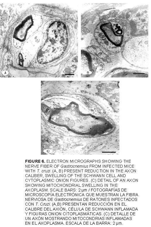
<P align="justify" style="word-spacing: 0; line-height: 100%"><FONT COLOR="000000" size="2" face="Verdana"> On the other hand, the capillary vessels damage in skeletal muscle of a  patient with advanced Chagas´disease with cardiac involvement, could eventually  lead to deficient blood perfusion through the muscle and cause anaerobic  metabolic changes affecting muscle function; other muscular abnormalities  are probably due to reduced oxygen in the abnormal muscle microvasculature.  These changes could affect oxygen extraction affecting the functional capacity  of the infected mice [18]. In the animals infected with DSM-strain<B> </B>the  nerve-muscle<I> </I>preparations exhibited marked changes, as the axonal degeneration  as shown by myelin digestion chambers, was present together with mitochondrial  swelling, signs of Schwann cells damage indicating a demyelinating process  and the presence of onion figures in the Schwann cells cytoplasm of the  infected nerves (<a href="#FIG6">FIG. 6 A</a>-<a href="#FIG6">C</a>). The nerve terminals from nerve-muscle preparations  showed changes in the distribution of subcellular organelles, axoplasm  becomes more electron dense and there was pronounced mitochondrial swelling.  Most of the synaptic vesicles were closely packed at the synaptic side,  near the active presynaptic zone and scarces in the axoplasma (<a href="#FIG7">FIG. 7 A</a>-<a href="#FIG7">C</a>).<B>  </B>None of the features described above could be observed in the non-infected  control group.<B> </B>Based on typical clinical findings related to decreased  motor activity, common symptoms observed in the all infected mice with  <I>T. cruzi</I>, included progressive neuromuscular junction disorders, which  may become associated with decreased muscular function, as consequence  of the micropathological changes of the intramuscular nerve fibers [31].  This alterations in NMJ of G affect the presynaptic membrane densities  in the active zones and the postsynaptic membrane densities become larger,  which shows, that in the normal adult mammal NMJ there is an activity-dependent  modulation of the neurotransmission-related structures in response to slight  physiological functional demands as observed in normal adult rats trained  to walk, indicanting a precise inverse relation between the amount of locomotor  activity and the complexity finally attained by the motor nerve terminals  [34]. In this study, it is possible that alterations in the skeletal muscle  fibers and motor nerve terminal of the NMJ at 40 day pi with DSM strain,  may be at least due in part to strain behavior [6], which produced irreversible  decrease of motor activity and paralysis in the rear limbs in the experimental  model used. It is possible that inflammation and parasites rupturing and  releasing antigen in infected skeletal muscle, become associated with the  membrane surface and development of inflammatory neuropathy [16, 17]. On  the other hand, it is possible the existence of plastic changes in the  branching pattern, as revealed by studies in the <I>extensor digitorum longus</I>  muscle of adult rats housed in metallic cages or trained to walk, indicating  a precise inverse relation between the amount of locomotor activity and  the complexity finally attained by the motor nerve terminals [15, 33]. </FONT></P>      ]]></body>
<body><![CDATA[<P align="center" style="word-spacing: 0; line-height: 100%"><a name="FIG6"><img border="0" src="/img/fbpe/rc/v16n6/art05fig6.jpg" width="517" height="773"></a></P>      
<P align="center" style="word-spacing: 0; line-height: 100%"><a name="FIG7"><img border="0" src="/img/fbpe/rc/v16n6/art05fig7.jpg" width="528" height="825"></a></P>      
<P align="justify" style="word-spacing: 0; line-height: 100%"><FONT COLOR="000000" size="2" face="Verdana"> Based in other findings, the results have showed that is possible that  clinical presentation findings during early <I>T. cruzi</I> infection, can be  at least in part due to both motor endplates and nerve terminals of the  neuromuscular junction disorders affecting neurological structures [23],  reflected in the progressive muscular dystrophy, with inactivation and  loss of mobility in the rear limbs of the infected mice, as occurred in  infected mice with DSM-strain isolated the wild mammalian that live in  the geographic area where Chagas´disease is endemic. </FONT></P>     <p style="word-spacing: 0; line-height: 100%" align="justify"><B><FONT COLOR="000000" size="2" face="Verdana"> CONCLUSIONS </FONT></B>      <P align="justify" style="word-spacing: 0; line-height: 100%"><FONT COLOR="000000" size="2" face="Verdana"> In this work, it was described the effect of <I>Trypanosoma cruzi </I>infection  with DSM-strain in the model murine, on microvasculature, neuromuscular  junction and motor nerve terminals lesions of <I>Gastrocnemius</I> muscle. The  common pathological clinical finding in mice acutely infected was the change  in the locomotor activity. The results of the present study show ultrastructurals  changes in the skeletal muscle fiber, affecting normal function of the  G muscle on infected mice.<I> </I>It determines the course of earlier stages of  pathological processes represented by irreversible decrease of the locomotor  activity and paralysis of the rear limbs of the mice during the course  of infection. </FONT></P>      <P align="justify" style="word-spacing: 0; line-height: 100%"><FONT COLOR="000000" size="2" face="Verdana"> This finding was attributable to presence of amastigotes and inflammatory  infiltrates associated with tissue destruction, as well as to capillary  alterations that could eventually lead to deficient blood perfusion through  the muscle and cause anaerobic metabolic changes affecting muscle function.  Other observed changes in the same muscle had been associated with morphologic  alterations of nerve terminal motor of the neuromuscular junction of G  muscle [15]. Particularly, the axonal elimination, swelling of the Schwann  cells, changes in synaptic morphological parameters as the presynaptic  membrane densities in the active zones, producing neuromuscular disorders  in skeletal muscle fiber modifying the motor activity of the mice with  acute Chagas infection. </FONT></P>      <P align="justify" style="word-spacing: 0; line-height: 100%"><FONT COLOR="000000" size="2" face="Verdana"> Emphasized the need for further studies of some aspects of the biology  and behavior of <I>T. cruzi </I>strains with high virulence and isolated from  the same geographic area, mainly in central region of Venezuela, where  <I>T. cruzi</I> frequently circulates between triatomine bugs, peridomestic animals  and the human population exposed to infection and to a clinical presentation  similar as showed by experimental animals used in this study. </FONT></P>      <P align="justify" style="word-spacing: 0; line-height: 100%"><B><FONT COLOR="000000" size="2" face="Verdana"> ACKNOWLEDGEMENTS </FONT></B></P>      <P align="justify" style="word-spacing: 0; line-height: 100%"><FONT COLOR="000000" size="2" face="Verdana"> The autors thanks by support to Fondo Nacional de Ciencia, Tecnología e  Innovación, (FONACIT). Caracas, Venezuela. Grant N<SUP>o</SUP>. S1-2002000500. </FONT></P>      <P align="justify" style="word-spacing: 0; line-height: 100%"><B><FONT COLOR="000000" size="2" face="Verdana"> BIBLIOGRAPHIC REFERENCES </FONT></B> </P>      ]]></body>
<body><![CDATA[<!-- ref --><P align="justify" style="word-spacing: 0; line-height: 100%"><font face="Verdana" size="2" color="000000">1. ANDRADE, Z.A.; CORREA, R.; SADIGURSKY, M.; FERRANS, V.J. Myocardial changes  in acute <I>Trypanosoma cruzi </I>infection: Ultrastructural evidence of immune  damage and the role of microangiopathy. <B>Am. J. Pathol.</B> 144:1403-1411. 1994.</font>&nbsp;&nbsp;&nbsp;&nbsp;&nbsp;&nbsp;&nbsp;&nbsp;[&#160;<a href="javascript:void(0);" onclick="javascript: window.open('/scielo.php?script=sci_nlinks&ref=1566494&pid=S0798-2259200600060000500001&lng=','','width=640,height=500,resizable=yes,scrollbars=1,menubar=yes,');">Links</a>&#160;]<!-- end-ref --><!-- ref --><P align="justify" style="word-spacing: 0; line-height: 100%"><font face="Verdana" size="2" color="000000">2. ANDRADE, Z.A.; ANDRADE, S.G.; SADIGURSKY, M.; WENTHOLD, R.J.; STEPHEN,  L.H.; FERRANS, V.J. The indeterminate phase of Chagas’ disease: Ultrastructural  characterization of cardiac changes in the canine model. <B>Am. J. Trop. Med.  Hyg. </B>57:328-336. 1997.</font>&nbsp;&nbsp;&nbsp;&nbsp;&nbsp;&nbsp;&nbsp;&nbsp;[&#160;<a href="javascript:void(0);" onclick="javascript: window.open('/scielo.php?script=sci_nlinks&ref=1566495&pid=S0798-2259200600060000500002&lng=','','width=640,height=500,resizable=yes,scrollbars=1,menubar=yes,');">Links</a>&#160;]<!-- end-ref --><!-- ref --><P align="justify" style="word-spacing: 0; line-height: 100%"><font face="Verdana" size="2" color="000000">3. ARAUJO, S. Alteraciones del sistema nervioso central de ratones con infección  aguda producida por <I>Trypanosoma cruzi.</I> Dpto de Biología, Facultad de Ciencias,  Universidad de los Andes, Mérida, Venezuela. (Trabajo Especial de Grado).  68 pp. 2002.</font>&nbsp;&nbsp;&nbsp;&nbsp;&nbsp;&nbsp;&nbsp;&nbsp;[&#160;<a href="javascript:void(0);" onclick="javascript: window.open('/scielo.php?script=sci_nlinks&ref=1566496&pid=S0798-2259200600060000500003&lng=','','width=640,height=500,resizable=yes,scrollbars=1,menubar=yes,');">Links</a>&#160;]<!-- end-ref --><!-- ref --><P align="justify" style="word-spacing: 0; line-height: 100%"><font face="Verdana" size="2" color="000000">4. BRENER, Z. Observações sobre a imunidade a superinfeccões em camundongos  experimentalmente inoculados con <I>Trypanosoma cruzi </I>e submetidos a tratamento.  <B>Rev. Inst. Med. Trop. Säo Paulo.</B> 4:119-123. 1962.</font>&nbsp;&nbsp;&nbsp;&nbsp;&nbsp;&nbsp;&nbsp;&nbsp;[&#160;<a href="javascript:void(0);" onclick="javascript: window.open('/scielo.php?script=sci_nlinks&ref=1566497&pid=S0798-2259200600060000500004&lng=','','width=640,height=500,resizable=yes,scrollbars=1,menubar=yes,');">Links</a>&#160;]<!-- end-ref --><!-- ref --><P align="justify" style="word-spacing: 0; line-height: 100%"><font face="Verdana" size="2" color="000000">5. GONZALEZ C, S.M.; SANZ, O.P.; MULLER, L.A.; MOLINA, H.A.; FERNANDEZ, J.;  RIMOLDI, M.T.; SICA, R.E.P. Peripheral nervous system damage in experimental  chronic Chagas’ disease. <B>Am. J. Trop. Med. Hyg.</B><I> </I>36:41-45. 1987.</font>&nbsp;&nbsp;&nbsp;&nbsp;&nbsp;&nbsp;&nbsp;&nbsp;[&#160;<a href="javascript:void(0);" onclick="javascript: window.open('/scielo.php?script=sci_nlinks&ref=1566498&pid=S0798-2259200600060000500005&lng=','','width=640,height=500,resizable=yes,scrollbars=1,menubar=yes,');">Links</a>&#160;]<!-- end-ref --><!-- ref --><P align="justify" style="word-spacing: 0; line-height: 100%"><font face="Verdana" size="2" color="000000">6. GONZALEZ C., S.M.; MIRKIN, G.A.; SOLANA, M.E.; TEKIEL, V.S. Patología por  <I>Trypanosoma cruzi</I>: ¿Cepa dependiente?. <B>Med</B>. 59:69-74. 1999.</font>&nbsp;&nbsp;&nbsp;&nbsp;&nbsp;&nbsp;&nbsp;&nbsp;[&#160;<a href="javascript:void(0);" onclick="javascript: window.open('/scielo.php?script=sci_nlinks&ref=1566499&pid=S0798-2259200600060000500006&lng=','','width=640,height=500,resizable=yes,scrollbars=1,menubar=yes,');">Links</a>&#160;]<!-- end-ref --><!-- ref --><P align="justify" style="word-spacing: 0; line-height: 100%"><font face="Verdana" size="2" color="000000">7. FACTOR, S.M.; CHO, S.; WITTNER, M.; TANOWITZ, H. Abnormalities of the microcirculation  in acute murine Chagas´disease. <B>Am. J. Trop. Med. Hyg.</B> 34:246-53. 1985.</font>&nbsp;&nbsp;&nbsp;&nbsp;&nbsp;&nbsp;&nbsp;&nbsp;[&#160;<a href="javascript:void(0);" onclick="javascript: window.open('/scielo.php?script=sci_nlinks&ref=1566500&pid=S0798-2259200600060000500007&lng=','','width=640,height=500,resizable=yes,scrollbars=1,menubar=yes,');">Links</a>&#160;]<!-- end-ref --><!-- ref --><P align="justify" style="word-spacing: 0; line-height: 100%"><font face="Verdana" size="2" color="000000">8. FELICIANGELI, M.D.; CAMPELL-LENDRUM, D.; MARTINEZ, D.; GONZALEZ, P.; DAVIES,  C. Chagas’ disease control in Venezuela lessons from the Andean region  and beyond. <B>Trends Parasitol. </B>19:44-49. 2003.</font>&nbsp;&nbsp;&nbsp;&nbsp;&nbsp;&nbsp;&nbsp;&nbsp;[&#160;<a href="javascript:void(0);" onclick="javascript: window.open('/scielo.php?script=sci_nlinks&ref=1566501&pid=S0798-2259200600060000500008&lng=','','width=640,height=500,resizable=yes,scrollbars=1,menubar=yes,');">Links</a>&#160;]<!-- end-ref --><!-- ref --><P align="justify" style="word-spacing: 0; line-height: 100%"><font face="Verdana" size="2" color="000000">9. GUILLEN-PERNIA, B.; LUGO DE Y., A.; MORENO, E. Dilatación del tracto digestivo  de ratones infectados con <I>Trypanosoma cruzi. </I><B>Kasmera.</B> 42:195-209. 2001.</font>&nbsp;&nbsp;&nbsp;&nbsp;&nbsp;&nbsp;&nbsp;&nbsp;[&#160;<a href="javascript:void(0);" onclick="javascript: window.open('/scielo.php?script=sci_nlinks&ref=1566502&pid=S0798-2259200600060000500009&lng=','','width=640,height=500,resizable=yes,scrollbars=1,menubar=yes,');">Links</a>&#160;]<!-- end-ref --><!-- ref --><P align="justify" style="word-spacing: 0; line-height: 100%"><font face="Verdana" size="2" color="000000">10. HIGUCHI, M.L.; DE BRITO, T.; REIS, M.M.; BARBOSA, A.; BELLOTTI, G.; PEREIRA-BARRETO,  A.C.; PILEGGI, F. Correlation between <I>Trypanosoma cruzi </I>parasitism and  myocardial inflammatory infiltrate in human chronic chagasic myocarditis:  Ligth microscopy and immunohistochemical findings. <B>Card.</B> <B>Pathol.</B><I> </I>2:101-106.  1993.</font>&nbsp;&nbsp;&nbsp;&nbsp;&nbsp;&nbsp;&nbsp;&nbsp;[&#160;<a href="javascript:void(0);" onclick="javascript: window.open('/scielo.php?script=sci_nlinks&ref=1566503&pid=S0798-2259200600060000500010&lng=','','width=640,height=500,resizable=yes,scrollbars=1,menubar=yes,');">Links</a>&#160;]<!-- end-ref --><!-- ref --><P align="justify" style="word-spacing: 0; line-height: 100%"><font face="Verdana" size="2" color="000000">11. HOARE, A. <B>The trypanosomes of mammals.</B> Blackwell Sci. Publ. Oxford &amp; Edinburg,  UK, 334 pp. 1972.</font>&nbsp;&nbsp;&nbsp;&nbsp;&nbsp;&nbsp;&nbsp;&nbsp;[&#160;<a href="javascript:void(0);" onclick="javascript: window.open('/scielo.php?script=sci_nlinks&ref=1566504&pid=S0798-2259200600060000500011&lng=','','width=640,height=500,resizable=yes,scrollbars=1,menubar=yes,');">Links</a>&#160;]<!-- end-ref --><!-- ref --><P align="justify" style="word-spacing: 0; line-height: 100%"><font face="Verdana" size="2" color="000000">12. KIM, C.T.; STROMMEN, J.A.; JOHNS, J.S.; WEISS, J.M; WEISS, L.D.; WILLIAMS,  F.H.; RASHBAUM, I.G. Neuromuscular rehabilitation and electrodiagnosis:  4. Pediatric issues. <B>Arch. Phys. Med. Rehabil. </B>86:28-32. 2005.</font>&nbsp;&nbsp;&nbsp;&nbsp;&nbsp;&nbsp;&nbsp;&nbsp;[&#160;<a href="javascript:void(0);" onclick="javascript: window.open('/scielo.php?script=sci_nlinks&ref=1566505&pid=S0798-2259200600060000500012&lng=','','width=640,height=500,resizable=yes,scrollbars=1,menubar=yes,');">Links</a>&#160;]<!-- end-ref --><!-- ref --><p align="justify" style="word-spacing: 0; line-height: 100%"><font face="Verdana" size="2" color="000000">13. LEON S, F.E.; PRADA, D.G.; BAYONA P, J.; VALDERRAMA, V.; GARCIA, I.; LEON,  M.E.; SUNNEMARK, D. Neurological effects of American tripanosomiasis: clinical  aspects. <B>Biomed.</B> 23:462-475.</font>&nbsp;&nbsp;&nbsp;&nbsp;&nbsp;&nbsp;&nbsp;&nbsp;[&#160;<a href="javascript:void(0);" onclick="javascript: window.open('/scielo.php?script=sci_nlinks&ref=1566506&pid=S0798-2259200600060000500013&lng=','','width=640,height=500,resizable=yes,scrollbars=1,menubar=yes,');">Links</a>&#160;]<!-- end-ref --><!-- ref --><p align="justify" style="word-spacing: 0; line-height: 100%"><font face="Verdana" size="2" color="000000">14. LOSAVIO, A.; JONES, M.C.; SANZ, O.P.; MIRKIN, G.; GONZALEZ-CAPPA, S.M.;  MUCNIK, S.; SICA, E.R.P. A sequential study of the peripheral nervous system  involvement in experimental Chagas’ disease. <B>Am. J. Trop. Med. Hyg</B>.<I> </I>41:59-547.  1989.</font>&nbsp;&nbsp;&nbsp;&nbsp;&nbsp;&nbsp;&nbsp;&nbsp;[&#160;<a href="javascript:void(0);" onclick="javascript: window.open('/scielo.php?script=sci_nlinks&ref=1566507&pid=S0798-2259200600060000500014&lng=','','width=640,height=500,resizable=yes,scrollbars=1,menubar=yes,');">Links</a>&#160;]<!-- end-ref --><!-- ref --><p align="justify" style="word-spacing: 0; line-height: 100%"><font face="Verdana" size="2" color="000000">15. LUGO DE Y., A.; GUTIERREZ, R.; COLASANTE, C. Ultrastructural changes in  neuromuscular junction of skeletal muscle of mice infected with <I>Trypanosoma  cruzi. </I><B>Rev. Ecol. Lat. Am. </B>6:1-11. 1999.</font>&nbsp;&nbsp;&nbsp;&nbsp;&nbsp;&nbsp;&nbsp;&nbsp;[&#160;<a href="javascript:void(0);" onclick="javascript: window.open('/scielo.php?script=sci_nlinks&ref=1566508&pid=S0798-2259200600060000500015&lng=','','width=640,height=500,resizable=yes,scrollbars=1,menubar=yes,');">Links</a>&#160;]<!-- end-ref --><!-- ref --><p align="justify" style="word-spacing: 0; line-height: 100%"><font face="Verdana" size="2" color="000000">16. MIRKIN, G.A.; JONES, M.; SANZ, O.P.; REY, R.; SICA, R.E.P.; GONZALEZ, S.M.  Experimental Chagas’ disease: Electrophysio logy and cell composition of  the neuromyopathic inflammatory lesions in mice infected with a myotropic  and a pantropic strain of <I>Trypanosoma cruzi</I>. <B>Clin. Immunol. Immunopathol.</B>  73:69-79. 1994.</font>&nbsp;&nbsp;&nbsp;&nbsp;&nbsp;&nbsp;&nbsp;&nbsp;[&#160;<a href="javascript:void(0);" onclick="javascript: window.open('/scielo.php?script=sci_nlinks&ref=1566509&pid=S0798-2259200600060000500016&lng=','','width=640,height=500,resizable=yes,scrollbars=1,menubar=yes,');">Links</a>&#160;]<!-- end-ref --><!-- ref --><p align="justify" style="word-spacing: 0; line-height: 100%"><font face="Verdana" size="2" color="000000">17. MOLINA, H.A.; CARDONA, R.L.; RIMOLDI, M.T. The neuromuscular pathology  of experimental Chagas’ disease. <B>J. Neurol. Sci. </B>81:287-300. 1987.</font>&nbsp;&nbsp;&nbsp;&nbsp;&nbsp;&nbsp;&nbsp;&nbsp;[&#160;<a href="javascript:void(0);" onclick="javascript: window.open('/scielo.php?script=sci_nlinks&ref=1566510&pid=S0798-2259200600060000500017&lng=','','width=640,height=500,resizable=yes,scrollbars=1,menubar=yes,');">Links</a>&#160;]<!-- end-ref --><!-- ref --><p align="justify" style="word-spacing: 0; line-height: 100%"><font face="Verdana" size="2" color="000000">18. MONTES DE O., M.; TORRES, S.H.; LOYO, J.G.; VASQUEZ, F.; HERNANDEZ, N.;  ANCHUSTEGUI, B.; PUIGBO, J.J. Exercise perfomance and skeletal muscle in  patients with advanced Chagas´disease. <B>Chest.</B> 125:1306-1314. 2004.</font>&nbsp;&nbsp;&nbsp;&nbsp;&nbsp;&nbsp;&nbsp;&nbsp;[&#160;<a href="javascript:void(0);" onclick="javascript: window.open('/scielo.php?script=sci_nlinks&ref=1566511&pid=S0798-2259200600060000500018&lng=','','width=640,height=500,resizable=yes,scrollbars=1,menubar=yes,');">Links</a>&#160;]<!-- end-ref --><!-- ref --><p align="justify" style="word-spacing: 0; line-height: 100%"><font face="Verdana" size="2" color="000000">19. MORENO, E., ANEZ, N., SCORZA, C., LUGO DE Y, A.; BORGES, R. Efecto de inóculos  bajos en la infección experimental por <I>Trypanosoma cruzi</I>. <B>Bol. Dir. Malariol.  San. Amb</B>. 39:70-77. 1999.</font>&nbsp;&nbsp;&nbsp;&nbsp;&nbsp;&nbsp;&nbsp;&nbsp;[&#160;<a href="javascript:void(0);" onclick="javascript: window.open('/scielo.php?script=sci_nlinks&ref=1566512&pid=S0798-2259200600060000500019&lng=','','width=640,height=500,resizable=yes,scrollbars=1,menubar=yes,');">Links</a>&#160;]<!-- end-ref --><!-- ref --><p align="justify" style="word-spacing: 0; line-height: 100%"><font face="Verdana" size="2" color="000000">20. MORENO, E.; GONZALEZ, N.; RIVERA, I.; GUILLEN-PERNIA, B.; LUGO DE Y, A.  Caracterización biológica e isoenzimática de aislados de <I>Trypanosoma cruzi</I>.  <B>Bol. Dir. Maral. San Amb. </B>52:17-28. 2002.</font>&nbsp;&nbsp;&nbsp;&nbsp;&nbsp;&nbsp;&nbsp;&nbsp;[&#160;<a href="javascript:void(0);" onclick="javascript: window.open('/scielo.php?script=sci_nlinks&ref=1566513&pid=S0798-2259200600060000500020&lng=','','width=640,height=500,resizable=yes,scrollbars=1,menubar=yes,');">Links</a>&#160;]<!-- end-ref --><!-- ref --><p align="justify" style="word-spacing: 0; line-height: 100%"><font face="Verdana" size="2" color="000000">21. MÜLLER, L.A.; ANASCO, N.; GONZALEZ CAPPA, S.M. <I>Trypanosoma cruzi</I>: isolated  dependence in the induction of lytic antibodies in the mouse and rabbits.  <B>Exp. Parasitol.</B> 61:284-293. 1986.</font>&nbsp;&nbsp;&nbsp;&nbsp;&nbsp;&nbsp;&nbsp;&nbsp;[&#160;<a href="javascript:void(0);" onclick="javascript: window.open('/scielo.php?script=sci_nlinks&ref=1566514&pid=S0798-2259200600060000500021&lng=','','width=640,height=500,resizable=yes,scrollbars=1,menubar=yes,');">Links</a>&#160;]<!-- end-ref --><!-- ref --><p align="justify" style="word-spacing: 0; line-height: 100%"><font face="Verdana" size="2" color="000000">22. PEREIRA, V.L.; ZAMORANO, M.M.B.; BOAINAIN, E. Estudo do comportamento biologico  de tres amostras de <I>Trypanosoma cruzi </I>isoladas de paciente de Instituto  Dvorak Pazzanese de Cardiología. <B>Rev. Inst. Med. Trop. Sao Paulo.</B> 29:155-161.  1987.</font>&nbsp;&nbsp;&nbsp;&nbsp;&nbsp;&nbsp;&nbsp;&nbsp;[&#160;<a href="javascript:void(0);" onclick="javascript: window.open('/scielo.php?script=sci_nlinks&ref=1566515&pid=S0798-2259200600060000500022&lng=','','width=640,height=500,resizable=yes,scrollbars=1,menubar=yes,');">Links</a>&#160;]<!-- end-ref --><!-- ref --><p align="justify" style="word-spacing: 0; line-height: 100%"><font color="000000" size="2" face="Verdana">23. PRAKASH, Y.S.; ZHAN, W.Z.; MIYATA, H.; SIECK, G.C. Adaptations of diaphragm  neuromuscular junction following inactivity. <B>Acta Anat.</B> 154:147-161. 1995.&nbsp; </font>&nbsp;&nbsp;&nbsp;&nbsp;&nbsp;&nbsp;&nbsp;&nbsp;[&#160;<a href="javascript:void(0);" onclick="javascript: window.open('/scielo.php?script=sci_nlinks&ref=1566516&pid=S0798-2259200600060000500023&lng=','','width=640,height=500,resizable=yes,scrollbars=1,menubar=yes,');">Links</a>&#160;]<!-- end-ref --><!-- ref --><p align="justify" style="word-spacing: 0; line-height: 100%"><font face="Verdana" size="2" color="000000">24. PETKOVA, S.B.; HUANG, H.; FACTOR, S.M.; PESTELL, R.G.; BOUZAHZAH, B.; JELICKS,  L.A.; WEISS, L.M.; DOUGLAS, S.A.; WITTNER, M.; TANOWITZ, H.B. The role  of endothelin in the pathogenesis of Chagas´disease. <B>Int J. Parasitol.</B>  31:499-511. 2001.</font>&nbsp;&nbsp;&nbsp;&nbsp;&nbsp;&nbsp;&nbsp;&nbsp;[&#160;<a href="javascript:void(0);" onclick="javascript: window.open('/scielo.php?script=sci_nlinks&ref=1566517&pid=S0798-2259200600060000500024&lng=','','width=640,height=500,resizable=yes,scrollbars=1,menubar=yes,');">Links</a>&#160;]<!-- end-ref --><!-- ref --><p align="justify" style="word-spacing: 0; line-height: 100%"><font face="Verdana" size="2" color="000000">25. QUIÑONES, M.E.; FINOL, H.J.; SUCRE, L.; GARCÍA, F.; ORTEGA, A. Anormalidades  en los capilares intramusculares del equino infectado con <I>Trypanosoma evansi  </I>(Steel, 1885).<B> Memorias de IV Jornadas de Microscopía Electrónica. SVME</B>.  Cumana, July 16-18. Venezuela, 10 pp. 1990.</font>&nbsp;&nbsp;&nbsp;&nbsp;&nbsp;&nbsp;&nbsp;&nbsp;[&#160;<a href="javascript:void(0);" onclick="javascript: window.open('/scielo.php?script=sci_nlinks&ref=1566518&pid=S0798-2259200600060000500025&lng=','','width=640,height=500,resizable=yes,scrollbars=1,menubar=yes,');">Links</a>&#160;]<!-- end-ref --><!-- ref --><p align="justify" style="word-spacing: 0; line-height: 100%"><font face="Verdana" size="2" color="000000">26. RIBERIO DOS SANTO, R.; HUDSON, L. <I>Trypanosoma cruzi </I>binding of parasite  antigens to mammalian cell membranes. <B>Parasit Immunol.</B><I> </I>2:1-10. 1980.</font>&nbsp;&nbsp;&nbsp;&nbsp;&nbsp;&nbsp;&nbsp;&nbsp;[&#160;<a href="javascript:void(0);" onclick="javascript: window.open('/scielo.php?script=sci_nlinks&ref=1566519&pid=S0798-2259200600060000500026&lng=','','width=640,height=500,resizable=yes,scrollbars=1,menubar=yes,');">Links</a>&#160;]<!-- end-ref --><!-- ref --><p align="justify" style="word-spacing: 0; line-height: 100%"><font face="Verdana" size="2" color="000000">27. ROSSI, M.A.; GONCALVES, S.; RIBEIRO DO SANTOS, R. Experimental <I>Trypanosoma  cruzi </I>cardiomyophaty<I> </I>in BALB/c mice: the potential role of intravascular  platelet aggregation in its genesis. <B>Am. J. Pathol.</B><I> </I>114:209-216. 1984.</font>&nbsp;&nbsp;&nbsp;&nbsp;&nbsp;&nbsp;&nbsp;&nbsp;[&#160;<a href="javascript:void(0);" onclick="javascript: window.open('/scielo.php?script=sci_nlinks&ref=1566520&pid=S0798-2259200600060000500027&lng=','','width=640,height=500,resizable=yes,scrollbars=1,menubar=yes,');">Links</a>&#160;]<!-- end-ref --><!-- ref --><p align="justify" style="word-spacing: 0; line-height: 100%"><font face="Verdana" size="2" color="000000">28. ROSSI, M.A. Microvascular changes as a cause of chronic cardiomyopathy  in Chagas’ disease. <B>Am. Heart. J. </B>120:233-236. 1990.</font>&nbsp;&nbsp;&nbsp;&nbsp;&nbsp;&nbsp;&nbsp;&nbsp;[&#160;<a href="javascript:void(0);" onclick="javascript: window.open('/scielo.php?script=sci_nlinks&ref=1566521&pid=S0798-2259200600060000500028&lng=','','width=640,height=500,resizable=yes,scrollbars=1,menubar=yes,');">Links</a>&#160;]<!-- end-ref --><!-- ref --><p align="justify" style="word-spacing: 0; line-height: 100%"><font face="Verdana" size="2" color="000000">29. ROSSI, M.A. Aortic endothelial cell changes in the acute septicemic phase  of experimental <I>Trypanosoma cruzi </I>infection in rats: scanning and transmission  electron microscopic study. <B>Am. J. Trop. Med. Hyg.</B><I> </I>57: 321-327. 1997.</font>&nbsp;&nbsp;&nbsp;&nbsp;&nbsp;&nbsp;&nbsp;&nbsp;[&#160;<a href="javascript:void(0);" onclick="javascript: window.open('/scielo.php?script=sci_nlinks&ref=1566522&pid=S0798-2259200600060000500029&lng=','','width=640,height=500,resizable=yes,scrollbars=1,menubar=yes,');">Links</a>&#160;]<!-- end-ref --><!-- ref --><p align="justify" style="word-spacing: 0; line-height: 100%"><font face="Verdana" size="2" color="000000">30. SICA, R.E.; FILIPINI, D.; PANIZZA, M.; FUMO, T.; BASO, S.; LAZZARI, J.;  MOLINA, H.A. Involvement of the peripheral sensory nervous system in human  Chagas’ disease<B>. Medicine</B> (B. Aires). 46:662-668. 1986.</font>&nbsp;&nbsp;&nbsp;&nbsp;&nbsp;&nbsp;&nbsp;&nbsp;[&#160;<a href="javascript:void(0);" onclick="javascript: window.open('/scielo.php?script=sci_nlinks&ref=1566523&pid=S0798-2259200600060000500030&lng=','','width=640,height=500,resizable=yes,scrollbars=1,menubar=yes,');">Links</a>&#160;]<!-- end-ref --><!-- ref --><p align="justify" style="word-spacing: 0; line-height: 100%"><font face="Verdana" size="2" color="000000">31. STROMMER, J.A.; JOHNS, J.S.; KIM, C.T.; WILLIAMS, F.H.; WEISS, L.D.; WEISS,  J.M.; RASHBAUM, I.G. Neuromuscular rehabilitation and electrodiagnosis: 3. Diseases of muscles and neuromuscular junction. <B>Arch. Phys. Med. Rehabil.</B>  86:18-27. 2005.</font>&nbsp;&nbsp;&nbsp;&nbsp;&nbsp;&nbsp;&nbsp;&nbsp;[&#160;<a href="javascript:void(0);" onclick="javascript: window.open('/scielo.php?script=sci_nlinks&ref=1566524&pid=S0798-2259200600060000500031&lng=','','width=640,height=500,resizable=yes,scrollbars=1,menubar=yes,');">Links</a>&#160;]<!-- end-ref --><!-- ref --><p align="justify" style="word-spacing: 0; line-height: 100%"><font face="Verdana" size="2" color="000000">32. TANOWITZ, H.B.; KAUL, D.K.; CHEN, B.; MORRIS, S.A.; FACTOR, S.M.; WEISS,  L.M.; WITTNER, M. Compromised microcirculation in acute murine <I>Trypanosoma  cruzi </I>infection. <B>J.</B> <B>Parasitol. </B>82:124-30. 1996.</font>&nbsp;&nbsp;&nbsp;&nbsp;&nbsp;&nbsp;&nbsp;&nbsp;[&#160;<a href="javascript:void(0);" onclick="javascript: window.open('/scielo.php?script=sci_nlinks&ref=1566525&pid=S0798-2259200600060000500032&lng=','','width=640,height=500,resizable=yes,scrollbars=1,menubar=yes,');">Links</a>&#160;]<!-- end-ref --><!-- ref --><p align="justify" style="word-spacing: 0; line-height: 100%"><font face="Verdana" size="2" color="000000">33. TOMAS, J.; FENOLL, R.; SANTAFE, M.; BATLLE, J.; MAYAYO, E. Motor nerve  terminal morphologic plasticity induced by small changes in the locomotor  activity of the adult rat. <B>Neurosci. Lett.</B> 106:137-140. 1989.</font>&nbsp;&nbsp;&nbsp;&nbsp;&nbsp;&nbsp;&nbsp;&nbsp;[&#160;<a href="javascript:void(0);" onclick="javascript: window.open('/scielo.php?script=sci_nlinks&ref=1566526&pid=S0798-2259200600060000500033&lng=','','width=640,height=500,resizable=yes,scrollbars=1,menubar=yes,');">Links</a>&#160;]<!-- end-ref --><!-- ref --><p align="justify" style="word-spacing: 0; line-height: 100%"><font face="Verdana" size="2" color="000000">34. TOMAS, J.; SANTAFE, M.; LANUZA, M.A.; FENOLL-BRUNET, M.R. Physiological  activity-dependent ultrastructural plasticity in normal adult rat neuromuscular  junctions. <B>Bio. Cell.</B> 89:19-28. 1997.</font>&nbsp;&nbsp;&nbsp;&nbsp;&nbsp;&nbsp;&nbsp;&nbsp;[&#160;<a href="javascript:void(0);" onclick="javascript: window.open('/scielo.php?script=sci_nlinks&ref=1566527&pid=S0798-2259200600060000500034&lng=','','width=640,height=500,resizable=yes,scrollbars=1,menubar=yes,');">Links</a>&#160;]<!-- end-ref --><!-- ref --><p align="justify" style="word-spacing: 0; line-height: 100%"><font face="Verdana" size="2" color="000000">35. TONINO, P.; FINOL, H.J.; CAMPO-AASEN, I. Un modelo murino para el estudio  de los efectos a distancia del <I>Toxoplasma gondii </I>sobre el músculo esquelético.  <B>Memorias del Primer Congreso Atlántico de Microscopía Electrónica. SVME</B>,  Mérida. 24-29 de mayo. Venezuela, 68-69 pp. 1992.</font>&nbsp;&nbsp;&nbsp;&nbsp;&nbsp;&nbsp;&nbsp;&nbsp;[&#160;<a href="javascript:void(0);" onclick="javascript: window.open('/scielo.php?script=sci_nlinks&ref=1566528&pid=S0798-2259200600060000500035&lng=','','width=640,height=500,resizable=yes,scrollbars=1,menubar=yes,');">Links</a>&#160;]<!-- end-ref --><!-- ref --><p align="justify" style="word-spacing: 0; line-height: 100%"><font face="Verdana" size="2" color="000000">36. TONINO, P.; FINOL, H.; MARQUEZ, A. Skeletal muscle capillary ultrastructure  in <I>Toxoplasma gondii </I>parasitized mice. <B>Acta Cientif Venezol.</B><I> </I>44:349-354.  1993.</font>&nbsp;&nbsp;&nbsp;&nbsp;&nbsp;&nbsp;&nbsp;&nbsp;[&#160;<a href="javascript:void(0);" onclick="javascript: window.open('/scielo.php?script=sci_nlinks&ref=1566529&pid=S0798-2259200600060000500036&lng=','','width=640,height=500,resizable=yes,scrollbars=1,menubar=yes,');">Links</a>&#160;]<!-- end-ref --><!-- ref --><p align="justify" style="word-spacing: 0; line-height: 100%"><font face="Verdana" size="2" color="000000">37. VIANNA, G. Contribucões para o estudo da anatomia patologica de Carlos  Chagas. <B>Mem. Inst Oswaldo Cruz</B>. 3:276-294. 1911.</font>&nbsp;&nbsp;&nbsp;&nbsp;&nbsp;&nbsp;&nbsp;&nbsp;[&#160;<a href="javascript:void(0);" onclick="javascript: window.open('/scielo.php?script=sci_nlinks&ref=1566530&pid=S0798-2259200600060000500037&lng=','','width=640,height=500,resizable=yes,scrollbars=1,menubar=yes,');">Links</a>&#160;]<!-- end-ref --><!-- ref --><p align="justify" style="word-spacing: 0; line-height: 100%"><font face="Verdana" size="2" color="000000">38. VICHI, F.L. Destruciao de neuronios motores na medulla espinal do ratos  na fase aguda da molestia de Chagas. <B>Rev. Inst. Med. Trop. Sao Paulo. </B>6:150-154.  1964.</font>&nbsp;&nbsp;&nbsp;&nbsp;&nbsp;&nbsp;&nbsp;&nbsp;[&#160;<a href="javascript:void(0);" onclick="javascript: window.open('/scielo.php?script=sci_nlinks&ref=1566531&pid=S0798-2259200600060000500038&lng=','','width=640,height=500,resizable=yes,scrollbars=1,menubar=yes,');">Links</a>&#160;]<!-- end-ref --><!-- ref --><p align="justify" style="word-spacing: 0; line-height: 100%"><font face="Verdana" size="2" color="000000">39. WORLD HEALTH ORGANIZATION. Chagas´disease. Thirteenth programme report  of the UNDP/World Bank/WHO special programme for research and training  in tropical disease. 112-123 pp. 1996.</font>&nbsp;&nbsp;&nbsp;&nbsp;&nbsp;&nbsp;&nbsp;&nbsp;[&#160;<a href="javascript:void(0);" onclick="javascript: window.open('/scielo.php?script=sci_nlinks&ref=1566532&pid=S0798-2259200600060000500039&lng=','','width=640,height=500,resizable=yes,scrollbars=1,menubar=yes,');">Links</a>&#160;]<!-- end-ref --> ]]></body>
<back>
<ref-list>
<ref id="B1">
<label>1</label><nlm-citation citation-type="journal">
<person-group person-group-type="author">
<name>
<surname><![CDATA[ANDRADE]]></surname>
<given-names><![CDATA[Z.A.]]></given-names>
</name>
<name>
<surname><![CDATA[CORREA]]></surname>
<given-names><![CDATA[R.]]></given-names>
</name>
<name>
<surname><![CDATA[SADIGURSKY]]></surname>
<given-names><![CDATA[M.]]></given-names>
</name>
<name>
<surname><![CDATA[FERRANS]]></surname>
<given-names><![CDATA[V.J]]></given-names>
</name>
</person-group>
<article-title xml:lang="en"><![CDATA[Myocardial changes in acute Trypanosoma cruzi infection: Ultrastructural evidence of immune damage and the role of microangiopathy]]></article-title>
<source><![CDATA[Am. J. Pathol.]]></source>
<year>1994</year>
<volume>144</volume>
<page-range>1403-1411</page-range></nlm-citation>
</ref>
<ref id="B2">
<label>2</label><nlm-citation citation-type="journal">
<person-group person-group-type="author">
<name>
<surname><![CDATA[ANDRADE]]></surname>
<given-names><![CDATA[Z.A.]]></given-names>
</name>
<name>
<surname><![CDATA[ANDRADE]]></surname>
<given-names><![CDATA[S.G.]]></given-names>
</name>
<name>
<surname><![CDATA[SADIGURSKY]]></surname>
<given-names><![CDATA[M.]]></given-names>
</name>
<name>
<surname><![CDATA[WENTHOLD]]></surname>
<given-names><![CDATA[R.J.]]></given-names>
</name>
<name>
<surname><![CDATA[STEPHEN]]></surname>
<given-names><![CDATA[L.H.]]></given-names>
</name>
<name>
<surname><![CDATA[FERRANS]]></surname>
<given-names><![CDATA[V.J]]></given-names>
</name>
</person-group>
<article-title xml:lang="en"><![CDATA[The indeterminate phase of Chagas’ disease: Ultrastructural characterization of cardiac changes in the canine model]]></article-title>
<source><![CDATA[Am. J. Trop. Med. Hyg.]]></source>
<year>1997</year>
<volume>57</volume>
<page-range>328-336</page-range></nlm-citation>
</ref>
<ref id="B3">
<label>3</label><nlm-citation citation-type="book">
<person-group person-group-type="author">
<name>
<surname><![CDATA[ARAUJO]]></surname>
<given-names><![CDATA[S]]></given-names>
</name>
</person-group>
<source><![CDATA[Alteraciones del sistema nervioso central de ratones con infección aguda producida por Trypanosoma cruzi]]></source>
<year>2002</year>
<page-range>68</page-range><publisher-loc><![CDATA[Mérida ]]></publisher-loc>
<publisher-name><![CDATA[Dpto de Biología, Facultad de Ciencias, Universidad de los Andes]]></publisher-name>
</nlm-citation>
</ref>
<ref id="B4">
<label>4</label><nlm-citation citation-type="journal">
<person-group person-group-type="author">
<name>
<surname><![CDATA[BRENER]]></surname>
<given-names><![CDATA[Z]]></given-names>
</name>
</person-group>
<article-title xml:lang="pt"><![CDATA[Observações sobre a imunidade a superinfeccões em camundongos experimentalmente inoculados con Trypanosoma cruzi e submetidos a tratamento]]></article-title>
<source><![CDATA[Rev. Inst. Med. Trop. Säo Paulo.]]></source>
<year>1962</year>
<volume>4</volume>
<page-range>119-123</page-range></nlm-citation>
</ref>
<ref id="B5">
<label>5</label><nlm-citation citation-type="journal">
<person-group person-group-type="author">
<name>
<surname><![CDATA[GONZALEZ C]]></surname>
<given-names><![CDATA[S.M.]]></given-names>
</name>
<name>
<surname><![CDATA[SANZ]]></surname>
<given-names><![CDATA[O.P.]]></given-names>
</name>
<name>
<surname><![CDATA[MULLER]]></surname>
<given-names><![CDATA[L.A.]]></given-names>
</name>
<name>
<surname><![CDATA[MOLINA]]></surname>
<given-names><![CDATA[H.A.]]></given-names>
</name>
<name>
<surname><![CDATA[FERNANDEZ]]></surname>
<given-names><![CDATA[J.]]></given-names>
</name>
<name>
<surname><![CDATA[RIMOLDI]]></surname>
<given-names><![CDATA[M.T.]]></given-names>
</name>
<name>
<surname><![CDATA[SICA]]></surname>
<given-names><![CDATA[R.E.P]]></given-names>
</name>
</person-group>
<article-title xml:lang="en"><![CDATA[Peripheral nervous system damage in experimental chronic Chagas’ disease]]></article-title>
<source><![CDATA[Am. J. Trop. Med. Hyg.]]></source>
<year>1987</year>
<volume>36</volume>
<page-range>41-45</page-range></nlm-citation>
</ref>
<ref id="B6">
<label>6</label><nlm-citation citation-type="journal">
<person-group person-group-type="author">
<name>
<surname><![CDATA[GONZALEZ C.]]></surname>
<given-names><![CDATA[S.M.]]></given-names>
</name>
<name>
<surname><![CDATA[MIRKIN]]></surname>
<given-names><![CDATA[G.A.]]></given-names>
</name>
<name>
<surname><![CDATA[SOLANA]]></surname>
<given-names><![CDATA[M.E.]]></given-names>
</name>
<name>
<surname><![CDATA[TEKIEL]]></surname>
<given-names><![CDATA[V.S]]></given-names>
</name>
</person-group>
<article-title xml:lang="es"><![CDATA[Patología por Trypanosoma cruzi: ¿Cepa dependiente?]]></article-title>
<source><![CDATA[Med.]]></source>
<year>1999</year>
<volume>59</volume>
<page-range>69-74</page-range></nlm-citation>
</ref>
<ref id="B7">
<label>7</label><nlm-citation citation-type="journal">
<person-group person-group-type="author">
<name>
<surname><![CDATA[FACTOR]]></surname>
<given-names><![CDATA[S.M.]]></given-names>
</name>
<name>
<surname><![CDATA[CHO]]></surname>
<given-names><![CDATA[S.]]></given-names>
</name>
<name>
<surname><![CDATA[WITTNER]]></surname>
<given-names><![CDATA[M.]]></given-names>
</name>
<name>
<surname><![CDATA[TANOWITZ]]></surname>
<given-names><![CDATA[H]]></given-names>
</name>
</person-group>
<article-title xml:lang="en"><![CDATA[Abnormalities of the microcirculation in acute murine Chagas´disease]]></article-title>
<source><![CDATA[Am. J. Trop. Med. Hyg.]]></source>
<year>1985</year>
<volume>34</volume>
<page-range>246-53</page-range></nlm-citation>
</ref>
<ref id="B8">
<label>8</label><nlm-citation citation-type="journal">
<person-group person-group-type="author">
<name>
<surname><![CDATA[FELICIANGELI]]></surname>
<given-names><![CDATA[M.D.]]></given-names>
</name>
<name>
<surname><![CDATA[CAMPELL-LENDRUM]]></surname>
<given-names><![CDATA[D.]]></given-names>
</name>
<name>
<surname><![CDATA[MARTINEZ]]></surname>
<given-names><![CDATA[D.]]></given-names>
</name>
<name>
<surname><![CDATA[GONZALEZ]]></surname>
<given-names><![CDATA[P.]]></given-names>
</name>
<name>
<surname><![CDATA[DAVIES]]></surname>
<given-names><![CDATA[C]]></given-names>
</name>
</person-group>
<article-title xml:lang="en"><![CDATA[Chagas’ disease control in Venezuela lessons from the Andean region and beyond]]></article-title>
<source><![CDATA[Trends Parasitol.]]></source>
<year>2003</year>
<volume>19</volume>
<page-range>44-49</page-range></nlm-citation>
</ref>
<ref id="B9">
<label>9</label><nlm-citation citation-type="journal">
<person-group person-group-type="author">
<name>
<surname><![CDATA[GUILLEN-PERNIA]]></surname>
<given-names><![CDATA[B.]]></given-names>
</name>
<name>
<surname><![CDATA[LUGO DE Y.]]></surname>
<given-names><![CDATA[A.]]></given-names>
</name>
<name>
<surname><![CDATA[MORENO]]></surname>
<given-names><![CDATA[E]]></given-names>
</name>
</person-group>
<article-title xml:lang="es"><![CDATA[Dilatación del tracto digestivo de ratones infectados con Trypanosoma cruzi]]></article-title>
<source><![CDATA[Kasmera.]]></source>
<year>2001</year>
<volume>42</volume>
<page-range>195-209</page-range></nlm-citation>
</ref>
<ref id="B10">
<label>10</label><nlm-citation citation-type="journal">
<person-group person-group-type="author">
<name>
<surname><![CDATA[HIGUCHI]]></surname>
<given-names><![CDATA[M.L.]]></given-names>
</name>
<name>
<surname><![CDATA[DE BRITO]]></surname>
<given-names><![CDATA[T.]]></given-names>
</name>
<name>
<surname><![CDATA[REIS]]></surname>
<given-names><![CDATA[M.M.]]></given-names>
</name>
<name>
<surname><![CDATA[BARBOSA]]></surname>
<given-names><![CDATA[A.]]></given-names>
</name>
<name>
<surname><![CDATA[BELLOTTI]]></surname>
<given-names><![CDATA[G.]]></given-names>
</name>
<name>
<surname><![CDATA[PEREIRA-BARRETO]]></surname>
<given-names><![CDATA[A.C.]]></given-names>
</name>
<name>
<surname><![CDATA[PILEGGI]]></surname>
<given-names><![CDATA[F]]></given-names>
</name>
</person-group>
<article-title xml:lang="en"><![CDATA[Correlation between Trypanosoma cruzi parasitism and myocardial inflammatory infiltrate in human chronic chagasic myocarditis: Ligth microscopy and immunohistochemical findings]]></article-title>
<source><![CDATA[Card. Pathol.]]></source>
<year>1993</year>
<volume>2</volume>
<page-range>101-106</page-range></nlm-citation>
</ref>
<ref id="B11">
<label>11</label><nlm-citation citation-type="book">
<person-group person-group-type="author">
<name>
<surname><![CDATA[HOARE]]></surname>
<given-names><![CDATA[A]]></given-names>
</name>
</person-group>
<source><![CDATA[The trypanosomes of mammals]]></source>
<year>1972</year>
<page-range>334</page-range><publisher-loc><![CDATA[^eUK UK]]></publisher-loc>
<publisher-name><![CDATA[Blackwell Sci. Publ. Oxford & Edinburg]]></publisher-name>
</nlm-citation>
</ref>
<ref id="B12">
<label>12</label><nlm-citation citation-type="journal">
<person-group person-group-type="author">
<name>
<surname><![CDATA[KIM]]></surname>
<given-names><![CDATA[C.T.]]></given-names>
</name>
<name>
<surname><![CDATA[STROMMEN]]></surname>
<given-names><![CDATA[J.A.]]></given-names>
</name>
<name>
<surname><![CDATA[JOHNS]]></surname>
<given-names><![CDATA[J.S.]]></given-names>
</name>
<name>
<surname><![CDATA[WEISS]]></surname>
<given-names><![CDATA[J.M]]></given-names>
</name>
<name>
<surname><![CDATA[WEISS]]></surname>
<given-names><![CDATA[L.D.]]></given-names>
</name>
<name>
<surname><![CDATA[WILLIAMS]]></surname>
<given-names><![CDATA[F.H.]]></given-names>
</name>
<name>
<surname><![CDATA[RASHBAUM]]></surname>
<given-names><![CDATA[I.G]]></given-names>
</name>
</person-group>
<article-title xml:lang="en"><![CDATA[Neuromuscular rehabilitation and electrodiagnosis: 4. Pediatric issues]]></article-title>
<source><![CDATA[Arch. Phys. Med. Rehabil.]]></source>
<year>2005</year>
<volume>86</volume>
<page-range>28-32</page-range></nlm-citation>
</ref>
<ref id="B13">
<label>13</label><nlm-citation citation-type="journal">
<person-group person-group-type="author">
<name>
<surname><![CDATA[LEON S]]></surname>
<given-names><![CDATA[F.E.]]></given-names>
</name>
<name>
<surname><![CDATA[PRADA]]></surname>
<given-names><![CDATA[D.G.]]></given-names>
</name>
<name>
<surname><![CDATA[BAYONA P]]></surname>
<given-names><![CDATA[J.]]></given-names>
</name>
<name>
<surname><![CDATA[VALDERRAMA]]></surname>
<given-names><![CDATA[V.]]></given-names>
</name>
<name>
<surname><![CDATA[GARCIA]]></surname>
<given-names><![CDATA[I.]]></given-names>
</name>
<name>
<surname><![CDATA[LEON]]></surname>
<given-names><![CDATA[M.E.]]></given-names>
</name>
<name>
<surname><![CDATA[SUNNEMARK]]></surname>
<given-names><![CDATA[D]]></given-names>
</name>
</person-group>
<article-title xml:lang="en"><![CDATA[Neurological effects of American tripanosomiasis: clinical aspects]]></article-title>
<source><![CDATA[Biomed.]]></source>
<year></year>
<volume>23</volume>
<page-range>462-475</page-range></nlm-citation>
</ref>
<ref id="B14">
<label>14</label><nlm-citation citation-type="journal">
<person-group person-group-type="author">
<name>
<surname><![CDATA[LOSAVIO]]></surname>
<given-names><![CDATA[A.]]></given-names>
</name>
<name>
<surname><![CDATA[JONES]]></surname>
<given-names><![CDATA[M.C.]]></given-names>
</name>
<name>
<surname><![CDATA[SANZ]]></surname>
<given-names><![CDATA[O.P.]]></given-names>
</name>
<name>
<surname><![CDATA[MIRKIN]]></surname>
<given-names><![CDATA[G.]]></given-names>
</name>
<name>
<surname><![CDATA[GONZALEZ-CAPPA]]></surname>
<given-names><![CDATA[S.M.]]></given-names>
</name>
<name>
<surname><![CDATA[MUCNIK]]></surname>
<given-names><![CDATA[S.]]></given-names>
</name>
<name>
<surname><![CDATA[SICA]]></surname>
<given-names><![CDATA[E.R.P]]></given-names>
</name>
</person-group>
<article-title xml:lang="en"><![CDATA[A sequential study of the peripheral nervous system involvement in experimental Chagas’ disease]]></article-title>
<source><![CDATA[Am. J. Trop. Med. Hyg.]]></source>
<year>1989</year>
<volume>41</volume>
<page-range>59-547</page-range></nlm-citation>
</ref>
<ref id="B15">
<label>15</label><nlm-citation citation-type="journal">
<person-group person-group-type="author">
<name>
<surname><![CDATA[LUGO DE Y.]]></surname>
<given-names><![CDATA[A.]]></given-names>
</name>
<name>
<surname><![CDATA[GUTIERREZ]]></surname>
<given-names><![CDATA[R.]]></given-names>
</name>
<name>
<surname><![CDATA[COLASANTE]]></surname>
<given-names><![CDATA[C]]></given-names>
</name>
</person-group>
<article-title xml:lang="en"><![CDATA[Ultrastructural changes in neuromuscular junction of skeletal muscle of mice infected with Trypanosoma cruzi]]></article-title>
<source><![CDATA[Rev. Ecol. Lat. Am.]]></source>
<year>1999</year>
<volume>6</volume>
<page-range>1-11</page-range></nlm-citation>
</ref>
<ref id="B16">
<label>16</label><nlm-citation citation-type="journal">
<person-group person-group-type="author">
<name>
<surname><![CDATA[MIRKIN]]></surname>
<given-names><![CDATA[G.A.]]></given-names>
</name>
<name>
<surname><![CDATA[JONES]]></surname>
<given-names><![CDATA[M.]]></given-names>
</name>
<name>
<surname><![CDATA[SANZ]]></surname>
<given-names><![CDATA[O.P.]]></given-names>
</name>
<name>
<surname><![CDATA[REY]]></surname>
<given-names><![CDATA[R.]]></given-names>
</name>
<name>
<surname><![CDATA[SICA]]></surname>
<given-names><![CDATA[R.E.P.]]></given-names>
</name>
<name>
<surname><![CDATA[GONZALEZ]]></surname>
<given-names><![CDATA[S.M]]></given-names>
</name>
</person-group>
<article-title xml:lang="en"><![CDATA[Experimental Chagas’ disease: Electrophysio logy and cell composition of the neuromyopathic inflammatory lesions in mice infected with a myotropic and a pantropic strain of Trypanosoma cruzi]]></article-title>
<source><![CDATA[Clin. Immunol. Immunopathol.]]></source>
<year>1994</year>
<volume>73</volume>
<page-range>69-79</page-range></nlm-citation>
</ref>
<ref id="B17">
<label>17</label><nlm-citation citation-type="journal">
<person-group person-group-type="author">
<name>
<surname><![CDATA[MOLINA]]></surname>
<given-names><![CDATA[H.A.]]></given-names>
</name>
<name>
<surname><![CDATA[CARDONA]]></surname>
<given-names><![CDATA[R.L.]]></given-names>
</name>
<name>
<surname><![CDATA[RIMOLDI]]></surname>
<given-names><![CDATA[M.T]]></given-names>
</name>
</person-group>
<article-title xml:lang="en"><![CDATA[The neuromuscular pathology of experimental Chagas’ disease]]></article-title>
<source><![CDATA[J. Neurol. Sci.]]></source>
<year>1987</year>
<volume>81</volume>
<page-range>287-300</page-range></nlm-citation>
</ref>
<ref id="B18">
<label>18</label><nlm-citation citation-type="journal">
<person-group person-group-type="author">
<name>
<surname><![CDATA[MONTES DE O.]]></surname>
<given-names><![CDATA[M.]]></given-names>
</name>
<name>
<surname><![CDATA[TORRES]]></surname>
<given-names><![CDATA[S.H.]]></given-names>
</name>
<name>
<surname><![CDATA[LOYO]]></surname>
<given-names><![CDATA[J.G.]]></given-names>
</name>
<name>
<surname><![CDATA[VASQUEZ]]></surname>
<given-names><![CDATA[F.]]></given-names>
</name>
<name>
<surname><![CDATA[HERNANDEZ]]></surname>
<given-names><![CDATA[N.]]></given-names>
</name>
<name>
<surname><![CDATA[ANCHUSTEGUI]]></surname>
<given-names><![CDATA[B.]]></given-names>
</name>
<name>
<surname><![CDATA[PUIGBO]]></surname>
<given-names><![CDATA[J.J]]></given-names>
</name>
</person-group>
<article-title xml:lang="en"><![CDATA[Exercise perfomance and skeletal muscle in patients with advanced Chagas´disease]]></article-title>
<source><![CDATA[Chest.]]></source>
<year>2004</year>
<volume>125</volume>
<page-range>1306-1314</page-range></nlm-citation>
</ref>
<ref id="B19">
<label>19</label><nlm-citation citation-type="journal">
<person-group person-group-type="author">
<name>
<surname><![CDATA[MORENO]]></surname>
<given-names><![CDATA[E]]></given-names>
</name>
<name>
<surname><![CDATA[ANEZ]]></surname>
<given-names><![CDATA[N]]></given-names>
</name>
<name>
<surname><![CDATA[SCORZA]]></surname>
<given-names><![CDATA[C]]></given-names>
</name>
<name>
<surname><![CDATA[LUGO DE Y]]></surname>
<given-names><![CDATA[A]]></given-names>
</name>
<name>
<surname><![CDATA[BORGES]]></surname>
<given-names><![CDATA[R]]></given-names>
</name>
</person-group>
<article-title xml:lang="es"><![CDATA[Efecto de inóculos bajos en la infección experimental por Trypanosoma cruzi]]></article-title>
<source><![CDATA[Bol. Dir. Malariol. San. Amb.]]></source>
<year>1999</year>
<volume>39</volume>
<page-range>70-77</page-range></nlm-citation>
</ref>
<ref id="B20">
<label>20</label><nlm-citation citation-type="journal">
<person-group person-group-type="author">
<name>
<surname><![CDATA[MORENO]]></surname>
<given-names><![CDATA[E.]]></given-names>
</name>
<name>
<surname><![CDATA[GONZALEZ]]></surname>
<given-names><![CDATA[N.]]></given-names>
</name>
<name>
<surname><![CDATA[RIVERA]]></surname>
<given-names><![CDATA[I.]]></given-names>
</name>
<name>
<surname><![CDATA[GUILLEN-PERNIA]]></surname>
<given-names><![CDATA[B.]]></given-names>
</name>
<name>
<surname><![CDATA[LUGO DE Y]]></surname>
<given-names><![CDATA[A]]></given-names>
</name>
</person-group>
<article-title xml:lang="es"><![CDATA[Caracterización biológica e isoenzimática de aislados de Trypanosoma cruzi]]></article-title>
<source><![CDATA[Bol. Dir. Maral. San Amb.]]></source>
<year>2002</year>
<volume>52</volume>
<page-range>17-28</page-range></nlm-citation>
</ref>
<ref id="B21">
<label>21</label><nlm-citation citation-type="journal">
<person-group person-group-type="author">
<name>
<surname><![CDATA[MÜLLER]]></surname>
<given-names><![CDATA[L.A.]]></given-names>
</name>
<name>
<surname><![CDATA[ANASCO]]></surname>
<given-names><![CDATA[N.]]></given-names>
</name>
<name>
<surname><![CDATA[GONZALEZ CAPPA]]></surname>
<given-names><![CDATA[S.M]]></given-names>
</name>
</person-group>
<article-title xml:lang="es"><![CDATA[Trypanosoma cruzi: isolated dependence in the induction of lytic antibodies in the mouse and rabbits]]></article-title>
<source><![CDATA[Exp. Parasitol.]]></source>
<year>1986</year>
<volume>61</volume>
<page-range>284-293</page-range></nlm-citation>
</ref>
<ref id="B22">
<label>22</label><nlm-citation citation-type="journal">
<person-group person-group-type="author">
<name>
<surname><![CDATA[PEREIRA]]></surname>
<given-names><![CDATA[V.L.]]></given-names>
</name>
<name>
<surname><![CDATA[ZAMORANO]]></surname>
<given-names><![CDATA[M.M.B.]]></given-names>
</name>
<name>
<surname><![CDATA[BOAINAIN]]></surname>
<given-names><![CDATA[E]]></given-names>
</name>
</person-group>
<article-title xml:lang="es"><![CDATA[Estudo do comportamento biologico de tres amostras de Trypanosoma cruzi isoladas de paciente de Instituto Dvorak Pazzanese de Cardiología]]></article-title>
<source><![CDATA[Rev. Inst. Med. Trop. Sao Paulo.]]></source>
<year>1987</year>
<volume>29</volume>
<page-range>155-161</page-range></nlm-citation>
</ref>
<ref id="B23">
<label>23</label><nlm-citation citation-type="journal">
<person-group person-group-type="author">
<name>
<surname><![CDATA[PRAKASH]]></surname>
<given-names><![CDATA[Y.S.]]></given-names>
</name>
<name>
<surname><![CDATA[ZHAN]]></surname>
<given-names><![CDATA[W.Z.]]></given-names>
</name>
<name>
<surname><![CDATA[MIYATA]]></surname>
<given-names><![CDATA[H.]]></given-names>
</name>
<name>
<surname><![CDATA[SIECK]]></surname>
<given-names><![CDATA[G.C]]></given-names>
</name>
</person-group>
<article-title xml:lang="en"><![CDATA[Adaptations of diaphragm neuromuscular junction following inactivity]]></article-title>
<source><![CDATA[Acta Anat.]]></source>
<year>1995</year>
<volume>154</volume>
<page-range>147-161</page-range></nlm-citation>
</ref>
<ref id="B24">
<label>24</label><nlm-citation citation-type="journal">
<person-group person-group-type="author">
<name>
<surname><![CDATA[PETKOVA]]></surname>
<given-names><![CDATA[S.B.]]></given-names>
</name>
<name>
<surname><![CDATA[HUANG]]></surname>
<given-names><![CDATA[H.]]></given-names>
</name>
<name>
<surname><![CDATA[FACTOR]]></surname>
<given-names><![CDATA[S.M.]]></given-names>
</name>
<name>
<surname><![CDATA[PESTELL]]></surname>
<given-names><![CDATA[R.G.]]></given-names>
</name>
<name>
<surname><![CDATA[BOUZAHZAH]]></surname>
<given-names><![CDATA[B.]]></given-names>
</name>
<name>
<surname><![CDATA[JELICKS]]></surname>
<given-names><![CDATA[L.A.]]></given-names>
</name>
<name>
<surname><![CDATA[WEISS]]></surname>
<given-names><![CDATA[L.M.]]></given-names>
</name>
<name>
<surname><![CDATA[DOUGLAS]]></surname>
<given-names><![CDATA[S.A.]]></given-names>
</name>
<name>
<surname><![CDATA[WITTNER]]></surname>
<given-names><![CDATA[M.]]></given-names>
</name>
<name>
<surname><![CDATA[TANOWITZ]]></surname>
<given-names><![CDATA[H.B]]></given-names>
</name>
</person-group>
<article-title xml:lang="en"><![CDATA[The role of endothelin in the pathogenesis of Chagas´disease]]></article-title>
<source><![CDATA[Int J. Parasitol.]]></source>
<year>2001</year>
<volume>31</volume>
<page-range>499-511</page-range></nlm-citation>
</ref>
<ref id="B25">
<label>25</label><nlm-citation citation-type="confpro">
<person-group person-group-type="author">
<name>
<surname><![CDATA[QUIÑONES]]></surname>
<given-names><![CDATA[M.E]]></given-names>
</name>
<name>
<surname><![CDATA[FINOL]]></surname>
<given-names><![CDATA[H.J]]></given-names>
</name>
<name>
<surname><![CDATA[SUCRE]]></surname>
<given-names><![CDATA[L]]></given-names>
</name>
<name>
<surname><![CDATA[GARCÍA]]></surname>
<given-names><![CDATA[F]]></given-names>
</name>
<name>
<surname><![CDATA[ORTEGA]]></surname>
<given-names><![CDATA[A]]></given-names>
</name>
</person-group>
<source><![CDATA[Anormalidades en los capilares intramusculares del equino infectado con Trypanosoma evansi (Steel, 1885)]]></source>
<year>1990</year>
<conf-name><![CDATA[IV Jornadas de Microscopía Electrónica]]></conf-name>
<conf-loc>Cumana </conf-loc>
<page-range>10</page-range></nlm-citation>
</ref>
<ref id="B26">
<label>26</label><nlm-citation citation-type="journal">
<person-group person-group-type="author">
<name>
<surname><![CDATA[RIBERIO DOS SANTO]]></surname>
<given-names><![CDATA[R.]]></given-names>
</name>
<name>
<surname><![CDATA[HUDSON]]></surname>
<given-names><![CDATA[L]]></given-names>
</name>
</person-group>
<article-title xml:lang="en"><![CDATA[Trypanosoma cruzi binding of parasite antigens to mammalian cell membranes]]></article-title>
<source><![CDATA[Parasit Immunol.]]></source>
<year>1980</year>
<volume>2</volume>
<page-range>1-10</page-range></nlm-citation>
</ref>
<ref id="B27">
<label>27</label><nlm-citation citation-type="journal">
<person-group person-group-type="author">
<name>
<surname><![CDATA[ROSSI]]></surname>
<given-names><![CDATA[M.A.]]></given-names>
</name>
<name>
<surname><![CDATA[GONCALVES]]></surname>
<given-names><![CDATA[S.]]></given-names>
</name>
<name>
<surname><![CDATA[RIBEIRO DO SANTOS]]></surname>
<given-names><![CDATA[R]]></given-names>
</name>
</person-group>
<article-title xml:lang="en"><![CDATA[Experimental Trypanosoma cruzi cardiomyophaty in BALB/c mice: the potential role of intravascular platelet aggregation in its genesis]]></article-title>
<source><![CDATA[Am. J. Pathol.]]></source>
<year>1984</year>
<volume>114</volume>
<page-range>209-216</page-range></nlm-citation>
</ref>
<ref id="B28">
<label>28</label><nlm-citation citation-type="journal">
<person-group person-group-type="author">
<name>
<surname><![CDATA[ROSSI]]></surname>
<given-names><![CDATA[M.A]]></given-names>
</name>
</person-group>
<article-title xml:lang="en"><![CDATA[Microvascular changes as a cause of chronic cardiomyopathy in Chagas’ disease]]></article-title>
<source><![CDATA[Am. Heart. J.]]></source>
<year>1990</year>
<volume>120</volume>
<page-range>233-236</page-range></nlm-citation>
</ref>
<ref id="B29">
<label>29</label><nlm-citation citation-type="journal">
<person-group person-group-type="author">
<name>
<surname><![CDATA[ROSSI]]></surname>
<given-names><![CDATA[M.A]]></given-names>
</name>
</person-group>
<article-title xml:lang="en"><![CDATA[Aortic endothelial cell changes in the acute septicemic phase of experimental Trypanosoma cruzi infection in rats: scanning and transmission electron microscopic study]]></article-title>
<source><![CDATA[Am. J. Trop. Med. Hyg.]]></source>
<year>1997</year>
<volume>57</volume>
<page-range>321-327</page-range></nlm-citation>
</ref>
<ref id="B30">
<label>30</label><nlm-citation citation-type="journal">
<person-group person-group-type="author">
<name>
<surname><![CDATA[SICA]]></surname>
<given-names><![CDATA[R.E.]]></given-names>
</name>
<name>
<surname><![CDATA[FILIPINI]]></surname>
<given-names><![CDATA[D.]]></given-names>
</name>
<name>
<surname><![CDATA[PANIZZA]]></surname>
<given-names><![CDATA[M.]]></given-names>
</name>
<name>
<surname><![CDATA[FUMO]]></surname>
<given-names><![CDATA[T.]]></given-names>
</name>
<name>
<surname><![CDATA[BASO]]></surname>
<given-names><![CDATA[S.]]></given-names>
</name>
<name>
<surname><![CDATA[LAZZARI]]></surname>
<given-names><![CDATA[J.]]></given-names>
</name>
<name>
<surname><![CDATA[MOLINA]]></surname>
<given-names><![CDATA[H.A]]></given-names>
</name>
</person-group>
<article-title xml:lang="en"><![CDATA[Involvement of the peripheral sensory nervous system in human Chagas’ disease]]></article-title>
<source><![CDATA[Medicine]]></source>
<year>1986</year>
<volume>46</volume>
<page-range>662-668</page-range><publisher-loc><![CDATA[B. Aires ]]></publisher-loc>
</nlm-citation>
</ref>
<ref id="B31">
<label>31</label><nlm-citation citation-type="journal">
<person-group person-group-type="author">
<name>
<surname><![CDATA[STROMMER]]></surname>
<given-names><![CDATA[J.A.]]></given-names>
</name>
<name>
<surname><![CDATA[JOHNS]]></surname>
<given-names><![CDATA[J.S.]]></given-names>
</name>
<name>
<surname><![CDATA[KIM]]></surname>
<given-names><![CDATA[C.T.]]></given-names>
</name>
<name>
<surname><![CDATA[WILLIAMS]]></surname>
<given-names><![CDATA[F.H.]]></given-names>
</name>
<name>
<surname><![CDATA[WEISS]]></surname>
<given-names><![CDATA[L.D.]]></given-names>
</name>
<name>
<surname><![CDATA[WEISS]]></surname>
<given-names><![CDATA[J.M.]]></given-names>
</name>
<name>
<surname><![CDATA[RASHBAUM]]></surname>
<given-names><![CDATA[I.G]]></given-names>
</name>
</person-group>
<article-title xml:lang="en"><![CDATA[Neuromuscular rehabilitation and electrodiagnosis: 3. Diseases of muscles and neuromuscular junction]]></article-title>
<source><![CDATA[Arch. Phys. Med. Rehabil.]]></source>
<year>2005</year>
<volume>86</volume>
<page-range>18-27</page-range></nlm-citation>
</ref>
<ref id="B32">
<label>32</label><nlm-citation citation-type="journal">
<person-group person-group-type="author">
<name>
<surname><![CDATA[TANOWITZ]]></surname>
<given-names><![CDATA[H.B.]]></given-names>
</name>
<name>
<surname><![CDATA[KAUL]]></surname>
<given-names><![CDATA[D.K.]]></given-names>
</name>
<name>
<surname><![CDATA[CHEN]]></surname>
<given-names><![CDATA[B.]]></given-names>
</name>
<name>
<surname><![CDATA[MORRIS]]></surname>
<given-names><![CDATA[S.A.]]></given-names>
</name>
<name>
<surname><![CDATA[FACTOR]]></surname>
<given-names><![CDATA[S.M.]]></given-names>
</name>
<name>
<surname><![CDATA[WEISS]]></surname>
<given-names><![CDATA[L.M.]]></given-names>
</name>
<name>
<surname><![CDATA[WITTNER]]></surname>
<given-names><![CDATA[M]]></given-names>
</name>
</person-group>
<article-title xml:lang="en"><![CDATA[Compromised microcirculation in acute murine Trypanosoma cruzi infection]]></article-title>
<source><![CDATA[J. Parasitol.]]></source>
<year>1996</year>
<volume>82</volume>
<page-range>124-30</page-range></nlm-citation>
</ref>
<ref id="B33">
<label>33</label><nlm-citation citation-type="journal">
<person-group person-group-type="author">
<name>
<surname><![CDATA[TOMAS]]></surname>
<given-names><![CDATA[J.]]></given-names>
</name>
<name>
<surname><![CDATA[FENOLL]]></surname>
<given-names><![CDATA[R.]]></given-names>
</name>
<name>
<surname><![CDATA[SANTAFE]]></surname>
<given-names><![CDATA[M.]]></given-names>
</name>
<name>
<surname><![CDATA[BATLLE]]></surname>
<given-names><![CDATA[J.]]></given-names>
</name>
<name>
<surname><![CDATA[MAYAYO]]></surname>
<given-names><![CDATA[E]]></given-names>
</name>
</person-group>
<article-title xml:lang="en"><![CDATA[Motor nerve terminal morphologic plasticity induced by small changes in the locomotor activity of the adult rat]]></article-title>
<source><![CDATA[Neurosci. Lett.]]></source>
<year>1989</year>
<volume>106</volume>
<page-range>137-140</page-range></nlm-citation>
</ref>
<ref id="B34">
<label>34</label><nlm-citation citation-type="journal">
<person-group person-group-type="author">
<name>
<surname><![CDATA[TOMAS]]></surname>
<given-names><![CDATA[J.]]></given-names>
</name>
<name>
<surname><![CDATA[SANTAFE]]></surname>
<given-names><![CDATA[M.]]></given-names>
</name>
<name>
<surname><![CDATA[LANUZA]]></surname>
<given-names><![CDATA[M.A.]]></given-names>
</name>
<name>
<surname><![CDATA[FENOLL-BRUNET]]></surname>
<given-names><![CDATA[M.R]]></given-names>
</name>
</person-group>
<article-title xml:lang="en"><![CDATA[Physiological activity-dependent ultrastructural plasticity in normal adult rat neuromuscular junctions]]></article-title>
<source><![CDATA[Bio. Cell.]]></source>
<year>1997</year>
<volume>89</volume>
<page-range>19-28</page-range></nlm-citation>
</ref>
<ref id="B35">
<label>35</label><nlm-citation citation-type="confpro">
<person-group person-group-type="author">
<name>
<surname><![CDATA[TONINO]]></surname>
<given-names><![CDATA[P]]></given-names>
</name>
<name>
<surname><![CDATA[FINOL]]></surname>
<given-names><![CDATA[H.J]]></given-names>
</name>
<name>
<surname><![CDATA[CAMPO-AASEN]]></surname>
<given-names><![CDATA[I]]></given-names>
</name>
</person-group>
<source><![CDATA[Un modelo murino para el estudio de los efectos a distancia del Toxoplasma gondii sobre el músculo esquelético]]></source>
<year>1992</year>
<conf-name><![CDATA[ Memorias del Primer Congreso Atlántico de Microscopía Electrónica]]></conf-name>
<conf-loc>Mérida </conf-loc>
<page-range>68-69</page-range></nlm-citation>
</ref>
<ref id="B36">
<label>36</label><nlm-citation citation-type="journal">
<person-group person-group-type="author">
<name>
<surname><![CDATA[TONINO]]></surname>
<given-names><![CDATA[P.]]></given-names>
</name>
<name>
<surname><![CDATA[FINOL]]></surname>
<given-names><![CDATA[H.]]></given-names>
</name>
<name>
<surname><![CDATA[MARQUEZ]]></surname>
<given-names><![CDATA[A]]></given-names>
</name>
</person-group>
<article-title xml:lang="en"><![CDATA[Skeletal muscle capillary ultrastructure in Toxoplasma gondii parasitized mice]]></article-title>
<source><![CDATA[Acta Cientif Venezol.]]></source>
<year>1993</year>
<volume>44</volume>
<page-range>349-354</page-range></nlm-citation>
</ref>
<ref id="B37">
<label>37</label><nlm-citation citation-type="journal">
<person-group person-group-type="author">
<name>
<surname><![CDATA[VIANNA]]></surname>
<given-names><![CDATA[G]]></given-names>
</name>
</person-group>
<article-title xml:lang="pt"><![CDATA[Contribucões para o estudo da anatomia patologica de Carlos Chagas]]></article-title>
<source><![CDATA[Mem. Inst Oswaldo Cruz.]]></source>
<year>1911</year>
<volume>3</volume>
<page-range>276-294</page-range></nlm-citation>
</ref>
<ref id="B38">
<label>38</label><nlm-citation citation-type="journal">
<person-group person-group-type="author">
<name>
<surname><![CDATA[VICHI]]></surname>
<given-names><![CDATA[F.L]]></given-names>
</name>
</person-group>
<article-title xml:lang="pt"><![CDATA[Destruciao de neuronios motores na medulla espinal do ratos na fase aguda da molestia de Chagas]]></article-title>
<source><![CDATA[Rev. Inst. Med. Trop. Sao Paulo.]]></source>
<year>1964</year>
<volume>6</volume>
<page-range>150-154</page-range></nlm-citation>
</ref>
<ref id="B39">
<label>39</label><nlm-citation citation-type="">
<collab>WORLD HEALTH ORGANIZATION</collab>
<source><![CDATA[Chagas´disease. Thirteenth programme report of the UNDP/World Bank/WHO special programme for research and training in tropical disease]]></source>
<year>1996</year>
<page-range>112-123</page-range></nlm-citation>
</ref>
</ref-list>
</back>
</article>