<?xml version="1.0" encoding="ISO-8859-1"?><article xmlns:mml="http://www.w3.org/1998/Math/MathML" xmlns:xlink="http://www.w3.org/1999/xlink" xmlns:xsi="http://www.w3.org/2001/XMLSchema-instance">
<front>
<journal-meta>
<journal-id>1690-3110</journal-id>
<journal-title><![CDATA[Revista Venezolana de Endocrinología y Metabolismo]]></journal-title>
<abbrev-journal-title><![CDATA[Rev. Venez. Endocrinol. Metab.]]></abbrev-journal-title>
<issn>1690-3110</issn>
<publisher>
<publisher-name><![CDATA[Sociedad Venezolana de Endocrinología y Metabolismo]]></publisher-name>
</publisher>
</journal-meta>
<article-meta>
<article-id>S1690-31102007000200013</article-id>
<title-group>
<article-title xml:lang="es"><![CDATA[Carcinoma bien diferenciado de tiroides]]></article-title>
</title-group>
<contrib-group>
<contrib contrib-type="author">
<name>
<surname><![CDATA[Benítez]]></surname>
<given-names><![CDATA[Isabel]]></given-names>
</name>
</contrib>
</contrib-group>
<aff id="A">
<institution><![CDATA[,  ]]></institution>
<addr-line><![CDATA[ ]]></addr-line>
</aff>
<pub-date pub-type="pub">
<day>00</day>
<month>06</month>
<year>2007</year>
</pub-date>
<pub-date pub-type="epub">
<day>00</day>
<month>06</month>
<year>2007</year>
</pub-date>
<volume>5</volume>
<numero>2</numero>
<fpage>35</fpage>
<lpage>37</lpage>
<copyright-statement/>
<copyright-year/>
<self-uri xlink:href="http://ve.scielo.org/scielo.php?script=sci_arttext&amp;pid=S1690-31102007000200013&amp;lng=en&amp;nrm=iso"></self-uri><self-uri xlink:href="http://ve.scielo.org/scielo.php?script=sci_abstract&amp;pid=S1690-31102007000200013&amp;lng=en&amp;nrm=iso"></self-uri><self-uri xlink:href="http://ve.scielo.org/scielo.php?script=sci_pdf&amp;pid=S1690-31102007000200013&amp;lng=en&amp;nrm=iso"></self-uri></article-meta>
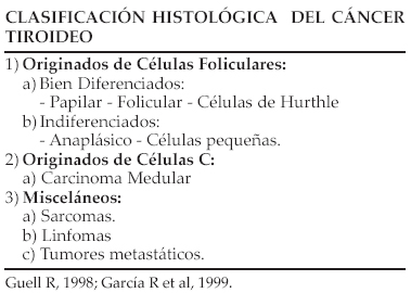
</front><body><![CDATA[ <p style="text-align: center; text-autospace; margin-top: 0px; margin-bottom: 0px" align="center"> <b><span style="color: rgb(35, 31, 32); font-family: Verdana">Carcinoma bien  diferenciado de tiroides</span></b></p>     <p style="text-align: center; text-autospace; margin-top: 0px; margin-bottom: 0px" align="center">&nbsp;</p>     <p style="text-align: center; text-autospace; margin-top: 0px; margin-bottom: 0px" align="center"> <b><span style="color: rgb(35, 31, 32); font-family: Verdana"><font size="2"> Dra. Isabel Benítez </font></span></b></p>     <p style="text-align: center; text-autospace; margin-top: 0px; margin-bottom: 0px" align="center">&nbsp;</p>     <p style="text-align: center; text-autospace; margin-top: 0px; margin-bottom: 0px" align="center"> <span style="color: rgb(35, 31, 32); font-family: Verdana"><font size="2"> Coordinadora de la Unidad de Diabetes y Endocrinología. Hospital “José Gregorio  Hernández” Trujillo, Venezuela.</font></span></p>     <p style="text-align: justify; text-autospace; margin-top: 0px; margin-bottom: 0px">&nbsp;</p>     <p style="text-align: justify; text-autospace; margin-top: 0px; margin-bottom: 0px"> <span style="color: rgb(35, 31, 32); font-family: Verdana"><font size="2">Aunque  la enfermedad nodular tiroidea es una patología bastante frecuente, los  carcinomas tiroideos son relativamente raros, estos constituyen el 1% de los  tumores malignos. La tasa de incidencia del carcinoma tiroideo varia a nivel  mundial entre 0,5 a 10 casos por 100.000 habitantes, sin embargo es la condición  maligna endocrina más frecuente (90%) y causa más muertes que todos los cánceres  endocrinos combinados, (64%). (AACE, 1997) </font></span></p>     <p style="text-align: justify; text-autospace; margin-top: 0px; margin-bottom: 0px">&nbsp;</p>     <p style="text-align: center; text-autospace; margin-top: 0px; margin-bottom: 0px"> <span style="color: rgb(35, 31, 32); font-family: Verdana"><font size="2"> <img src="/img/fbpe/rvdem/v5n2/art13img1.jpg"></font></span></p>     
<p style="text-align: justify; text-autospace; margin-top: 0px; margin-bottom: 0px">&nbsp;</p>     ]]></body>
<body><![CDATA[<p style="text-align: center; text-autospace; margin-top: 0px; margin-bottom: 0px"> <span style="color: rgb(35, 31, 32); font-family: Verdana"><font size="2"> <img src="/img/fbpe/rvdem/v5n2/art13img2.jpg"></font></span></p>     
<p style="text-align: center; text-autospace; margin-top: 0px; margin-bottom: 0px"> &nbsp;</p>     <p style="text-align: center; text-autospace; margin-top: 0px; margin-bottom: 0px"> <span style="color: rgb(35, 31, 32); font-family: Verdana"><font size="2"> <img src="/img/fbpe/rvdem/v5n2/art13img3.jpg"></font></span></p>     
<p style="text-align: justify; text-autospace; margin-top: 0px; margin-bottom: 0px">&nbsp;</p>     <p style="text-align: justify; text-autospace; margin-top: 0px; margin-bottom: 0px"> <b><span style="color: rgb(35, 31, 32); font-family: Verdana"><font size="2"> Tratamiento del carcinoma bien diferenciado de tiroides metas del tratamiento  inicial </font></span></b></p>     <p style="text-align: justify; text-autospace; margin-top: 0px; margin-bottom: 0px">&nbsp;</p>     <p style="text-align: justify; text-autospace; margin-top: 0px; margin-bottom: 0px"> <span style="color: rgb(35, 31, 32); font-family: Verdana"><font size="2"> Excéresis completa del tejido patológico existente en cuello (tumor primario,  invasión fuera de la cápsula y nódulos linfáticos afectados). </font></span></p>     <p style="text-align: justify; text-autospace; margin-top: 0px; margin-bottom: 0px"> <span style="color: rgb(35, 31, 32); font-family: Verdana"><font size="2"> Minimizar la morbilidad relacionada con la enfermedad y efectos del tratamiento.</font></span></p>     <p style="text-align: justify; text-autospace; margin-top: 0px; margin-bottom: 0px"> <span style="color: rgb(35, 31, 32); font-family: Verdana"><font size="2"> Permitir una exacta estratificación de la enfermedad. Facilitar el tratamiento  postoperatorio con <sup>131</sup>I. </font></span></p>     <p style="text-align: justify; text-autospace; margin-top: 0px; margin-bottom: 0px"> <span style="color: rgb(35, 31, 32); font-family: Verdana"><font size="2"> Permitir una adecuada vigilancia para recurrencia de la enfermedad. </font> </span></p>     ]]></body>
<body><![CDATA[<p style="text-align: justify; text-autospace; margin-top: 0px; margin-bottom: 0px"> <span style="color: rgb(35, 31, 32); font-family: Verdana"><font size="2"> Disminuir el riesgo de recurrencia y metástasis. </font></span></p>     <p style="text-align: justify; text-autospace; margin-top: 0px; margin-bottom: 0px"> <span style="color: rgb(35, 31, 32); font-family: Verdana"><font size="2">Con la  finalidad de lograr estas metas se recomienda un estudio de extensión previo que  permita plantear el tipo de intervención ideal. Este podría ser realizado con  una gammagrafía con yodo-talio ó sólo con talio y la ecografía cervical más  citología de la adenopatía sospechosa. </font></span></p>     <p style="text-align: justify; text-autospace; margin-top: 0px; margin-bottom: 0px">&nbsp;</p>     <p style="text-align: justify; text-autospace; margin-top: 0px; margin-bottom: 0px"> <b><span style="color: rgb(35, 31, 32); font-family: Verdana"><font size="2"> Tratamiento quirúrgico </font></span></b></p>     <p style="text-align: justify; text-autospace; margin-top: 0px; margin-bottom: 0px">&nbsp;</p>     <p style="text-align: justify; text-autospace; margin-top: 0px; margin-bottom: 0px"> <span style="color: rgb(35, 31, 32); font-family: Verdana"><font size="2">La  curación del carcinoma diferenciado de tiroides se inicia en quirófano con un  planteamiento correcto y la técnica quirúrgica adecuada.Las opciones quirúrgicas  están limitadas a dos posibilidades: </font></span></p>     <p style="text-align: justify; text-autospace; margin-top: 0px; margin-bottom: 0px">&nbsp;</p>     <p style="text-align: justify; text-autospace; margin-top: 0px; margin-bottom: 0px"> <span style="color: rgb(35, 31, 32); font-family: Verdana"><font size="2"> Tiroidectomía casi total: se remueve todo el tejido tiroideo visible dejando  sólo una pequeña cantidad (&lt;1gr) cerca de la inserción del nervio laríngeo  recurrente.</font></span></p>     <p style="text-align: justify; text-autospace; margin-top: 0px; margin-bottom: 0px">&nbsp;</p>     <p style="text-align: justify; text-autospace; margin-top: 0px; margin-bottom: 0px"> <span style="color: rgb(35, 31, 32); font-family: Verdana"><font size="2"> Tiroidectomía total. La lobectomía es considerada como el único tratamiento  necesario sólo en los casos de carcinoma papilar intratiroideo, aislado,  pequeño, en ausencia de metástasis a ganglios cervicales y en pacientes de bajo  riesgo. </font></span></p>     ]]></body>
<body><![CDATA[<p style="text-align: justify; text-autospace; margin-top: 0px; margin-bottom: 0px">&nbsp;</p>     <p style="text-align: justify; text-autospace; margin-top: 0px; margin-bottom: 0px"> <b><span style="color: rgb(35, 31, 32); font-family: Verdana"><font size="2"> Justificación de la Tiroidectomía total: </font></span></b></p>     <p style="text-align: justify; text-autospace; margin-top: 0px; margin-bottom: 0px"> <span style="color: rgb(35, 31, 32); font-family: Verdana"><font size="2"> Permite el uso del <sup>131</sup>I: ablación de restos y diagnóstico y  tratamiento de persistencias, recidivas y mt. </font></span></p>     <p style="text-align: justify; text-autospace; margin-top: 0px; margin-bottom: 0px"> <span style="color: rgb(35, 31, 32); font-family: Verdana"><font size="2"> Permite el uso de Tg para seguimiento. </font></span></p>     <p style="text-align: justify; text-autospace; margin-top: 0px; margin-bottom: 0px"> <span style="color: rgb(35, 31, 32); font-family: Verdana"><font size="2">Evita  recidivas en el lóbulo contralateral. </font></span></p>     <p style="text-align: justify; text-autospace; margin-top: 0px; margin-bottom: 0px"> <span style="color: rgb(35, 31, 32); font-family: Verdana"><font size="2">Evita  la transformación en carcinoma anaplásico. </font></span></p>     <p style="text-align: justify; text-autospace; margin-top: 0px; margin-bottom: 0px"> <span style="color: rgb(35, 31, 32); font-family: Verdana"><font size="2"> Aumenta la supervivencia en Tu &gt; 1 cm. </font></span></p>     <p style="text-align: justify; text-autospace; margin-top: 0px; margin-bottom: 0px"> <span style="color: rgb(35, 31, 32); font-family: Verdana"><font size="2"> Disminuye la necesidad de reintervención. </font></span></p>     <p style="text-align: justify; text-autospace; margin-top: 0px; margin-bottom: 0px">&nbsp;</p>     <p style="text-align: justify; text-autospace; margin-top: 0px; margin-bottom: 0px"> <span style="color: rgb(35, 31, 32); font-family: Verdana"><font size="2">En  cuanto a la actuación sobre ganglios linfáticos es recomendable la disección de  los ganglios del compartimiento central y la disección ipsilateral ó bilateral  radical modificada (compartimiento II-V) si se demuestra afectación ganglionar y  en todos los pacientes con alto riesgo. </font></span></p>     ]]></body>
<body><![CDATA[<p style="text-align: justify; text-autospace; margin-top: 0px; margin-bottom: 0px">&nbsp;</p>     <p style="text-align: justify; text-autospace; margin-top: 0px; margin-bottom: 0px"> <span style="color: rgb(35, 31, 32); font-family: Verdana"><font size="2">Una  vez realizada la cirugía, debe hacerse la estratificación del paciente, ya que  nos permite establecer: </font></span></p>     <p style="text-align: justify; text-autospace; margin-top: 0px; margin-bottom: 0px">&nbsp;</p>     <p style="text-align: justify; text-autospace; margin-top: 0px; margin-bottom: 0px"> <span style="color: rgb(35, 31, 32); font-family: Verdana"><font size="2">El  pronóstico individual de cada paciente. </font></span></p>     <p style="text-align: justify; text-autospace; margin-top: 0px; margin-bottom: 0px"> <span style="color: rgb(35, 31, 32); font-family: Verdana"><font size="2">La  terapia postoperatoria adecuada: <sup>131</sup>I y supresión de TSH. </font> </span></p>     <p style="text-align: justify; text-autospace; margin-top: 0px; margin-bottom: 0px"> <span style="color: rgb(35, 31, 32); font-family: Verdana"><font size="2"> Determinar la secuencia y la intensidad del seguimiento. </font></span></p>     <p style="text-align: justify; text-autospace; margin-top: 0px; margin-bottom: 0px"> <span style="color: rgb(35, 31, 32); font-family: Verdana"><font size="2">La  unificación de criterios sobre el tratamiento en el equipo médico. </font> </span></p>     <p style="text-align: justify; text-autospace; margin-top: 0px; margin-bottom: 0px">&nbsp;</p>     <p style="text-align: justify; text-autospace; margin-top: 0px; margin-bottom: 0px"> <span style="color: rgb(35, 31, 32); font-family: Verdana"><font size="2"> Existen muchos sistemas de clasificación, pero el sistema TNM es el más  recomendado para todos los tumores incluido el cáncer de tiroides, por  considerar que identifica mejor los factores necesarios para la clasificación  individual de riesgo de cada paciente. </font></span></p>     <p style="text-align: justify; text-autospace; margin-top: 0px; margin-bottom: 0px">&nbsp;</p>     ]]></body>
<body><![CDATA[<p style="text-align: justify; text-autospace; margin-top: 0px; margin-bottom: 0px"> <b><span style="color: rgb(35, 31, 32); font-family: Verdana"><font size="2"> Tratamiento con Radioyodo </font></span></b></p>     <p style="text-align: justify; text-autospace; margin-top: 0px; margin-bottom: 0px">&nbsp;</p>     <p style="text-align: justify; text-autospace; margin-top: 0px; margin-bottom: 0px"> <span style="color: rgb(35, 31, 32); font-family: Verdana"><font size="2">Se ha  considerado como parte del tratamiento obligatorio del cáncer diferenciado de  tiroides, sin embargo actualmente representa un tema controversial en cuanto a  sus beneficios en ciertas situaciones y la necesidad de aplicación a todos los  pacientes, pues actualmente algunos autores sugieren obviar la ablación en  sujetos de bajo riesgo (Tu &lt; 1,5cm y sin afectación ganglionar, en pacientes  jóvenes y con tumores pequeños). Se ha visto además que no existen diferencias  en las recidivas a 30 años tras la utilización de dosis bajas (29 a 50 mCi) y  altas (51 a 200 mCi). (Rubial M.A., et al, 2005) Por el contrario hay quienes  sugieren que la terapia de ablación del tejido remanente con <sup>131</sup>I  debería ser usada en todos los pacientes ya que destruye los restos  microscópicos del cáncer, facilita la detección de enfermedad persistente ó  recurrente con rastreos corporales y mejora la sensibilidad de la Tg para el  seguimiento </font></span></p>     <p style="text-align: justify; text-autospace; margin-top: 0px; margin-bottom: 0px">&nbsp;</p>     <p style="text-align: justify; text-autospace; margin-top: 0px; margin-bottom: 0px"> <b><span style="color: rgb(35, 31, 32); font-family: Verdana"><font size="2"> Protocolo para el tratamiento ablativo con Radioyodo </font></span></b></p>     <p style="text-align: justify; text-autospace; margin-top: 0px; margin-bottom: 0px">&nbsp;</p>     <p style="text-align: justify; text-autospace; margin-top: 0px; margin-bottom: 0px"> <span style="color: rgb(35, 31, 32); font-family: Verdana"><font size="2">Para  todos se recomienda una dieta pobre en yodo (&lt; 50ug/día) 2 semanas antes del  tratamiento. </font></span></p>     <p style="text-align: justify; text-autospace; margin-top: 0px; margin-bottom: 0px"> <span style="color: rgb(35, 31, 32); font-family: Verdana"><font size="2">Con  Retiro de T4: Retiro de la toma de LT4 durante 4 a 6 semanas ó Retiro de toma de  LT4 e inicio de toma de T3 50ug/día durante 3 semanas. </font></span></p>     <p style="text-align: justify; text-autospace; margin-top: 0px; margin-bottom: 0px"> <span style="color: rgb(35, 31, 32); font-family: Verdana"><font size="2">Retiro  de toma de T3 durante 2 semanas. </font></span></p>     <p style="text-align: justify; text-autospace; margin-top: 0px; margin-bottom: 0px"> <span style="color: rgb(35, 31, 32); font-family: Verdana"><font size="2"> Demostración de elevación de TSH &gt; 30 mU/l. </font></span></p>     ]]></body>
<body><![CDATA[<p style="text-align: justify; text-autospace; margin-top: 0px; margin-bottom: 0px"> <span style="color: rgb(35, 31, 32); font-family: Verdana"><font size="2">Toma  de muestra para TSH, Tg y Ac-Tg y administración de <sup>131</sup>I. </font> </span></p>     <p style="text-align: justify; text-autospace; margin-top: 0px; margin-bottom: 0px"> <span style="color: rgb(35, 31, 32); font-family: Verdana"><font size="2"> Reinicio del tratamiento con LT4 entre el día 2 y 5 del <sup>131</sup>I.</font></span><span style="color: black; font-family: Verdana"><font size="2"> </font></span></p>     <p style="text-align: justify; text-autospace; margin-top: 0px; margin-bottom: 0px"> <span style="color: rgb(35, 31, 32); font-family: Verdana"><font size="2"> Realización de rastreo de 3 a 10 días después de la administración del <sup>131</sup>I. </font></span></p>     <p style="text-align: justify; text-autospace; margin-top: 0px; margin-bottom: 0px">&nbsp;</p>     <p style="text-align: justify; text-autospace; margin-top: 0px; margin-bottom: 0px"> <b><span style="color: rgb(35, 31, 32); font-family: Verdana"><font size="2">Con  TSH recombinante humana: </font></span></b></p>     <p style="text-align: justify; text-autospace; margin-top: 0px; margin-bottom: 0px"> <span style="color: rgb(35, 31, 32); font-family: Verdana"><font size="2">La  administración de TSHrh puede utilizarse cuando: no se tolera el hipotiroidismo,  las metástasis se encuentran en sitios potencialmente peligrosos o cuando hay  imposibilidad para aumentar satisfactoriamente TSH después de la supresión de  tiroxina. </font></span></p>     <p style="text-align: justify; text-autospace; margin-top: 0px; margin-bottom: 0px">&nbsp;</p>     <p style="text-align: justify; text-autospace; margin-top: 0px; margin-bottom: 0px"> <span style="color: rgb(35, 31, 32); font-family: Verdana"><font size="2">Día 1:  toma de muestra para TSH, Tg y Ac-Tg, previa administración de 0,9 mg de TSHrh  VIM. </font></span></p>     <p style="text-align: justify; text-autospace; margin-top: 0px; margin-bottom: 0px"> <span style="color: rgb(35, 31, 32); font-family: Verdana"><font size="2">Día 2:  se administra una nueva dosis de 0,9 mg de TSHrh VIM. </font></span></p>     <p style="text-align: justify; text-autospace; margin-top: 0px; margin-bottom: 0px"> <span style="color: rgb(35, 31, 32); font-family: Verdana"><font size="2">Día 3  ó 4: administración de dosis terapéutica de <sup>131</sup>I, con determinación  de TSH, Tg y Ac-Tg </font></span></p>     ]]></body>
<body><![CDATA[<p style="text-align: justify; text-autospace; margin-top: 0px; margin-bottom: 0px"> <span style="color: rgb(35, 31, 32); font-family: Verdana"><font size="2"> Realización de rastreo de 3 a 10 días después de la administración del <sup>131</sup>I. </font></span></p>     <p style="text-align: justify; text-autospace; margin-top: 0px; margin-bottom: 0px"> <span style="color: rgb(35, 31, 32); font-family: Verdana"><font size="2">La  ablación satisfactoria de restos tiroideos es definida como la ausencia de  captación en estudios diagnósticos posteriores con <sup>131</sup>I. </font> </span></p>     <p style="text-align: justify; text-autospace; margin-top: 0px; margin-bottom: 0px">&nbsp;</p>     <p style="text-align: justify; text-autospace; margin-top: 0px; margin-bottom: 0px"> <b><span style="color: rgb(35, 31, 32); font-family: Verdana"><font size="2"> Terapia supresiva con TSH </font></span></b></p>     <p style="text-align: justify; text-autospace; margin-top: 0px; margin-bottom: 0px">&nbsp;</p>     <p style="text-align: justify; text-autospace; margin-top: 0px; margin-bottom: 0px"> <span style="color: rgb(35, 31, 32); font-family: Verdana"><font size="2">Debido  al aumento de la tasa de crecimiento celular como respuesta a la estimulación de  TSH en las células del carcinoma tiroideo diferenciado, la administración de  dosis suprafisiológicas de LT4 para suprimir TSH es utilizada comúnmente en el  tratamiento de este tipo de pacientes. Dosis: </font></span></p>     <p style="text-align: justify; text-autospace; margin-top: 0px; margin-bottom: 0px">&nbsp;</p>     <p style="text-align: justify; text-autospace; margin-top: 0px; margin-bottom: 0px"> <span style="color: rgb(35, 31, 32); font-family: Verdana"><font size="2">- En  pacientes de alto riesgo: LT4: 2,2 a 2,8ug/Kg/día, ajustando el tratamiento  individual con el objetivo de lograr TSH &lt; 0,1uU/ml. </font></span></p>     <p style="text-align: justify; text-autospace; margin-top: 0px; margin-bottom: 0px"> <span style="color: rgb(35, 31, 32); font-family: Verdana"><font size="2">- En  pacientes de bajo riesgo: se usan dosis sustitutivas de LT4 para obtener TSH de  0,4 a 2uU/ml. </font></span></p>     <p style="text-align: justify; text-autospace; margin-top: 0px; margin-bottom: 0px">&nbsp;</p>     ]]></body>
<body><![CDATA[<p style="text-align: justify; text-autospace; margin-top: 0px; margin-bottom: 0px"> <b><span style="color: rgb(35, 31, 32); font-family: Verdana"><font size="2"> Seguimiento del carcinoma diferenciado de tiroides </font></span></b></p>     <p style="text-align: justify; text-autospace; margin-top: 0px; margin-bottom: 0px">&nbsp;</p>     <p style="text-align: justify; text-autospace; margin-top: 0px; margin-bottom: 0px"> <span style="color: rgb(35, 31, 32); font-family: Verdana"><font size="2">En  pacientes con tiroidectomía total o casi total, el criterio de ausencia de  enfermedad está dado por lo siguiente: no evidencia clínica de tumor, no  evidencia de tumor por estudios de imagen, valores de Tg circulante  indetectables con TSH &gt; 25uU/ml y con Ac- Tg negativos. El seguimiento es  diferente para cada paciente según el riesgo que posea de tener persistencia o  recurrencia de la enfermedad. </font></span></p>     <p style="text-align: justify; text-autospace; margin-top: 0px; margin-bottom: 0px">&nbsp;</p>     <p style="text-align: justify; text-autospace; margin-top: 0px; margin-bottom: 0px"> <span style="color: rgb(35, 31, 32); font-family: Verdana"><font size="2">1) </font></span><b><span style="color: rgb(35, 31, 32); font-family: Verdana"> <font size="2">Seguimiento a corto plazo</font></span></b><span style="color: rgb(35, 31, 32); font-family: Verdana"><font size="2">: </font></span></p>     <p style="text-align: justify; text-autospace; margin-top: 0px; margin-bottom: 0px">&nbsp;</p>     <p style="text-align: justify; text-autospace; margin-top: 0px; margin-bottom: 0px"> <span style="color: rgb(35, 31, 32); font-family: Verdana"><font size="2">A los  6 o 12 meses del tratamiento quirúrgico y con radioyodo se valoran: ultrasonido  cervical, Tg y Ac-Tg con TSH &gt; 25 uU/ml, con retiro previo de la terapia  supresita: </font></span></p>     <p style="text-align: justify; text-autospace; margin-top: 0px; margin-bottom: 0px">&nbsp;</p>     <p style="text-align: justify; text-autospace; margin-top: 0px; margin-bottom: 0px"> <span style="color: rgb(35, 31, 32); font-family: Verdana"><font size="2">a) El  resultado de Tg indetectable con Ac-Tg negativos y TSH elevada indica remisión  de la enfermedad. Después de un año de tratamiento y con iguales resultados los  pacientes deben ser reevaluados en 1 a 3 años. </font></span></p>     <p style="text-align: justify; text-autospace; margin-top: 0px; margin-bottom: 0px"> <span style="color: rgb(35, 31, 32); font-family: Verdana"><font size="2">b) Tg  igual o menor a 2 ng/ml en Tu de bajo riesgo y con Ac-Tg negativos y TSH elevada  también puede considerarse como remisión. </font></span></p>     ]]></body>
<body><![CDATA[<p style="text-align: justify; text-autospace; margin-top: 0px; margin-bottom: 0px"> <span style="color: rgb(35, 31, 32); font-family: Verdana"><font size="2">c) Tg  &gt;2 ng/ml en iguales condiciones para Tu de cualquier nivel de riesgo indica  persistencia de enfermedad y justifica estudios de localización de tumor y una  nueva dosis ablativo de <sup>131</sup>I. </font></span></p>     <p style="text-align: justify; text-autospace; margin-top: 0px; margin-bottom: 0px"> <span style="color: rgb(35, 31, 32); font-family: Verdana"><font size="2">d) Tg  &gt; 10 ng/ml en paciente con tiroidectomía uni o bilateral, con o sin terapia  ablativo de remanente tiroideo, indica la necesidad de otros estudios  diagnósticos. </font></span></p>     <p style="text-align: justify; text-autospace; margin-top: 0px; margin-bottom: 0px">&nbsp;</p>     <p style="text-align: justify; text-autospace; margin-top: 0px; margin-bottom: 0px"> <span style="color: rgb(35, 31, 32); font-family: Verdana"><font size="2">2) </font></span><b><span style="color: rgb(35, 31, 32); font-family: Verdana"> <font size="2">Seguimiento a largo plazo</font></span></b><span style="color: rgb(35, 31, 32); font-family: Verdana"><font size="2">: </font></span></p>     <p style="text-align: justify; text-autospace; margin-top: 0px; margin-bottom: 0px">&nbsp;</p>     <p style="text-align: justify; text-autospace; margin-top: 0px; margin-bottom: 0px"> <span style="color: rgb(35, 31, 32); font-family: Verdana"><font size="2">a)  Luego de 2 años con diagnóstico de remisión se disminuirá la dosis de LT4 hasta  una dosis sustitutiva que permita valores de TSH no &gt; 2uU/ ml. Cada 6 a 12 meses  se realizará seguimiento con Tg, Ac-Tg y TSH, examen físico y ecografía de  cuello. Confirmada la remisión en otros dos años se valorará sólo Tg basal bajo  tratamiento sustitutivo con LT4. Un posterior aumento de Tg es indicativo de  medición de Tg con TSH elevada. </font></span></p>     <p style="text-align: justify; text-autospace; margin-top: 0px; margin-bottom: 0px"> <span style="color: rgb(35, 31, 32); font-family: Verdana"><font size="2">b) En  tumores de riesgo moderado o alto en remisión, se cuantificará Tg con TSH  elevada cada 2 años durante 8 a 10 años. </font></span></p>     <p style="text-align: justify; text-autospace; margin-top: 0px; margin-bottom: 0px"> <span style="color: rgb(35, 31, 32); font-family: Verdana"><font size="2">c) En  tumores sin remisión se cuantificará Tg con TSH suprimida y ecografía de cuello  de alta resolución cada 3 meses. La presencia de masas tumorales con captación &gt;  2% de <sup>131</sup>I y accesibles a cirugía son candidatas a tratamiento  quirúrgico previo al tratamiento ablativo con radioyodo. Puede requerirse de  otros métodos: TAC, RM, FDG-PET, en casos de Tg elevada y rastreo corporal  negativo. El hallazgo de imágenes sospechosas requiere la confirmación con  citología por punción. </font></span></p>     <p style="text-align: justify; text-autospace; margin-top: 0px; margin-bottom: 0px"> <span style="color: rgb(35, 31, 32); font-family: Verdana"><font size="2">d)  Estudio morfológico negativo, con PAAF negativa y Tg &lt; 10ng/ml con TSH elevada  requiere la determinación de Tg cada 3 a 6 meses por 2 años y luego anual si es  confirmada la remisión. </font></span></p>     <p style="text-align: justify; text-autospace; margin-top: 0px; margin-bottom: 0px"> <span style="color: rgb(35, 31, 32); font-family: Verdana"><font size="2">e)  Estudio morfológico sospechoso, confirmado con PAAF o Tg&gt; o igual a 10 ng/ml con  TSH suprimida condiciona dos posibles conductas: cirugía si es posible y/ó nueva  dosis de radioyodo. La persistencia de Tg elevada excluye nuevas dosis de  radioyodo. </font></span></p>     ]]></body>
<body><![CDATA[<p style="text-align: justify; text-autospace; margin-top: 0px; margin-bottom: 0px"> <span style="color: rgb(35, 31, 32); font-family: Verdana"><font size="2">f) Si  la presencia de enfermedad sin criterios de cirugía es demostrada después de una  dosis empírica de <sup>131</sup>I, y hay evidencias de disminución del tumor, la  terapia debe ser repetida hasta la erradicación del tumor o hasta que no  responda más al tratamiento. Debe evaluarse el riesgo de dosis frecuentes de <sup>131</sup>I.</font></span><span style="color: black; font-family: Verdana"><font size="2"> </font></span></p>     <p style="text-align: justify; text-autospace; margin-top: 0px; margin-bottom: 0px">&nbsp;</p>     <p style="text-align: justify; text-autospace; margin-top: 0px; margin-bottom: 0px"> <b><span style="color: rgb(35, 31, 32); font-family: Verdana"><font size="2"> Otras opciones terapéuticas en el carcinoma diferenciado de tiroides </font> </span></b></p>     <p style="text-align: justify; text-autospace; margin-top: 0px; margin-bottom: 0px">&nbsp;</p>     <p style="text-align: justify; text-autospace; margin-top: 0px; margin-bottom: 0px"> <b><span style="color: rgb(35, 31, 32); font-family: Verdana"><font size="2"> Radioterapia Externa</font></span></b><span style="color: rgb(35, 31, 32); font-family: Verdana"><font size="2">:  es útil en el manejo de la enfermedad cervical irresecable, metástasis dolorosas  en hueso o localizadas en sitios críticos ó ante la presencia de síntomas  compresivos sin criterio quirúrgico. (Cooper D., et al 2006) </font></span></p>     <p style="text-align: justify; text-autospace; margin-top: 0px; margin-bottom: 0px">&nbsp;</p>     <p style="text-align: justify; text-autospace; margin-top: 0px; margin-bottom: 0px"> <b><span style="color: rgb(35, 31, 32); font-family: Verdana"><font size="2"> Quimioterapia</font></span></b><span style="color: rgb(35, 31, 32); font-family: Verdana"><font size="2">:  podría ser de discreta utilidad en lesiones resistentes al tratamiento con  radioyodo. La Doxorubicina ha demostrado ser relativamente eficaz en la  estabilización de la enfermedad cuando se usa a dosis: 60 – 75 mg/m2 cada 3  semanas. </font></span></p>     <p style="text-align: justify; text-autospace; margin-top: 0px; margin-bottom: 0px">&nbsp;</p>     <p style="text-align: justify; text-autospace; margin-top: 0px; margin-bottom: 0px"> <b><span style="color: rgb(35, 31, 32); font-family: Verdana"><font size="2"> Nuevas Terapias</font></span></b><span style="color: rgb(35, 31, 32); font-family: Verdana"><font size="2">:  Inhibidores de oncogenes. Moduladores de crecimiento o apoptosis. Inhibidores de  la angiogénesis. Inmunomoduladores. Terapia génica.</font></span></p>     <p style="text-align: justify; text-autospace; margin-top: 0px; margin-bottom: 0px">&nbsp;</p>     ]]></body>
<body><![CDATA[<p style="text-align: justify; text-autospace; margin-top: 0px; margin-bottom: 0px"> <b><span style="color: rgb(35, 31, 32); font-family: Verdana"><font size="2"> Referencias bibliográficas</font></span></b></p>     <p style="text-align: justify; text-autospace; margin-top: 0px; margin-bottom: 0px">&nbsp;</p>     <!-- ref --><p style="text-align: justify; text-autospace; margin-top: 0px; margin-bottom: 0px"> <span style="color: rgb(35, 31, 32); font-family: Verdana"><font size="2">1.  Cobhin, Rhoda H., Gharib, Hossein. </font></span> <span style="color: rgb(35, 31, 32); font-family: Verdana" lang="EN-GB"> <font size="2">Management of Thyroid Carcinoma. AACE/AAES Medical/Surgical  Guidelines for Clinical Practice. Thyroid Carcinoma Task Force. Endocrine  Practice. 7:3, 2001. </font></span>&nbsp;&nbsp;&nbsp;&nbsp;&nbsp;&nbsp;&nbsp;&nbsp;[&#160;<a href="javascript:void(0);" onclick="javascript: window.open('/scielo.php?script=sci_nlinks&ref=3060763&pid=S1690-3110200700020001300001&lng=','','width=640,height=500,resizable=yes,scrollbars=1,menubar=yes,');">Links</a>&#160;]<!-- end-ref --><p style="text-align: justify; text-autospace; margin-top: 0px; margin-bottom: 0px">&nbsp;</p>     <!-- ref --><p style="text-align: justify; text-autospace; margin-top: 0px; margin-bottom: 0px"> <span style="color: rgb(35, 31, 32); font-family: Verdana" lang="EN-GB"> <font size="2">2. Cooper, David S., Doherty, Gerard M., Haugen Bryan R., Kools  Richard T., Lee Sthephanie L., mandel Susan J., Mazzaferri Ernest L., McIver  Bryan, Sherman Steven I., and Tuttle R. Michael. Management Guidelines for  Patients with Thyroid Nodules and Differentiated Thyroid Cancer. The American  Thyroid Association Guidelines Taskforce. 16:2, 2006.</font></span>&nbsp;&nbsp;&nbsp;&nbsp;&nbsp;&nbsp;&nbsp;&nbsp;[&#160;<a href="javascript:void(0);" onclick="javascript: window.open('/scielo.php?script=sci_nlinks&ref=3060765&pid=S1690-3110200700020001300002&lng=','','width=640,height=500,resizable=yes,scrollbars=1,menubar=yes,');">Links</a>&#160;]<!-- end-ref --><p style="text-align: justify; text-autospace; margin-top: 0px; margin-bottom: 0px">&nbsp;</p>     <!-- ref --><p style="text-align: justify; text-autospace; margin-top: 0px; margin-bottom: 0px"> <span style="color: rgb(35, 31, 32); font-family: Verdana" lang="EN-GB"> <font size="2">3. De Groot leslie J., Kaplan Edwin L., McCormick Maureen and  Status Francis H. Natural history, treatment and course of papillary thyroid  carcinoma. J Clin Endocrinol Metab. </font></span> <span style="color: rgb(35, 31, 32); font-family: Verdana"><font size="2">1990.  71(2): 414-424.</font></span>&nbsp;&nbsp;&nbsp;&nbsp;&nbsp;&nbsp;&nbsp;&nbsp;[&#160;<a href="javascript:void(0);" onclick="javascript: window.open('/scielo.php?script=sci_nlinks&ref=3060767&pid=S1690-3110200700020001300003&lng=','','width=640,height=500,resizable=yes,scrollbars=1,menubar=yes,');">Links</a>&#160;]<!-- end-ref --><p style="text-align: justify; text-autospace; margin-top: 0px; margin-bottom: 0px">&nbsp;</p>     <!-- ref --><p style="text-align: justify; text-autospace; margin-top: 0px; margin-bottom: 0px"> <span style="color: rgb(35, 31, 32); font-family: Verdana"><font size="2">4.  García González R., Sanz González de Suso I., Cuesta Roca C. Anatomía  Patológica: Citología e Histopatología. En: Dieguez C., Pavia C., Yturriaga R.  Actualizaciones en Endocrinología: Tiroides. 1ra. Edición. España: McGraw-  Hill-Interamericana. 1999. p.p.145-164.</font></span>&nbsp;&nbsp;&nbsp;&nbsp;&nbsp;&nbsp;&nbsp;&nbsp;[&#160;<a href="javascript:void(0);" onclick="javascript: window.open('/scielo.php?script=sci_nlinks&ref=3060769&pid=S1690-3110200700020001300004&lng=','','width=640,height=500,resizable=yes,scrollbars=1,menubar=yes,');">Links</a>&#160;]<!-- end-ref --><p style="text-align: justify; text-autospace; margin-top: 0px; margin-bottom: 0px">&nbsp;</p>     ]]></body>
<body><![CDATA[<!-- ref --><p style="text-align: justify; text-autospace; margin-top: 0px; margin-bottom: 0px"> <span style="color: rgb(35, 31, 32); font-family: Verdana"><font size="2">5.  Gómez Sáez J.M. Cáncer de Tiroides. En: Dieguez C., Pavia C., Yturriaga R.  Actualizaciones en Endocrinología: Tiroides. 1ra. Edición. España: McGraw-Hill-  Interamericana. 1999. p.p. 271-282.</font></span>&nbsp;&nbsp;&nbsp;&nbsp;&nbsp;&nbsp;&nbsp;&nbsp;[&#160;<a href="javascript:void(0);" onclick="javascript: window.open('/scielo.php?script=sci_nlinks&ref=3060771&pid=S1690-3110200700020001300005&lng=','','width=640,height=500,resizable=yes,scrollbars=1,menubar=yes,');">Links</a>&#160;]<!-- end-ref --><p style="text-align: justify; text-autospace; margin-top: 0px; margin-bottom: 0px">&nbsp;</p>     <!-- ref --><p style="text-align: justify; text-autospace; margin-top: 0px; margin-bottom: 0px"> <span style="color: rgb(35, 31, 32); font-family: Verdana"><font size="2">6.  Gómez J.M. Futuro del Tratamiento del carcinoma de Tiroides. Endocrinología y  Nutrición. 2005; 52(Supl 1) 45–50. </font></span>&nbsp;&nbsp;&nbsp;&nbsp;&nbsp;&nbsp;&nbsp;&nbsp;[&#160;<a href="javascript:void(0);" onclick="javascript: window.open('/scielo.php?script=sci_nlinks&ref=3060773&pid=S1690-3110200700020001300006&lng=','','width=640,height=500,resizable=yes,scrollbars=1,menubar=yes,');">Links</a>&#160;]<!-- end-ref --><p style="text-align: justify; text-autospace; margin-top: 0px; margin-bottom: 0px">&nbsp;</p>     <!-- ref --><p style="text-align: justify; text-autospace; margin-top: 0px; margin-bottom: 0px"> <span style="color: rgb(35, 31, 32); font-family: Verdana"><font size="2">7.  Güell Ricardo. La Glándula Tiroides: Embriología y Fisiopatología. En:  Enfermedades del Tiroides en Niños y Adolescentes. Barcelona: Permanyer  Publications. 1998. p.p. 3-19</font></span>&nbsp;&nbsp;&nbsp;&nbsp;&nbsp;&nbsp;&nbsp;&nbsp;[&#160;<a href="javascript:void(0);" onclick="javascript: window.open('/scielo.php?script=sci_nlinks&ref=3060775&pid=S1690-3110200700020001300007&lng=','','width=640,height=500,resizable=yes,scrollbars=1,menubar=yes,');">Links</a>&#160;]<!-- end-ref --><p style="text-align: justify; text-autospace; margin-top: 0px; margin-bottom: 0px">&nbsp;</p>     <!-- ref --><p style="text-align: justify; text-autospace; margin-top: 0px; margin-bottom: 0px"> <span style="color: rgb(35, 31, 32); font-family: Verdana"><font size="2">8.  Larrad Jiménez A., Sitges Serra A. Avances en el tratamiento Quirúrgico del  Cáncer Diferenciado de Tiroides. Endocrinología y Nutrición. 2005; 52(Supl 1)  11- 17.</font></span>&nbsp;&nbsp;&nbsp;&nbsp;&nbsp;&nbsp;&nbsp;&nbsp;[&#160;<a href="javascript:void(0);" onclick="javascript: window.open('/scielo.php?script=sci_nlinks&ref=3060777&pid=S1690-3110200700020001300008&lng=','','width=640,height=500,resizable=yes,scrollbars=1,menubar=yes,');">Links</a>&#160;]<!-- end-ref --><p style="text-align: justify; text-autospace; margin-top: 0px; margin-bottom: 0px">&nbsp;</p>     <!-- ref --><p style="text-align: justify; text-autospace; margin-top: 0px; margin-bottom: 0px"> <span style="color: rgb(35, 31, 32); font-family: Verdana"><font size="2">9.  Lope Carvajal V., Pollan Santamaría A. M. Epidemiología del Cáncer Diferenciado  de Tiroides. Endocrinología y Nutrición. 2005; 52(Supl 1) 2-10.</font></span>&nbsp;&nbsp;&nbsp;&nbsp;&nbsp;&nbsp;&nbsp;&nbsp;[&#160;<a href="javascript:void(0);" onclick="javascript: window.open('/scielo.php?script=sci_nlinks&ref=3060779&pid=S1690-3110200700020001300009&lng=','','width=640,height=500,resizable=yes,scrollbars=1,menubar=yes,');">Links</a>&#160;]<!-- end-ref --><p style="text-align: justify; text-autospace; margin-top: 0px; margin-bottom: 0px">&nbsp;</p>     ]]></body>
<body><![CDATA[<!-- ref --><p style="text-align: justify; text-autospace; margin-top: 0px; margin-bottom: 0px"> <span style="color: rgb(35, 31, 32); font-family: Verdana"><font size="2">10.  Rubial Morell A., Pubul Nuñez V., Cabezas Agrícola J.M., y Bernabeu Morón I.  Actualización del Tratamiento con Radioyodo: Utilidad de la TSH Recombinante  Humana. Endocrinología y Nutrición. 2005; 52(Supl 1) 18-22.</font></span>&nbsp;&nbsp;&nbsp;&nbsp;&nbsp;&nbsp;&nbsp;&nbsp;[&#160;<a href="javascript:void(0);" onclick="javascript: window.open('/scielo.php?script=sci_nlinks&ref=3060781&pid=S1690-3110200700020001300010&lng=','','width=640,height=500,resizable=yes,scrollbars=1,menubar=yes,');">Links</a>&#160;]<!-- end-ref --><p style="text-align: justify; text-autospace; margin-top: 0px; margin-bottom: 0px">&nbsp;</p>     <!-- ref --><p style="text-align: justify; text-autospace; margin-top: 0px; margin-bottom: 0px"> <span style="color: rgb(35, 31, 32); font-family: Verdana" lang="EN-GB"> <font size="2">11. Samaan Naguib A., Schultz Pamela N., Hickey Robert C.,  Goepfert Helmuth, Haynie Thomas P., Johnston Dennis A. and Ordonez Nelson G. The  results of various modalities of treatment of well-defferentited thyroid  carcinomas: A restrospective review of 1599 patients. </font></span> <span style="color: rgb(35, 31, 32); font-family: Verdana"><font size="2">J Clin  Endocrinol Metab. 1992. 75: 714-720.</font></span>&nbsp;&nbsp;&nbsp;&nbsp;&nbsp;&nbsp;&nbsp;&nbsp;[&#160;<a href="javascript:void(0);" onclick="javascript: window.open('/scielo.php?script=sci_nlinks&ref=3060783&pid=S1690-3110200700020001300011&lng=','','width=640,height=500,resizable=yes,scrollbars=1,menubar=yes,');">Links</a>&#160;]<!-- end-ref --><p style="text-align: justify; text-autospace; margin-top: 0px; margin-bottom: 0px">&nbsp;</p>     <!-- ref --><p style="text-align: justify; text-autospace; margin-top: 0px; margin-bottom: 0px"> <span style="color: rgb(35, 31, 32); font-family: Verdana"><font size="2">12.  Sánchez franco F. Directrices para el Tratamiento del Carcinoma Diferenciado de  Tiroides. Endocrinología y </font></span> <span style="color: rgb(35, 31, 32); font-family: Verdana" lang="EN-GB"> <font size="2">Nutrición. 2005; 52(Supl 1) 23-31.</font></span>&nbsp;&nbsp;&nbsp;&nbsp;&nbsp;&nbsp;&nbsp;&nbsp;[&#160;<a href="javascript:void(0);" onclick="javascript: window.open('/scielo.php?script=sci_nlinks&ref=3060785&pid=S1690-3110200700020001300012&lng=','','width=640,height=500,resizable=yes,scrollbars=1,menubar=yes,');">Links</a>&#160;]<!-- end-ref --><p style="text-align: justify; text-autospace; margin-top: 0px; margin-bottom: 0px">&nbsp;</p>     <!-- ref --><p style="text-align: justify; text-autospace; margin-top: 0px; margin-bottom: 0px"> <span style="color: rgb(35, 31, 32); font-family: Verdana" lang="EN-GB"> <font size="2">13. Tennvall J., Biörklund A., Möller T., Ranstam J., and Akerman  M. Prognostic factors of papillary, follicular and medullary carcinomas of the  thyroid gland. Restrospective multivariate análisis of 216 patients with a  median follow-up of 11 years. Acta Radiological Oncology. 1985. 24 (fasc. 1):  17-24.</font></span>&nbsp;&nbsp;&nbsp;&nbsp;&nbsp;&nbsp;&nbsp;&nbsp;[&#160;<a href="javascript:void(0);" onclick="javascript: window.open('/scielo.php?script=sci_nlinks&ref=3060787&pid=S1690-3110200700020001300013&lng=','','width=640,height=500,resizable=yes,scrollbars=1,menubar=yes,');">Links</a>&#160;]<!-- end-ref --> ]]></body>
<back>
<ref-list>
<ref id="B1">
<label>1</label><nlm-citation citation-type="journal">
<person-group person-group-type="author">
<name>
<surname><![CDATA[Cobhin]]></surname>
<given-names><![CDATA[Rhoda H]]></given-names>
</name>
<name>
<surname><![CDATA[Gharib]]></surname>
<given-names><![CDATA[Hossein]]></given-names>
</name>
</person-group>
<article-title xml:lang="en"><![CDATA[Management of Thyroid Carcinoma: AACE/AAES Medical/Surgical Guidelines for Clinical Practice. Thyroid Carcinoma Task Force.]]></article-title>
<source><![CDATA[Endocrine Practice]]></source>
<year>2001</year>
<volume>7</volume>
<page-range>3</page-range></nlm-citation>
</ref>
<ref id="B2">
<label>2</label><nlm-citation citation-type="book">
<person-group person-group-type="author">
<name>
<surname><![CDATA[Cooper]]></surname>
<given-names><![CDATA[David S]]></given-names>
</name>
<name>
<surname><![CDATA[Doherty]]></surname>
<given-names><![CDATA[Gerard M]]></given-names>
</name>
<name>
<surname><![CDATA[Haugen Bryan]]></surname>
<given-names><![CDATA[R]]></given-names>
</name>
<name>
<surname><![CDATA[Kools]]></surname>
<given-names><![CDATA[Richard T]]></given-names>
</name>
<name>
<surname><![CDATA[Lee]]></surname>
<given-names><![CDATA[Sthephanie L]]></given-names>
</name>
<name>
<surname><![CDATA[mandel]]></surname>
<given-names><![CDATA[Susan J]]></given-names>
</name>
<name>
<surname><![CDATA[Mazzaferri]]></surname>
<given-names><![CDATA[Ernest L]]></given-names>
</name>
<name>
<surname><![CDATA[McIver]]></surname>
<given-names><![CDATA[Bryan]]></given-names>
</name>
<name>
<surname><![CDATA[Sherman]]></surname>
<given-names><![CDATA[Steven I]]></given-names>
</name>
<name>
<surname><![CDATA[Tuttle R]]></surname>
<given-names><![CDATA[Michael]]></given-names>
</name>
</person-group>
<source><![CDATA[Management Guidelines for Patients with Thyroid Nodules and Differentiated Thyroid Cancer.]]></source>
<year>2006</year>
<volume>16</volume>
<page-range>2</page-range><publisher-name><![CDATA[The American Thyroid Association Guidelines Taskforce]]></publisher-name>
</nlm-citation>
</ref>
<ref id="B3">
<label>3</label><nlm-citation citation-type="journal">
<person-group person-group-type="author">
<name>
<surname><![CDATA[De]]></surname>
<given-names><![CDATA[Groot leslie J.]]></given-names>
</name>
<name>
<surname><![CDATA[Kaplan]]></surname>
<given-names><![CDATA[Edwin L.]]></given-names>
</name>
<name>
<surname><![CDATA[McCormick]]></surname>
<given-names><![CDATA[Maureen]]></given-names>
</name>
<name>
<surname><![CDATA[Status]]></surname>
<given-names><![CDATA[Francis H.]]></given-names>
</name>
</person-group>
<article-title xml:lang="en"><![CDATA[Natural history, treatment and course of papillary thyroid carcinoma]]></article-title>
<source><![CDATA[J Clin Endocrinol Metab]]></source>
<year>1990</year>
<volume>71</volume>
<numero>2</numero>
<issue>2</issue>
<page-range>414-424</page-range></nlm-citation>
</ref>
<ref id="B4">
<label>4</label><nlm-citation citation-type="book">
<person-group person-group-type="author">
<name>
<surname><![CDATA[García González]]></surname>
<given-names><![CDATA[R]]></given-names>
</name>
<name>
<surname><![CDATA[Sanz González de]]></surname>
<given-names><![CDATA[Suso I]]></given-names>
</name>
<name>
<surname><![CDATA[Cuesta Roca]]></surname>
<given-names><![CDATA[C]]></given-names>
</name>
</person-group>
<article-title xml:lang="es"><![CDATA[Anatomía Patológica: Citología e Histopatología]]></article-title>
<person-group person-group-type="editor">
<name>
<surname><![CDATA[Dieguez]]></surname>
<given-names><![CDATA[C]]></given-names>
</name>
<name>
<surname><![CDATA[Pavia]]></surname>
<given-names><![CDATA[C]]></given-names>
</name>
<name>
<surname><![CDATA[Yturriaga]]></surname>
<given-names><![CDATA[R.]]></given-names>
</name>
</person-group>
<source><![CDATA[Actualizaciones en Endocrinología: Tiroides]]></source>
<year>1999</year>
<edition>1ra. Edición</edition>
<page-range>145-164</page-range><publisher-name><![CDATA[McGraw- Hill-Interamericana]]></publisher-name>
</nlm-citation>
</ref>
<ref id="B5">
<label>5</label><nlm-citation citation-type="book">
<person-group person-group-type="author">
<name>
<surname><![CDATA[Gómez Sáez]]></surname>
<given-names><![CDATA[J.M]]></given-names>
</name>
</person-group>
<article-title xml:lang="es"><![CDATA[Cáncer de Tiroides]]></article-title>
<person-group person-group-type="editor">
<name>
<surname><![CDATA[Dieguez]]></surname>
<given-names><![CDATA[C]]></given-names>
</name>
<name>
<surname><![CDATA[Pavia]]></surname>
<given-names><![CDATA[C]]></given-names>
</name>
<name>
<surname><![CDATA[Yturriaga]]></surname>
<given-names><![CDATA[R]]></given-names>
</name>
</person-group>
<source><![CDATA[Actualizaciones en Endocrinología: Tiroides]]></source>
<year>1999</year>
<edition>1ra. Edición</edition>
<page-range>271-282</page-range><publisher-name><![CDATA[McGraw-Hill- Interamericana]]></publisher-name>
</nlm-citation>
</ref>
<ref id="B6">
<label>6</label><nlm-citation citation-type="book">
<person-group person-group-type="author">
<name>
<surname><![CDATA[Gómez]]></surname>
<given-names><![CDATA[J.M.]]></given-names>
</name>
</person-group>
<source><![CDATA[Futuro del Tratamiento del carcinoma de Tiroides.]]></source>
<year>2005</year>
<volume>52</volume>
<page-range>45-50</page-range><publisher-name><![CDATA[Endocrinología y Nutrición]]></publisher-name>
</nlm-citation>
</ref>
<ref id="B7">
<label>7</label><nlm-citation citation-type="book">
<person-group person-group-type="author">
<name>
<surname><![CDATA[Güell]]></surname>
<given-names><![CDATA[Ricardo]]></given-names>
</name>
</person-group>
<source><![CDATA[La Glándula Tiroides: Embriología y Fisiopatología]]></source>
<year>1998</year>
<page-range>3-19</page-range><publisher-loc><![CDATA[Barcelona ]]></publisher-loc>
<publisher-name><![CDATA[Permanyer Publications]]></publisher-name>
</nlm-citation>
</ref>
<ref id="B8">
<label>8</label><nlm-citation citation-type="book">
<person-group person-group-type="author">
<name>
<surname><![CDATA[Larrad Jiménez]]></surname>
<given-names><![CDATA[A]]></given-names>
</name>
<name>
<surname><![CDATA[Sitges Serra]]></surname>
<given-names><![CDATA[A]]></given-names>
</name>
</person-group>
<source><![CDATA[Avances en el tratamiento Quirúrgico del Cáncer Diferenciado de Tiroides.]]></source>
<year>2005</year>
<volume>52</volume>
<page-range>11- 17</page-range><publisher-name><![CDATA[Endocrinología y Nutrición]]></publisher-name>
</nlm-citation>
</ref>
<ref id="B9">
<label>9</label><nlm-citation citation-type="book">
<person-group person-group-type="author">
<name>
<surname><![CDATA[Lope Carvajal]]></surname>
<given-names><![CDATA[V]]></given-names>
</name>
<name>
<surname><![CDATA[Pollan Santamaría]]></surname>
<given-names><![CDATA[A. M.]]></given-names>
</name>
</person-group>
<source><![CDATA[Epidemiología del Cáncer Diferenciado de Tiroides]]></source>
<year>2005</year>
<volume>52</volume>
<page-range>2-10</page-range><publisher-name><![CDATA[Endocrinología y Nutrición.]]></publisher-name>
</nlm-citation>
</ref>
<ref id="B10">
<label>10</label><nlm-citation citation-type="book">
<person-group person-group-type="author">
<name>
<surname><![CDATA[Rubial Morell]]></surname>
<given-names><![CDATA[A]]></given-names>
</name>
<name>
<surname><![CDATA[Pubul Nuñez]]></surname>
<given-names><![CDATA[V]]></given-names>
</name>
<name>
<surname><![CDATA[Cabezas Agrícola]]></surname>
<given-names><![CDATA[J.M]]></given-names>
</name>
<name>
<surname><![CDATA[Bernabeu Morón]]></surname>
<given-names><![CDATA[I.]]></given-names>
</name>
</person-group>
<source><![CDATA[Actualización del Tratamiento con Radioyodo: Utilidad de la TSH Recombinante Humana]]></source>
<year>2005</year>
<volume>52</volume>
<page-range>18-22</page-range><publisher-name><![CDATA[Endocrinología y Nutrición]]></publisher-name>
</nlm-citation>
</ref>
<ref id="B11">
<label>11</label><nlm-citation citation-type="journal">
<person-group person-group-type="author">
<name>
<surname><![CDATA[Samaan Naguib]]></surname>
<given-names><![CDATA[A]]></given-names>
</name>
<name>
<surname><![CDATA[Schultz Pamela]]></surname>
<given-names><![CDATA[N]]></given-names>
</name>
<name>
<surname><![CDATA[Hickey Robert]]></surname>
<given-names><![CDATA[C]]></given-names>
</name>
<name>
<surname><![CDATA[Goepfert]]></surname>
<given-names><![CDATA[Helmuth]]></given-names>
</name>
<name>
<surname><![CDATA[Haynie]]></surname>
<given-names><![CDATA[Thomas P]]></given-names>
</name>
<name>
<surname><![CDATA[Johnston]]></surname>
<given-names><![CDATA[Dennis A]]></given-names>
</name>
<name>
<surname><![CDATA[Ordonez]]></surname>
<given-names><![CDATA[Nelson G.]]></given-names>
</name>
</person-group>
<source><![CDATA[J Clin Endocrinol Metab]]></source>
<year>1992</year>
<volume>75</volume>
<page-range>714-720</page-range></nlm-citation>
</ref>
<ref id="B12">
<label>12</label><nlm-citation citation-type="journal">
<person-group person-group-type="author">
<name>
<surname><![CDATA[Sánchez]]></surname>
<given-names><![CDATA[franco F.]]></given-names>
</name>
</person-group>
<article-title xml:lang="es"><![CDATA[Directrices para el Tratamiento del Carcinoma Diferenciado de Tiroides]]></article-title>
<source><![CDATA[Endocrinología y Nutrición]]></source>
<year>2005</year>
<volume>52</volume>
<numero>Supl 1</numero>
<issue>Supl 1</issue>
<page-range>23-31</page-range></nlm-citation>
</ref>
<ref id="B13">
<label>13</label><nlm-citation citation-type="journal">
<person-group person-group-type="author">
<name>
<surname><![CDATA[Tennvall]]></surname>
<given-names><![CDATA[J.]]></given-names>
</name>
<name>
<surname><![CDATA[Biörklund]]></surname>
<given-names><![CDATA[A.]]></given-names>
</name>
<name>
<surname><![CDATA[Möller]]></surname>
<given-names><![CDATA[T.]]></given-names>
</name>
<name>
<surname><![CDATA[Ranstam]]></surname>
<given-names><![CDATA[J.]]></given-names>
</name>
<name>
<surname><![CDATA[and]]></surname>
<given-names><![CDATA[Akerman M. Prognostic factors of papillary]]></given-names>
</name>
<name>
<surname><![CDATA[follicular]]></surname>
<given-names><![CDATA[and medullary carcinomas of the thyroid gland]]></given-names>
</name>
</person-group>
<article-title xml:lang="es"><![CDATA[Restrospective multivariate análisis of 216 patients with a median follow-up of 11 years]]></article-title>
<source><![CDATA[Acta Radiological Oncology]]></source>
<year>1985</year>
<volume>24</volume>
<numero>fasc. 1</numero>
<issue>fasc. 1</issue>
<page-range>17-24</page-range></nlm-citation>
</ref>
</ref-list>
</back>
</article>
