<?xml version="1.0" encoding="ISO-8859-1"?><article xmlns:mml="http://www.w3.org/1998/Math/MathML" xmlns:xlink="http://www.w3.org/1999/xlink" xmlns:xsi="http://www.w3.org/2001/XMLSchema-instance">
<front>
<journal-meta>
<journal-id>0084-5906</journal-id>
<journal-title><![CDATA[Acta Botánica Venezuelica]]></journal-title>
<abbrev-journal-title><![CDATA[Acta Bot. Venez.]]></abbrev-journal-title>
<issn>0084-5906</issn>
<publisher>
<publisher-name><![CDATA[Fundación Instituto Botánico de Venezuela Dr. Tobías Lasser]]></publisher-name>
</publisher>
</journal-meta>
<article-meta>
<article-id>S0084-59062010000100007</article-id>
<title-group>
<article-title xml:lang="en"><![CDATA[Morphology and anatomy of flowers of Dalechampia stipulacea Müll.Arg. (Euphorbiaceae)]]></article-title>
<article-title xml:lang="es"><![CDATA[Morfología y anatomía de flores de Dalechampia stipulacea Müll.Arg. (Euphorbiaceae)]]></article-title>
</title-group>
<contrib-group>
<contrib contrib-type="author">
<name>
<surname><![CDATA[Souza]]></surname>
<given-names><![CDATA[Luiz Antonio de]]></given-names>
</name>
<xref ref-type="aff" rid="A01"/>
</contrib>
<contrib contrib-type="author">
<name>
<surname><![CDATA[Silva]]></surname>
<given-names><![CDATA[Aparecido Caetano da]]></given-names>
</name>
<xref ref-type="aff" rid="A01"/>
</contrib>
<contrib contrib-type="author">
<name>
<surname><![CDATA[Moscheta]]></surname>
<given-names><![CDATA[Ismar Sebastião]]></given-names>
</name>
<xref ref-type="aff" rid="A01"/>
</contrib>
</contrib-group>
<aff id="A01">
<institution><![CDATA[,Universidade Estadual de Maringá Departamento de Biologia ]]></institution>
<addr-line><![CDATA[Maringá Paraná]]></addr-line>
<country>Brasil</country>
</aff>
<pub-date pub-type="pub">
<day>00</day>
<month>06</month>
<year>2010</year>
</pub-date>
<pub-date pub-type="epub">
<day>00</day>
<month>06</month>
<year>2010</year>
</pub-date>
<volume>33</volume>
<numero>1</numero>
<fpage>103</fpage>
<lpage>117</lpage>
<copyright-statement/>
<copyright-year/>
<self-uri xlink:href="http://ve.scielo.org/scielo.php?script=sci_arttext&amp;pid=S0084-59062010000100007&amp;lng=en&amp;nrm=iso"></self-uri><self-uri xlink:href="http://ve.scielo.org/scielo.php?script=sci_abstract&amp;pid=S0084-59062010000100007&amp;lng=en&amp;nrm=iso"></self-uri><self-uri xlink:href="http://ve.scielo.org/scielo.php?script=sci_pdf&amp;pid=S0084-59062010000100007&amp;lng=en&amp;nrm=iso"></self-uri><abstract abstract-type="short" xml:lang="es"><p><![CDATA[Las flores de Dalechampia han sido reportadas como modelo para los estudios de evolución floral. Sin embargo, la literatura registra escasos estudios sobre la anatomía floral de estas plantas. El análisis estructural de las inflorescencias y flores de Dalechampia stipulacea es el objetivo del trabajo. El pseudanto consiste en inflorescencias masculinas y femeninas con dos brácteas grandes y flores monoclamídeas. En la inflorescencia masculina hay una glándula resinosa. En las brácteas y flores se encuentran tricomas no glandulares, tricomas glandulares y glándulas. Las brácteas y el perianto no presentan parénquima en empalizada. La antera es tetrasporangiada con la pared formada por epidermis, endotecio, capa media y tapete secretor. En el ovario se observaron dos meristemas, el adaxial con origen epidérmico y otro intermedio proveniente del mesófilo. El óvulo es anátropo, bitegumentado y crasinucelar. El estilo tiene el tejido transmisor central con una hendidura reducida. Los resultados anatómicos obtenidos son comparados con las flores e inflorescencias de otras especies del género y discutidos en relación con la ecología de la polinización.]]></p></abstract>
<abstract abstract-type="short" xml:lang="en"><p><![CDATA[The flowers of Dalechampia species have been reported as a model for the study of floral evolution. However, the literature does not record floral anatomical studies of these plants. The structural analyses of inflorescences and flowers of Dalechampia stipulacea was the objective of this paper. The pseudanthium consists of male and female inflorescences with two large bracts and monochlamydeous flowers. In the male inflorescence there is a resinous gland. In the bracts and flowers occurred nonglandular, glandular trichomes and glandular emergencies. The bracts and perianth without palisade parenchyma. The anther is tetrasporangiate with wall formed by epidermis, endothecium, middle layer and secretory tapetum. In the ovary wall meristematic regions were observed in the epidermis and mesophyll regions. The ovule is anatropous, bitegmic and crassinucellate. The style has central transmitting tissue with a reduced rift. The obtained anatomical results are compared with flowers of other species of the genus and discussed in relation to pollination ecology.]]></p></abstract>
<kwd-group>
<kwd lng="es"><![CDATA[Bráctea]]></kwd>
<kwd lng="es"><![CDATA[Dalechampia]]></kwd>
<kwd lng="es"><![CDATA[Euphorbiaceae]]></kwd>
<kwd lng="es"><![CDATA[flor femenina]]></kwd>
<kwd lng="es"><![CDATA[flor masculina]]></kwd>
<kwd lng="es"><![CDATA[perigonio]]></kwd>
<kwd lng="es"><![CDATA[pseudanto]]></kwd>
<kwd lng="en"><![CDATA[Bract]]></kwd>
<kwd lng="en"><![CDATA[Dalechampia]]></kwd>
<kwd lng="en"><![CDATA[Euphorbiaceae]]></kwd>
<kwd lng="en"><![CDATA[female flower]]></kwd>
<kwd lng="en"><![CDATA[male flower]]></kwd>
<kwd lng="en"><![CDATA[perigone]]></kwd>
<kwd lng="en"><![CDATA[pseudanthium]]></kwd>
</kwd-group>
</article-meta>
</front><body><![CDATA[ <p ALIGN="center" style="line-height: 100%"><b><font face="Verdana" size="3"><span style="mso-fareast-font-family: Times New Roman; mso-ansi-language: EN-US; mso-fareast-language: ES; mso-bidi-language: AR-SA" lang="EN-US"><span style="mso-fareast-font-family: Times New Roman; mso-bidi-font-family: Times New Roman; mso-ansi-language: EN-US; mso-fareast-language: ES; mso-bidi-language: AR-SA">Morphology and anatomy of flowers of</span><span style="mso-fareast-font-family: Times New Roman; mso-ansi-language: EN-US; mso-fareast-language: ES; mso-bidi-language: AR-SA"> </span><i><span style="mso-fareast-font-family: Times New Roman; mso-bidi-font-family: Times New Roman; mso-ansi-language: EN-US; mso-fareast-language: ES; mso-bidi-language: AR-SA">Dalechampia stipulacea </span></i><span style="mso-fareast-font-family: Times New Roman; mso-bidi-font-family: Times New Roman; mso-ansi-language: EN-US; mso-fareast-language: ES; mso-bidi-language: AR-SA">Müll.Arg. (Euphorbiaceae)</span></span></font></p>     <p ALIGN="center" style="line-height: 100%"><font size="3"><font face="Verdana">Morfología y anatomía de flores de <i>Dalechampia stipulacea </i>Müll.Arg.</font></font></b><font face="Verdana" size="3"> </font><b><font face="Verdana" size="3">(Euphorbiaceae)</font></p>     <p ALIGN="center" style="line-height: 100%"><font face="Verdana" size="2">Luiz Antonio de Souza, Aparecido Caetano da Silva</font></b><font face="Verdana" size="2"> </font><b><font size="2"><font face="Verdana">e Ismar Sebastião Moscheta</font></font></p> </b>     <p ALIGN="justify" style="line-height: 100%"><font size="2"><font face="Verdana">Universidade Estadual de Maringá, Departamento de Biologia,</font></font><font face="Verdana" size="2"> Avenida Colombo, 5790, Maringá, Paraná, Brasil <a href="mailto:lasouza@uem.br">lasouza@uem.br</a></font></p> <b>     <p ALIGN="justify" style="line-height: 100%"><font face="Verdana" size="2">RESUMEN</font></p> </b>     <p ALIGN="justify" style="line-height: 100%"><font face="Verdana" size="2">Las flores de <i>Dalechampia </i>han sido reportadas como modelo para los estudios de</font> <font face="Verdana" size="2">evolución floral. Sin embargo, la literatura registra escasos estudios sobre la anatomía floral</font> <font face="Verdana" size="2">de estas plantas. El análisis estructural de las inflorescencias y flores de </font><i><font face="Verdana" size="2">Dalechampia stipulacea</font></i> <font face="Verdana" size="2">es el objetivo del trabajo. El pseudanto consiste en inflorescencias masculinas y femeninas</font> <font face="Verdana" size="2">con dos brácteas grandes y flores monoclamídeas. En la inflorescencia masculina hay</font> <font face="Verdana" size="2">una glándula resinosa. En las brácteas y flores se encuentran tricomas no glandulares, tricomas</font> <font face="Verdana" size="2">glandulares y glándulas. Las brácteas y el perianto no presentan parénquima en empalizada.</font> <font face="Verdana" size="2">La antera es tetrasporangiada con la pared formada por epidermis, endotecio, capa</font> <font face="Verdana" size="2">media y tapete secretor. En el ovario se observaron dos meristemas, el adaxial con origen</font> <font face="Verdana" size="2">epidérmico y otro intermedio proveniente del mesófilo. El óvulo es anátropo, bitegumentado</font> <font face="Verdana" size="2">y crasinucelar. El estilo tiene el tejido transmisor central con una hendidura reducida. Los</font> <font face="Verdana" size="2">resultados anatómicos obtenidos son comparados con las flores e inflorescencias de otras</font> <font face="Verdana" size="2">especies del género y discutidos en relación con la ecología de la polinización.</font></p> <b>     <p ALIGN="justify" style="line-height: 100%"><font face="Verdana" size="2">Palabras clave: </font></b><font face="Verdana" size="2">Bráctea, <i>Dalechampia</i>, Euphorbiaceae, flor femenina, flor masculina, perigonio, pseudanto</font></p> <b>     <p ALIGN="justify" style="line-height: 100%"><font face="Verdana" size="2">ABSTRACT</font></p> </b>     <p ALIGN="justify" style="line-height: 100%"><font face="Verdana" size="2">The flowers of <i>Dalechampia </i>species have been reported as a model for the study of</font> <font face="Verdana" size="2">floral evolution. However, the literature does not record floral anatomical studies of these</font> <font face="Verdana" size="2">plants. The structural analyses of inflorescences and flowers of </font><i><font face="Verdana" size="2">Dalechampia stipulacea</font></i> <font face="Verdana" size="2">was the objective of this paper. The pseudanthium consists of male and female inflorescences</font> <font face="Verdana" size="2">with two large bracts and monochlamydeous flowers. In the male inflorescence there is</font> <font face="Verdana" size="2">a resinous gland. In the bracts and flowers occurred nonglandular, glandular trichomes and</font> <font face="Verdana" size="2">glandular emergencies. The bracts and perianth without palisade parenchyma. The anther is</font> <font face="Verdana" size="2">tetrasporangiate with wall formed by epidermis, endothecium, middle layer and secretory</font> <font face="Verdana" size="2">tapetum. In the ovary wall meristematic regions were observed in the epidermis and mesophyll</font> <font face="Verdana" size="2">regions. The ovule is anatropous, bitegmic and crassinucellate. The style has central</font> <font face="Verdana" size="2">transmitting tissue with a reduced rift. The obtained anatomical results are compared with</font> <font face="Verdana" size="2">flowers of other species of the genus and discussed in relation to pollination ecology.</font></p> <b>     <p ALIGN="justify" style="line-height: 100%"><font face="Verdana" size="2">Key words: </font></b><font face="Verdana" size="2">Bract, <i>Dalechampia</i>, Euphorbiaceae, female flower, male flower, perigone, pseudanthium</font></p>     ]]></body>
<body><![CDATA[<p ALIGN="justify" style="line-height: 100%"><font face="Verdana" size="2">Recibido: 03/12/2008</font></p>     <p ALIGN="justify" style="line-height: 100%"><font face="Verdana" size="2">Aceptado: 30/11/2009</font></p> <b>     <p ALIGN="justify" style="line-height: 100%"><font face="Verdana" size="2">INTRODUCTION</font></p> </b>     <p ALIGN="justify" style="line-height: 100%"><font face="Verdana" size="2">The neotropical species of <i>Dalechampia </i>Plum. ex L. (Euphorbiaceae) constitute</font> <font face="Verdana" size="2">a model system for the study of floral evolution. This system was chosen</font> <font face="Verdana" size="2">because it comprises a tractable number of closely related species exhibiting considerable</font> <font face="Verdana" size="2">variation in pollination ecology. The pseudanthia of most </font><i><font face="Verdana" size="2">Dalechampia</font></i> <font face="Verdana" size="2">species secrete a triterpene resin that is collected by bees that use resin in</font> <font face="Verdana" size="2">nest construction. The resin-reward system apparently evolved by modification</font> <font face="Verdana" size="2">of preexisting resin-secretion system that defended the flowers from herbivores</font> <font face="Verdana" size="2">(Armbruster 1996).</font></p>     <p ALIGN="justify" style="line-height: 100%"><font face="Verdana" size="2">Most of the <i>Dalechampia </i>species are also noteworthy for the presence of</font> <font face="Verdana" size="2">stinging trichomes in the flowers and other plant organs, for which they are commonly</font> <font face="Verdana" size="2">called “pó-de-mico”, “cipó-urtiga”, “urtiga” and “urtiga-de-boi”. The pseudanthium</font> <font face="Verdana" size="2">inflorescence that occurs in the genus is also considered unique among</font> <font face="Verdana" size="2">Euphorbiaceae (Maia <i>et al</i>. 2002).</font></p>     <p ALIGN="justify" style="line-height: 100%"><font face="Verdana" size="2">The flower morphology of <i>Dalechampia </i>species has been presented in several</font> <font face="Verdana" size="2">investigations of taxonomic nature, in pollination studies or floral biology</font> <font face="Verdana" size="2">(Graner 1942; Capinpin &amp; Bruce 1955; Webster &amp; Webster 1972; Armbruster &amp;</font> <font face="Verdana" size="2">Webster 1979; Armbruster 1996; Sazima <i>et al</i>. 1985; Armbruster <i>et al</i>. 1992; Freitas</font> <font face="Verdana" size="2"><i>et al</i></font><font face="Verdana" size="2">. 2001; Silva <i>et al</i>. 2001; Maia <i>et al</i>. 2002). Anatomical studies of flowers</font> <font face="Verdana" size="2">of Euphorbiaceae are scarce, but include notably studies of: </font><i><font face="Verdana" size="2">Manihot utilissima</font></i> <font face="Verdana" size="2">Pohl (<i>Manihot esculenta </i>Crantz) microsporogenesis and megasporogenesis (Graner</font> <font face="Verdana" size="2">1935); ontogenetic and structural analysis of the <i>Manihot utilissima </i>flowers</font> <font face="Verdana" size="2">(calyx, stamen and gynoecium) (Toledo 1963); ovule integuments, chalaza and</font> <font face="Verdana" size="2">vascularization of Crotonoideae, Euphorbioideae and Acalyphoideae (Tokuoka &amp;</font> <font face="Verdana" size="2">Tobe 1998, 2002, 2003); and <i>Croton sarcopetalus </i>Müll.Arg. floral and extrafloral</font> <font face="Verdana" size="2">nectaries (Freitas <i>et al</i>. 2001). Therefore, the present investigation has as objective</font> <font face="Verdana" size="2">the morphological and anatomical analysis of the </font><i><font face="Verdana" size="2">Dalechampia stipulacea</font></i> <font face="Verdana" size="2">Müll.Arg. inflorescence which is a liana that occurs at high frequency in forest</font> <font face="Verdana" size="2">remnants of the Parana, Brazil.</font></p> <b>     <p ALIGN="justify" style="line-height: 100%"><font face="Verdana" size="2">MATERIAL AND METHODS</font></p> </b>     <p ALIGN="justify" style="line-height: 100%"><font face="Verdana" size="2">Inflorescences of two <i>Dalechampia stipulacea </i>were collected at the forest</font> <font face="Verdana" size="2">remnant (“Bosque dos Pioneiros”), in Maringa, Brazil (state of Parana). Voucher</font> <font face="Verdana" size="2">materials were deposited at the UEM Herbarium, collection number: K.S. Mourão</font> <font face="Verdana" size="2">11720 and L.A. Souza 11746.</font></p>     <p ALIGN="justify" style="line-height: 100%"><font face="Verdana" size="2">Morphological analyses of the inflorescences, flowers and floral buds were</font> <font face="Verdana" size="2">conducted with fresh and fixed material. The terminology used here follows that of</font> <font face="Verdana" size="2">Sazima <i>et al</i>. (1985), Weberling (1992), Armbruster (1996) and Maia <i>et al</i>. (2002).</font></p>     <p ALIGN="justify" style="line-height: 100%"><font face="Verdana" size="2">The Foster’s (1950) clearing technique was used for study of venation pattern</font> <font face="Verdana" size="2">in the bracts. The terminology of venation pattern was based on Hickey (1979).</font></p>     ]]></body>
<body><![CDATA[<p ALIGN="justify" style="line-height: 100%"><font face="Verdana" size="2">Anatomical preparations of inflorescences were made from fixed material</font> <font face="Verdana" size="2">in FAA (formalin, acetic acid, ethanol), dehydrated through alcohol series, embedded</font> <font face="Verdana" size="2">in hydroxymethacrylate (Gerrits 1991), sectioned via rotary microtome</font> <font face="Verdana" size="2">(cross- and longitudinal sections), and stained following the toluidine blue 0,05%</font> <font face="Verdana" size="2">in phosphate buffer pH 4,7 technique (O’Brien <i>et al</i>. 1964).</font></p>     <p ALIGN="justify" style="line-height: 100%"><font face="Verdana" size="2">Photomicrographs were prepared using an Olympus BX50 optical microscope</font> <font face="Verdana" size="2">and Nova Optical Systems stereoscope fitted with a digital Canon Power Shot</font> <font face="Verdana" size="2">A95 camera and subsequently prepared using software Zoom Browser EX 4.6.</font> <b><font face="Verdana" size="2">RESULTS AND DISCUSSION</font></p>     <p ALIGN="justify" style="line-height: 100%"><font face="Verdana" size="2">Inflorescence and flower morphology</font></p> </b>     <p ALIGN="justify" style="line-height: 100%"><font face="Verdana" size="2">The monoecious plant of <i>D. stipulacea </i>possesses a pseudanthium inflorescence</font> <font face="Verdana" size="2">with two white or yellow-greenish bracts which subtend the staminate and</font> <font face="Verdana" size="2">pistillate cymes. Bracts are ovate with round and slightly cordate bases, and 3,</font> <font face="Verdana" size="2">4-lobed apices (<a href="#fig1">Fig. 1a</a>). The bracts have actinodromous venation with seven primary</font> <font face="Verdana" size="2">veins (five thick and two fine veins) diverging radially from a single point;</font> <font face="Verdana" size="2">the veins have basal position and marginal development (<a href="#fig1">Fig. 1a</a>). The bracts have</font> <font face="Verdana" size="2">trichomes and stalked glands.</font></p>     <p ALIGN="center" style="line-height: 100%"><a name="fig1"><img border="0" src="/img/fbpe/abv/v33n1/art07fig1.gif" width="495" height="568"></a></p>     
<p ALIGN="justify" style="line-height: 100%"><font face="Verdana" size="2">In <i>Dalechampia </i>the inflorescence is a bilaterally symmetrical pseudanthium,</font> <font face="Verdana" size="2">a unique configuration within the Euphorbiaceae. The sequence of changes</font> <font face="Verdana" size="2">in color and position of the bracts during inflorescence development reflects successive</font> <font face="Verdana" size="2">adaptation to protection of flowers (before anthesis), to attraction of pollinators</font> <font face="Verdana" size="2">(at anthesis) and to protection of capsules (after anthesis) (Webster &amp;</font> <font face="Verdana" size="2">Webster 1972).</font></p>     <p ALIGN="justify" style="line-height: 100%"><font face="Verdana" size="2">The male inflorescence (<a href="#fig1">Fig. 1b, d</a>) was considered by Sazima <i>et al</i>. (1985)</font> <font face="Verdana" size="2">as a pleiochasium. It has six pedicellate, monochlamydeous flowers with green</font> <font face="Verdana" size="2">sepaloid perianth, an androphore and numerous stamens (<a href="#fig1">Fig. 1c</a>). The anthers</font> <font face="Verdana" size="2">are bithecate and rimose. The inflorescences presented two small, obovate and</font> <font face="Verdana" size="2">pilose bracts.</font></p>     <p ALIGN="justify" style="line-height: 100%"><font face="Verdana" size="2">The male inflorescence includes a laminar gland (<a href="#fig1">Fig. 1b</a>) which exudes a</font> <font face="Verdana" size="2">highly sticky compound. Armbruster &amp; Webster (1979) recorded that this gland</font> <font face="Verdana" size="2">complex in <i>Dalechampia </i>secretes resin as a reward for insect visits. Only female</font> <font face="Verdana" size="2">bees of <i>Euglossa melanotricha</i>, Apidae, Euglossini, are attracted to the </font><i><font face="Verdana" size="2">D. stipulacea</font></i> <font face="Verdana" size="2">inflorescences, where they gather resin; these euglossine bee species are considered</font> <font face="Verdana" size="2">as the most effective pollinator in <i>D. stipulacea </i>(Sazima <i>et al</i>. 1985).</font></p>     <p ALIGN="justify" style="line-height: 100%"><font face="Verdana" size="2">The female cyme inflorescence (<a href="#fig1">Fig. 1b, d</a>) consists of three flowers with two</font> <font face="Verdana" size="2">short, obovate, pilose and white bracts. The pistillate flowers are monochlamydeous</font> <font face="Verdana" size="2">with green perianth, trilobate and pilose ovary, long pilose style, and clavate stigma</font> <font face="Verdana" size="2">with a central cavity. The perianth possesses trichomes and stalked glands.</font></p> <b>     <p ALIGN="justify" style="line-height: 100%"><font face="Verdana" size="2">Inflorescence and flower anatomy</font></p> </b>     ]]></body>
<body><![CDATA[<p ALIGN="justify" style="line-height: 100%"><font face="Verdana" size="2">The pseudanthium presents nonglandular and glandular trichomes, and</font> <font face="Verdana" size="2">glands in the bracts and flowers (<a href="#fig2">Fig. 2</a>). Nonglandular trichomes are unicellular,</font> <font face="Verdana" size="2">thick-walled and pointed (<a href="#fig2">Fig. 2a</a>). Unicellular glandular trichomes (<a href="#fig2">Fig. 2c</a>) occur</font> <font face="Verdana" size="2">sparsely on the perianth; these trichomes have wide bases and pointed extremities;</font> <font face="Verdana" size="2">the literature does not record unicellular glandular trichomes for <i>Dalechampia</i>.</font> <font face="Verdana" size="2">Pluricellular glandular trichomes (<a href="#fig2">Fig. 2b</a>) present a central cell armed with a</font> <font face="Verdana" size="2">sharp-pointed crystal, surrounded by several epidermal jacket-cells.</font></p>     <p ALIGN="center" style="line-height: 100%"><a name="fig2"><img border="0" src="/img/fbpe/abv/v33n1/art07fig2.gif" width="494" height="742"></a></p>     
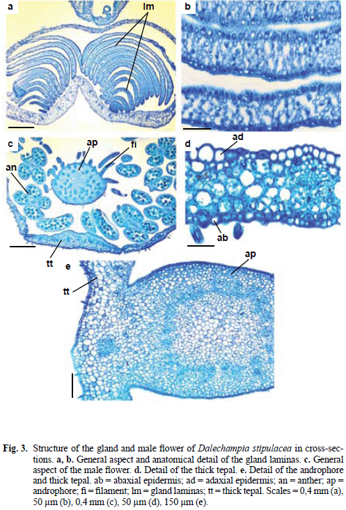
<p ALIGN="justify" style="line-height: 100%"><font face="Verdana" size="2">Pluricellular glandular trichomes of <i>D. stipulacea </i>are similar to the stinging</font> <font face="Verdana" size="2">hairs of <i>Tragia </i>(Thurston 1976) and <i>Dalechampia scandens </i>L. hairs (Webster &amp;</font> <font face="Verdana" size="2">Webster 1972). The trichomes of these species can be irritating to the skin. Maia</font> <font face="Verdana" size="2"><i>et al</i></font><font face="Verdana" size="2">. (2002) made note of these stinging trichomes, considering them to be an important</font> <font face="Verdana" size="2">character in the recognition of most of the <i>Dalechampia </i>species.</font></p>     <p ALIGN="justify" style="line-height: 100%"><font face="Verdana" size="2">The glands (<a href="#fig2">Fig. 2d</a>) consist of a long stalk with uniseriate epidermis that</font> <font face="Verdana" size="2">presents nonglandular trichomes and stomata, parenchyma with chloroplasts, and</font> <font face="Verdana" size="2">a collateral vascular bundle. The head of each gland (<a href="#fig2">Fig. 2d, e</a>) consists of a palisade-like secretory epidermis subtended by parenchyma.</font></p>     <p ALIGN="justify" style="line-height: 100%"><font face="Verdana" size="2">The <i>D. stipulacea </i>stalked glands have structural similarity to special outgrowths</font> <font face="Verdana" size="2">(nectaries) on the teeth of leaf margins (<i>Prunus </i>and <i>Ailanthus</i>) or on</font> <font face="Verdana" size="2">different parts of the floral organs (Fahn 1990). Studies of floral biology of </font><font face="Verdana" size="2"><i>D.</i></font> <font face="Verdana" size="2"><i>stipulacea </i></font><font face="Verdana" size="2">(Sazima <i>et al</i>. 1985) and of other <i>Dalechampia </i>species (Armbruster</font> <font face="Verdana" size="2">&amp; Webster 1979, 1981) make no reference to the nectaries in pseudantha. On the</font> <font face="Verdana" size="2">contrary, in these investigations the emergencies are denominated improperly as</font> <font face="Verdana" size="2">glandular hairs that occur in the female flower perianth. It is possible that the </font><font face="Verdana" size="2"><i>D.</i></font> <font face="Verdana" size="2"><i>stipulacea </i></font><font face="Verdana" size="2">glands have an important role in the pollination, but that were not considered</font> <font face="Verdana" size="2">by workers of floral biology. The perianth and bract glands of </font><i><font face="Verdana" size="2">D. stipulacea</font></i> <font face="Verdana" size="2">could be considered as perigonal nectaries and extrafloral ones, respectively</font> <font face="Verdana" size="2">according to Fahn (1990) classification. <i>Croton sarcopetalus </i>outer nectary of female</font> <font face="Verdana" size="2">flower (Freitas <i>et al</i>. 2001) is similar to the <i>D. stipulacea </i>one, consisting of</font> <font face="Verdana" size="2">a secretory epidermis (column-shaped cells) subtended by secretory parenchyma</font> <font face="Verdana" size="2">and ground parenchyma.</font></p>     <p ALIGN="justify" style="line-height: 100%"><font face="Verdana" size="2">The two pseudanthium bracts (<a href="#fig2">Fig. 2f</a>) present uniseriate epidermis with</font> <font face="Verdana" size="2">trichomes, glands and stomata. The ordinary epidermal cells possess sinuous anticlinal</font> <font face="Verdana" size="2">walls and thick outer periclinal walls. Mesophyll possesses uniform photosynthetic</font> <font face="Verdana" size="2">parenchyma.</font></p>     <p ALIGN="justify" style="line-height: 100%"><font face="Verdana" size="2">Pseudanthium bracts of <i>D. stipulacea </i>have external structure similar to</font> <font face="Verdana" size="2">dicotyledonous foliage leaves, but they do not have differentiated palisade and</font> <font face="Verdana" size="2">spongy parenchyma. The striking structural features of <i>D. stipulacea </i>bracts, such</font> <font face="Verdana" size="2">as the uniform mesophyll with chloroplasts and stinging trichomes, should be related</font> <font face="Verdana" size="2">to the photosynthetic function and the anti-herbivore mechanism.</font></p>     <p ALIGN="justify" style="line-height: 100%"><font face="Verdana" size="2">The female flower bracts (<a href="#fig1">Fig. 1d</a>) present uniseriate epidermis with stomata</font> <font face="Verdana" size="2">on both surfaces. In bracts nonglandular trichomes, stinging trichomes and</font> <font face="Verdana" size="2">glands were also observed. Mesophyll consists of uniform parenchyma with few</font> <font face="Verdana" size="2">chloroplasts. The number of mesophyll layers is higher in the bract base, while</font> <font face="Verdana" size="2">towards the apex this number decreases gradually. In the bract tip the volume of</font> <font face="Verdana" size="2">intercellular spaces is greater than at the base. The vascular bundles are collateral</font> <font face="Verdana" size="2">and occur in the mesophyll proper.</font></p>     <p ALIGN="justify" style="line-height: 100%"><font face="Verdana" size="2">The anatomical studies of bracts remain relatively poor in the literature. It</font> <font face="Verdana" size="2">is probable that the most of species present bracts with uniform mesophyll, as observed</font> <font face="Verdana" size="2">in <i>D. stipulacea </i>inflorescences. Bracts with dorsiventral mesophyll occur</font> <font face="Verdana" size="2">in Piperaceae (Iwazaki <i>et al</i>. 2006).</font></p>     <p ALIGN="justify" style="line-height: 100%"><font face="Verdana" size="2">Glands of the male inflorescence of <i>D. stipulacea </i>(<a href="#fig1">Fig. 1b</a>) consist of two</font> <font face="Verdana" size="2">tightly appressed groups of laminas with variable dimensions (<a href="#fig3">Fig. 3a</a>). In </font><i><font face="Verdana" size="2">D. scandens</font></i> <font face="Verdana" size="2">there is a similar gland which was interpreted by Webster &amp; Webster (1972) as</font> <font face="Verdana" size="2">modified bractlets with secretory epithelium. Each lamina (<a href="#fig3">Fig. 3a, b</a>) is formed by</font> <font face="Verdana" size="2">parenchyma and glabrous uniseriate epidermis. The laminas close to male flowers</font> <font face="Verdana" size="2">are larger and show epidermal cells with thick-walls and spongy parenchyma; successive laminas have thinner epidermal cells and fewer intercellular spaces among</font> <font face="Verdana" size="2">the parenchyma cells. The lamina parenchyma sometimes contains druses.</font></p>     ]]></body>
<body><![CDATA[<p ALIGN="center" style="line-height: 100%"><a name="fig3"><img border="0" src="/img/fbpe/abv/v33n1/art07fig3.gif" width="497" height="732"></a></p>     
<p ALIGN="justify" style="line-height: 100%"><font face="Verdana" size="2">The male flower perianth (<a href="#fig3">Fig. 3c, d</a>) consists of four tepals; one of these is</font> <font face="Verdana" size="2">thicker (top to bottom) and narrower (side to side). This thicker tepal consists of</font> <font face="Verdana" size="2">uniseriate epidermis with stomata on abaxial surface only and uniform parenchyma</font> <font face="Verdana" size="2">with chloroplasts and druse idioblasts.</font></p>     <p ALIGN="justify" style="line-height: 100%"><font face="Verdana" size="2">In angiospermous, coloured perianth elements usually have poorly developed</font> <font face="Verdana" size="2">vascular system, veins without sclerenchyma, and mesophyll with spongy</font> <font face="Verdana" size="2">parenchyma in which the cells may have chloroplasts or pigments in the cell sap,</font> <font face="Verdana" size="2">or both (Fahn 1990). The male flower perianth of <i>Dalechampia stipulacea </i>lacks</font> <font face="Verdana" size="2">pigments in the cell sap but otherwise shows structure similar to that described by</font> <font face="Verdana" size="2">Fahn (1990).</font></p>     <p ALIGN="justify" style="line-height: 100%"><font face="Verdana" size="2">The androphore is fused to the base of the thicker/narrower tepal (<a href="#fig3">Fig. 3c, e</a>).</font> <font face="Verdana" size="2">The androphore has pilose uniseriate epidermis, parenchyma, and a cylinder of collateral</font> <font face="Verdana" size="2">bundles which encloses a parenchymatous pith. Anthers are tetrasporangiate;</font> <font face="Verdana" size="2">and, when immature, anther walls consist of a uniseriate epidermis, an endothecium</font> <font face="Verdana" size="2">with thin-walled cells, one middle layer and secretory tapetum (<a href="#fig4">Fig. 4a</a>).</font></p>     <p ALIGN="center" style="line-height: 100%"><a name="fig4"><img border="0" src="/img/fbpe/abv/v33n1/art07fig4.gif" width="494" height="697"></a></p>     
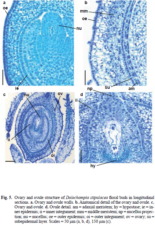
<p ALIGN="justify" style="line-height: 100%"><font face="Verdana" size="2">Davis (1966) defined four types of anther wall based on a survey of 80 dicotyledonous</font> <font face="Verdana" size="2">families: basic, dicotyledonous, monocotyledonous and reduced.</font> <font face="Verdana" size="2">Anthers of <i>D. stipulacea </i>should belong to the dicotyledonous type in which only</font> <font face="Verdana" size="2">cells of the outer secondary parietal layer divide, giving rise to the endothelium</font> <font face="Verdana" size="2">and the single middle layer, while the cells of the inner layer develop directly into</font> <font face="Verdana" size="2">tapetum. Alternatively, but less probable, the <i>D. stipulacea </i>anther could belong to</font> <font face="Verdana" size="2">the Monocotyledonous type that also has a single middle layer. Therefore, studies</font> <font face="Verdana" size="2">about details of anther wall formation and definition of the type of </font><i><font face="Verdana" size="2">D. stipulacea</font></i> <font face="Verdana" size="2">are necessary.</font></p>     <p ALIGN="justify" style="line-height: 100%"><font face="Verdana" size="2">In maturity, the anther wall (<a href="#fig4">Fig. 4b</a>) consists of discontinuous epidermis</font> <font face="Verdana" size="2">with tabular or lenticular cells, endothecium with thick-walled cells, and secretory</font> <font face="Verdana" size="2">tapetum with binucleate cells. In certain areas the tapetum is discontinuous</font> <font face="Verdana" size="2">and shows cell projections that penetrate between the developing pollen grains,</font> <font face="Verdana" size="2">imitating an amoeboid type of tapetum. However, Carniel (1963) reported that the</font> <font face="Verdana" size="2">secretory tapetum can be confused with amoeboid or periplasmodial tapetum in</font> <font face="Verdana" size="2">final stage of degeneration.</font></p>     <p ALIGN="justify" style="line-height: 100%"><font face="Verdana" size="2">The female flower perianth (<a href="#fig4">Fig. 4c, d</a>) has a single epidermis with small</font> <font face="Verdana" size="2">and cube-shaped cells, nonglandular trichomes, and unicellular and pluricellular</font> <font face="Verdana" size="2">glandular trichomes. In the abaxial surface and leaf margin there are glands, notably</font> <font face="Verdana" size="2">numerous. Stomata are absent. In the mesophyll occur two layers of photosynthetic</font> <font face="Verdana" size="2">parenchymas: the abaxial surface parenchyma is formed by large and isodiametric</font> <font face="Verdana" size="2">cells, and the adaxial surface parenchyma shows tangentially elongated</font> <font face="Verdana" size="2">cells. Vascularization is made by collateral bundles immersed in the mesophyll.</font> <font face="Verdana" size="2">The female flower perianth is different from that of the male flower, not only in</font> <font face="Verdana" size="2">shape, but also for exhibiting unicellular glandular trichomes, glands and heterogeneous</font> <font face="Verdana" size="2">parenchymatous mesophyll.</font></p>     <p ALIGN="justify" style="line-height: 100%"><font face="Verdana" size="2">Prior to anthesis, the ovary wall surface (<a href="#fig5">Fig. 5a</a>) consists of outer epidermis,</font> <font face="Verdana" size="2">with small and cuboidal cells containing dense cytoplasm. The mesophyll is</font> <font face="Verdana" size="2">parenchymatous and consists of four or more cell layers. The subepidermal mesophyll</font> <font face="Verdana" size="2">layer and inner epidermis are constituted by shortly elongated cells.</font></p>     <p ALIGN="center" style="line-height: 100%"><a name="fig5"><img border="0" src="/img/fbpe/abv/v33n1/art07fig5.gif" width="495" height="717"></a></p>     
]]></body>
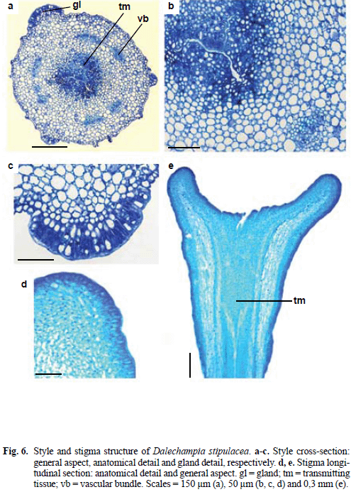
<body><![CDATA[<p ALIGN="justify" style="line-height: 100%"><font face="Verdana" size="2">Still prior to anthesis (<a href="#fig5">Fig. 5b</a>), the inner epidermis and middle cell layers divide</font> <font face="Verdana" size="2">periclinally forming two meristematic regions in the ovary wall, the adaxial</font> <font face="Verdana" size="2">meristem and the middle meristem. Between the meristematic regions a layer of</font> <font face="Verdana" size="2">radially elongated cells can be discerned. In this stage the mesophyll cells became</font> <font face="Verdana" size="2">vacuolized; trichomes began the differentiation in the inner epidermis.</font></p>     <p ALIGN="justify" style="line-height: 100%"><font face="Verdana" size="2">The presence of meristems in the ovary wall (preanthesis or postanthesis</font> <font face="Verdana" size="2">stage) is frequent in plants with follicles, legumes or capsules (Roth 1977; Souza</font> <font face="Verdana" size="2">2006). Toledo (1963) reported parenchyma in the <i>Manihot utilissima </i>(</font><font face="Verdana" size="2"><i>Manihot</i></font> <font face="Verdana" size="2"><i>esculenta</i></font><font face="Verdana" size="2">) ovary in which cell layers close to locule differentiate in endocarp. </font><font face="Verdana" size="2"><i>Mahihot</i></font> <font face="Verdana" size="2"><i>caerulescens </i></font><font face="Verdana" size="2">Pohl. and <i>Manihot tripartita </i>Müll.Arg. form adaxial meristem,</font> <font face="Verdana" size="2">not in the flower, but in the developing fruit (Oliveira 2007). </font><i><font face="Verdana" size="2">Dalechampia stipulacea</font></i> <font face="Verdana" size="2">did not just develop a meristem, which usually is verified in ovaries and/or</font> <font face="Verdana" size="2">fruits, but two meristems, an adaxial meristem and other middle one.</font></p>     <p ALIGN="justify" style="line-height: 100%"><font face="Verdana" size="2">The ovary mesophyll usually has uniform parenchyma. Differently of most</font> <font face="Verdana" size="2">of the studied species, <i>D. stipulacea </i>possess mesophyll in preanthesis with different</font> <font face="Verdana" size="2">tissues, mainly for the meristem differentiation. Other Euphorbiaceae species,</font> <font face="Verdana" size="2">as <i>Manihot utilissima </i>(<i>Manihot esculenta</i>) (Toledo 1963), </font><i><font face="Verdana" size="2">Manihot caerulescens</font></i> <font face="Verdana" size="2">and <i>Manihot tripartita </i>(Oliveira 2007) have heterogeneous mesophyll.</font> <font face="Verdana" size="2">Besides, Erythroxylaceae species belonging to Malpighiales presented subadaxial</font> <font face="Verdana" size="2">and subabaxial meristems, and parenchyma in the mesophyll (Nakamura 2003).</font></p> <i>     <p ALIGN="justify" style="line-height: 100%"><font face="Verdana" size="2">Dalechampia stipulacea </font></i><font face="Verdana" size="2">ovules (<a href="#fig5">Fig. 5c, d</a>) are anatropous, bitegmic and</font> <font face="Verdana" size="2">crassinucellate. The integuments are multi-layered, with vascular tissue just in</font> <font face="Verdana" size="2">the outer integument, and micropyle occluded by the nucellar beak; there is a hypostase</font> <font face="Verdana" size="2">characterized by thick-walled cells. The hypostase is a cup-shaped region</font> <font face="Verdana" size="2">of thick-walled cells occurring at the chalazal end of the ovule, abutting directly</font> <font face="Verdana" size="2">with embryo sac and partially surrounding its chalazal end (Tiwari 1983). The</font> <font face="Verdana" size="2">hypostase probably has an integral function in the translocation of nutrients into</font> <font face="Verdana" size="2">the megagametophyte and, after fertilization, into the embryo sac (Tilton 1980);</font> <font face="Verdana" size="2">as reported in Euphorbiaceae (Bouman 1984). Ovule structure has systematic implications</font> <font face="Verdana" size="2">in Euphorbiaceae (Tokuoka &amp; Tobe 2003); these authors published a</font> <font face="Verdana" size="2">series of papers that deal with Euphorbiaceae ovule and considered all 20 tribes of</font> <font face="Verdana" size="2">Acalyphoideae examined with bitegmic and non-vascularized inner integument</font> <font face="Verdana" size="2">ovules. The inner integument of the ovule of <i>D. stipulacea </i>is non-vascularized,</font> <font face="Verdana" size="2">but Silva &amp; Souza (2009) registered in the seed of this species that the basal part</font> <font face="Verdana" size="2">of the tegmen develops by intercalary growth, and few vascular bundles extend</font> <font face="Verdana" size="2">from the chalaza by postchalazal branching.</font></p>     <p ALIGN="justify" style="line-height: 100%"><font face="Verdana" size="2">The style (<a href="#fig1">Fig. 1b, d</a>, <a href="#fig6"> 6a, b</a>) has uniseriate epidermis presenting nonglandular</font> <font face="Verdana" size="2">trichomes and stomata, multi-layered parenchyma, six collateral vascular bundles</font> <font face="Verdana" size="2">and central transmitting tissue. In the surface of the style there is a gland with</font> <font face="Verdana" size="2">radially elongated cells disposed as a palisade similar to the epidermal cells in the</font> <font face="Verdana" size="2">head of stalked glands. The style glands could have the nectary function, although</font> <font face="Verdana" size="2">this was not mentioned by Armbruster &amp; Webster (1979, 1981) nor Sazima <i>et al</i>.</font> <font face="Verdana" size="2">(1985) in floral biology studies of <i>D. stipulacea</i>.</font></p>     <p ALIGN="center" style="line-height: 100%"><a name="fig6"><img border="0" src="/img/fbpe/abv/v33n1/art07fig6.gif" width="495" height="693"></a></p>     
<p ALIGN="justify" style="line-height: 100%"><font face="Verdana" size="2">Styles of <i>D. stipulacea </i>are intermediate between the commonly encountered</font> <font face="Verdana" size="2">states of hollow or solid (Fahn 1990). It presents central rifts (<a href="#fig6">Fig. 6a, b</a>) that</font> <font face="Verdana" size="2">makes possible to identify the transmitting tissue of each carpel.</font></p>     <p ALIGN="justify" style="line-height: 100%"><font face="Verdana" size="2">The enlarged stigma consists of a central cavity of circular contour which</font> <font face="Verdana" size="2">is covered by secretory epidermis with non-papillose elongated cells (<a href="#fig6">Fig. 6d, e</a>).</font> <font face="Verdana" size="2">Armbruster (1996) described different types of stigmas of <i>Dalechampia</i>, without</font> <font face="Verdana" size="2">mentioning <i>D. stipulacea</i>. The author considered that the expanded stigma appears</font> <font face="Verdana" size="2">to be associated with fragrance secretion throughout the genus, which suggests</font> <font face="Verdana" size="2">that the large stigmatic surface evolved to promote fragrance production for</font> <font face="Verdana" size="2">attraction of pollinators.</font></p>     <p ALIGN="justify" style="line-height: 100%"><font face="Verdana" size="2">Some striking structural features of the inflorescence and flower of <i>D. stipulacea</i>,</font> <font face="Verdana" size="2">such as pseudanthium with a unique configuration within the Euphorbiaceae,</font> <font face="Verdana" size="2">laminar gland in the male inflorescence, stalked glands in the bracts and male and</font> <font face="Verdana" size="2">female inflorescences, stinging trichomes, adaxial and middle meristems formed</font> <font face="Verdana" size="2">in the ovary wall, and micropyle occluded by the nucellar beak, are important for</font> <font face="Verdana" size="2">taxonomic and pollination ecology studies of the genus <i>Dalechampia</i>. On the other</font> <font face="Verdana" size="2">hand, the <i>Dalechampia </i>literature makes no reference to the encountered features in</font> <font face="Verdana" size="2"><i>D. stipulacea</i></font><font face="Verdana" size="2">, such as unicellular glandular trichomes and the style glands.</font></p> <b>     <p ALIGN="justify" style="line-height: 100%"><font face="Verdana" size="2">ACKNOWLEDGMENTS</font></p> </b>     ]]></body>
<body><![CDATA[<p ALIGN="justify" style="line-height: 100%"><font face="Verdana" size="2">We thank Dr. Olavo A. Guimarães of the Universidade Federal do Paraná</font> <font face="Verdana" size="2">for the identification of studied species. We thank CNPq (Conselho Nacional de</font> <font face="Verdana" size="2">Desenvolvimento Científico e Tecnológico, Brazil) for funding this research.</font></p> <b>     <p ALIGN="justify" style="line-height: 100%"><font face="Verdana" size="2">BIBLIOGRAPHY</font></p> </b>     <!-- ref --><p ALIGN="justify" style="line-height: 100%"><font face="Verdana" size="2">1. Armbruster, W.S. 1996. Evolution of floral morphology and function: an integrative</font> <font face="Verdana" size="2">approach to adaptation, constraint, and compromise in </font><i><font face="Verdana" size="2">Dalechampia</font></i> <font face="Verdana" size="2">(Euphorbiaceae). In: Lloyd, D.G. &amp; S.C.H. Barrett (eds.).</font> <font face="Verdana" size="2"><i>Floral biology studies on floral evolution in animal-pollinated plants</i></font><font face="Verdana" size="2">,</font> <font face="Verdana" size="2">pp. 241-272. Chapman &amp; Hall, New York.</font>&nbsp;&nbsp;&nbsp;&nbsp;&nbsp;&nbsp;&nbsp;&nbsp;[&#160;<a href="javascript:void(0);" onclick="javascript: window.open('/scielo.php?script=sci_nlinks&ref=037409&pid=S0084-5906201000010000700001&lng=','','width=640,height=500,resizable=yes,scrollbars=1,menubar=yes,');">Links</a>&#160;]<!-- end-ref --><!-- ref --><p ALIGN="justify" style="line-height: 100%"><font face="Verdana" size="2">2. Armbruster, W.S., A.L. Herzig &amp; T.P. Clausen. 1992. Pollination of two sympatric</font> <font face="Verdana" size="2">species of <i>Dalechampia </i>(Euphorbiaceae) in Suriname by male euglossine</font> <font face="Verdana" size="2">bees. <i>Amer. J. Bot</i>. 79: 1374-1381.</font>&nbsp;&nbsp;&nbsp;&nbsp;&nbsp;&nbsp;&nbsp;&nbsp;[&#160;<a href="javascript:void(0);" onclick="javascript: window.open('/scielo.php?script=sci_nlinks&ref=037410&pid=S0084-5906201000010000700002&lng=','','width=640,height=500,resizable=yes,scrollbars=1,menubar=yes,');">Links</a>&#160;]<!-- end-ref --><!-- ref --><p ALIGN="justify" style="line-height: 100%"><font size="2"><font face="Verdana">3. Armbruster, W.S. &amp; G.L. Webster. 1979. Pollination of two species of </font></font><i><font face="Verdana" size="2">Dalechampia</font></i> <font face="Verdana" size="2">(Euphorbiaceae) in Mexico by Euglossine bees. <i>Biotropica </i>11:278-283.</font>&nbsp;&nbsp;&nbsp;&nbsp;&nbsp;&nbsp;&nbsp;&nbsp;[&#160;<a href="javascript:void(0);" onclick="javascript: window.open('/scielo.php?script=sci_nlinks&ref=037411&pid=S0084-5906201000010000700003&lng=','','width=640,height=500,resizable=yes,scrollbars=1,menubar=yes,');">Links</a>&#160;]<!-- end-ref --><!-- ref --><p ALIGN="justify" style="line-height: 100%"><font face="Verdana" size="2">4. Armbruster, W.S. &amp; G.L. Webster. 1981. Sistemas de polinização de duas espé</font> <font face="Verdana" size="2">cies simpátricas de <i>Dalechampia </i>(Euphorbiaceae) no Amazonas, Brasil.</font> <font face="Verdana" size="2"><i>Acta Amazon</i></font><font face="Verdana" size="2">. 11: 13-17.</font>&nbsp;&nbsp;&nbsp;&nbsp;&nbsp;&nbsp;&nbsp;&nbsp;[&#160;<a href="javascript:void(0);" onclick="javascript: window.open('/scielo.php?script=sci_nlinks&ref=037412&pid=S0084-5906201000010000700004&lng=','','width=640,height=500,resizable=yes,scrollbars=1,menubar=yes,');">Links</a>&#160;]<!-- end-ref --><!-- ref --><p ALIGN="justify" style="line-height: 100%"><font size="2"><font face="Verdana">5. Bouman, F. 1984. The ovule. In: Johri, B.M. (ed.). </font></font><i><font face="Verdana" size="2">Embryology of angiosperms,</font></i> <font face="Verdana" size="2">pp. 123-157. Springer-Verlag, New York.</font>&nbsp;&nbsp;&nbsp;&nbsp;&nbsp;&nbsp;&nbsp;&nbsp;[&#160;<a href="javascript:void(0);" onclick="javascript: window.open('/scielo.php?script=sci_nlinks&ref=037413&pid=S0084-5906201000010000700005&lng=','','width=640,height=500,resizable=yes,scrollbars=1,menubar=yes,');">Links</a>&#160;]<!-- end-ref --><!-- ref --><p ALIGN="justify" style="line-height: 100%"><font face="Verdana" size="2">6. Capinpin, J.M. &amp; V.C. Bruce. 1955. Floral biology and cytology of <i>Manihot utilissima</i>.</font> <font face="Verdana" size="2"><i>Philip. Agric</i></font><font face="Verdana" size="2">. 39: 306-316.</font>&nbsp;&nbsp;&nbsp;&nbsp;&nbsp;&nbsp;&nbsp;&nbsp;[&#160;<a href="javascript:void(0);" onclick="javascript: window.open('/scielo.php?script=sci_nlinks&ref=037414&pid=S0084-5906201000010000700006&lng=','','width=640,height=500,resizable=yes,scrollbars=1,menubar=yes,');">Links</a>&#160;]<!-- end-ref --><!-- ref --><p ALIGN="justify" style="line-height: 100%"><font face="Verdana" size="2">7. Carniel, K. 1963. Das Antherentapetum. <i>Österr. Bot. Z</i>. 110: 45-176.</font>&nbsp;&nbsp;&nbsp;&nbsp;&nbsp;&nbsp;&nbsp;&nbsp;[&#160;<a href="javascript:void(0);" onclick="javascript: window.open('/scielo.php?script=sci_nlinks&ref=037415&pid=S0084-5906201000010000700007&lng=','','width=640,height=500,resizable=yes,scrollbars=1,menubar=yes,');">Links</a>&#160;]<!-- end-ref --><!-- ref --><p ALIGN="justify" style="line-height: 100%"><font face="Verdana" size="2">8. Davis, G.L. 1966. <i>Systematic embryology of the angiosperms</i>. John Wiley &amp;</font> <font face="Verdana" size="2">Sons, New York.</font>&nbsp;&nbsp;&nbsp;&nbsp;&nbsp;&nbsp;&nbsp;&nbsp;[&#160;<a href="javascript:void(0);" onclick="javascript: window.open('/scielo.php?script=sci_nlinks&ref=037416&pid=S0084-5906201000010000700008&lng=','','width=640,height=500,resizable=yes,scrollbars=1,menubar=yes,');">Links</a>&#160;]<!-- end-ref --><!-- ref --><p ALIGN="justify" style="line-height: 100%"><font face="Verdana" size="2">9. Fahn, A. 1990. <i>Plant anatomy</i>. Pergamon Press, Oxford.</font>&nbsp;&nbsp;&nbsp;&nbsp;&nbsp;&nbsp;&nbsp;&nbsp;[&#160;<a href="javascript:void(0);" onclick="javascript: window.open('/scielo.php?script=sci_nlinks&ref=037417&pid=S0084-5906201000010000700009&lng=','','width=640,height=500,resizable=yes,scrollbars=1,menubar=yes,');">Links</a>&#160;]<!-- end-ref --><!-- ref --><p ALIGN="justify" style="line-height: 100%"><font face="Verdana" size="2">10. Foster, A.S. 1950. Techniques for the study of venation patterns in the leaves</font> <font face="Verdana" size="2">of angiosperms. </font><font face="Verdana" size="2"><i>Proceedings 7th International Botanical Congress</i></font> <font face="Verdana" size="2"><i>Stockholm</i></font><font face="Verdana" size="2">.</font>&nbsp;&nbsp;&nbsp;&nbsp;&nbsp;&nbsp;&nbsp;&nbsp;[&#160;<a href="javascript:void(0);" onclick="javascript: window.open('/scielo.php?script=sci_nlinks&ref=037418&pid=S0084-5906201000010000700010&lng=','','width=640,height=500,resizable=yes,scrollbars=1,menubar=yes,');">Links</a>&#160;]<!-- end-ref --><!-- ref --><p ALIGN="justify" style="line-height: 100%"><font face="Verdana" size="2">11. Freitas, L., G. Bernardello, L. Galetto &amp; A.A.S. Paoli. 2001. Nectaries and reproductive</font> <font face="Verdana" size="2">biology of <i>Croton sarcopetalus </i>(Euphorbiaceae). </font><font face="Verdana" size="2"><i>Bot. J. Linn.</i></font> <font face="Verdana" size="2"><i>Soc</i></font><font face="Verdana" size="2">. 136: 267-277.</font>&nbsp;&nbsp;&nbsp;&nbsp;&nbsp;&nbsp;&nbsp;&nbsp;[&#160;<a href="javascript:void(0);" onclick="javascript: window.open('/scielo.php?script=sci_nlinks&ref=037419&pid=S0084-5906201000010000700011&lng=','','width=640,height=500,resizable=yes,scrollbars=1,menubar=yes,');">Links</a>&#160;]<!-- end-ref --><!-- ref --><p ALIGN="justify" style="line-height: 100%"><font size="2"><font face="Verdana">12. Gerrits, P.O. 1991. </font></font><font face="Verdana" size="2"><i>The application of glycol methacrylate in histotechnology</i></font> <font face="Verdana" size="2"><i>(some fundamental principles)</i></font><font face="Verdana" size="2">. Leica Gmbh., Germany.</font>&nbsp;&nbsp;&nbsp;&nbsp;&nbsp;&nbsp;&nbsp;&nbsp;[&#160;<a href="javascript:void(0);" onclick="javascript: window.open('/scielo.php?script=sci_nlinks&ref=037420&pid=S0084-5906201000010000700012&lng=','','width=640,height=500,resizable=yes,scrollbars=1,menubar=yes,');">Links</a>&#160;]<!-- end-ref --><!-- ref --><p ALIGN="justify" style="line-height: 100%"><font size="2"><font face="Verdana">13. Graner, E.A. 1935. Contribuição para o estudo citológico da mandioca. </font></font><font face="Verdana" size="2"><i>Public.</i></font> <font face="Verdana" size="2"><i>Esc. Sup. Agric. Luiz de Queiroz </i></font><font face="Verdana" size="2">1-28.</font>&nbsp;&nbsp;&nbsp;&nbsp;&nbsp;&nbsp;&nbsp;&nbsp;[&#160;<a href="javascript:void(0);" onclick="javascript: window.open('/scielo.php?script=sci_nlinks&ref=037421&pid=S0084-5906201000010000700013&lng=','','width=640,height=500,resizable=yes,scrollbars=1,menubar=yes,');">Links</a>&#160;]<!-- end-ref --><!-- ref --><p ALIGN="justify" style="line-height: 100%"><font size="2"><font face="Verdana">14. Graner, E.A. 1942. Notas sobre florescimento e frutificação da mandioca. </font></font><i><font face="Verdana" size="2">Bragantia</font></i> <font face="Verdana" size="2">2: 1-12.</font>&nbsp;&nbsp;&nbsp;&nbsp;&nbsp;&nbsp;&nbsp;&nbsp;[&#160;<a href="javascript:void(0);" onclick="javascript: window.open('/scielo.php?script=sci_nlinks&ref=037422&pid=S0084-5906201000010000700014&lng=','','width=640,height=500,resizable=yes,scrollbars=1,menubar=yes,');">Links</a>&#160;]<!-- end-ref --><!-- ref --><p ALIGN="justify" style="line-height: 100%"><font face="Verdana" size="2">15. Hickey, L.J. 1979. A revised classification of the architecture of dicotyledonous</font> <font face="Verdana" size="2">leaves. In: Metcalfe, C.R. &amp; L. Chalk (eds.). <i>Anatomy of the dicotyledons</i>,</font> <font face="Verdana" size="2">pp. 25-39. Clarendon Press, Oxford.</font>&nbsp;&nbsp;&nbsp;&nbsp;&nbsp;&nbsp;&nbsp;&nbsp;[&#160;<a href="javascript:void(0);" onclick="javascript: window.open('/scielo.php?script=sci_nlinks&ref=037423&pid=S0084-5906201000010000700015&lng=','','width=640,height=500,resizable=yes,scrollbars=1,menubar=yes,');">Links</a>&#160;]<!-- end-ref --><!-- ref --><p ALIGN="justify" style="line-height: 100%"><font face="Verdana" size="2">16. Iwazaki, M.C., L.A. Souza &amp; J.H.G. Oliveira. 2006. Morfo-anatomia comparativa</font> <font face="Verdana" size="2">da flor de <i>Peperomia dahlstedtii </i>C. DC., <i>Ottonia martiana </i>Miq. e</font> <font face="Verdana" size="2"><i>Piper gaudichaudianum </i></font><font face="Verdana" size="2">Kunth (Piperaceae). <i>Hoehnea </i>33: 545-558.</font>&nbsp;&nbsp;&nbsp;&nbsp;&nbsp;&nbsp;&nbsp;&nbsp;[&#160;<a href="javascript:void(0);" onclick="javascript: window.open('/scielo.php?script=sci_nlinks&ref=037424&pid=S0084-5906201000010000700016&lng=','','width=640,height=500,resizable=yes,scrollbars=1,menubar=yes,');">Links</a>&#160;]<!-- end-ref --><!-- ref --><p ALIGN="justify" style="line-height: 100%"><font face="Verdana" size="2">17. Maia, V.C.R., M. Emmerich &amp; L.S. Valle. 2002. <i>Dalechampia </i>Plum. ex L. (Euphorbiaceae)</font> <font face="Verdana" size="2">- Taxonomia das espécies ocorrentes nas restingas do Estado</font> <font face="Verdana" size="2">do Rio de Janeiro, Brasil. <i>Bol. Mus. Nac. (Bot.) </i>119: 1-29.</font>&nbsp;&nbsp;&nbsp;&nbsp;&nbsp;&nbsp;&nbsp;&nbsp;[&#160;<a href="javascript:void(0);" onclick="javascript: window.open('/scielo.php?script=sci_nlinks&ref=037425&pid=S0084-5906201000010000700017&lng=','','width=640,height=500,resizable=yes,scrollbars=1,menubar=yes,');">Links</a>&#160;]<!-- end-ref --><!-- ref --><p ALIGN="justify" style="line-height: 100%"><font face="Verdana" size="2">18. Nakamura, A.T. 2003. Morfologia e anatomia dos frutos e sementes de três espécies</font> <font face="Verdana" size="2">de <i>Erythroxylum </i>P.Browne (Erythroxylaceae) de cerrado do Estado</font> <font face="Verdana" size="2">de São Paulo. Dissertação de Mestrado. Universidade Estadual</font> <font face="Verdana" size="2">Paulista. Botucatu (Brasil).</font>&nbsp;&nbsp;&nbsp;&nbsp;&nbsp;&nbsp;&nbsp;&nbsp;[&#160;<a href="javascript:void(0);" onclick="javascript: window.open('/scielo.php?script=sci_nlinks&ref=037426&pid=S0084-5906201000010000700018&lng=','','width=640,height=500,resizable=yes,scrollbars=1,menubar=yes,');">Links</a>&#160;]<!-- end-ref --><!-- ref --><p ALIGN="justify" style="line-height: 100%"><font face="Verdana" size="2">19. O’Brien, T.P., N. Feder &amp; M.E. McCully. 1964. Polychromatic staining of plant</font> <font face="Verdana" size="2">cell walls by toluidine blue O. <i>Protoplasma </i>59: 368-373.</font>&nbsp;&nbsp;&nbsp;&nbsp;&nbsp;&nbsp;&nbsp;&nbsp;[&#160;<a href="javascript:void(0);" onclick="javascript: window.open('/scielo.php?script=sci_nlinks&ref=037427&pid=S0084-5906201000010000700019&lng=','','width=640,height=500,resizable=yes,scrollbars=1,menubar=yes,');">Links</a>&#160;]<!-- end-ref --><!-- ref --><p ALIGN="justify" style="line-height: 100%"><font face="Verdana" size="2">20. Oliveira, J.H.G. 2007. Morfologia, anatomia e desenvolvimento do fruto e semente</font> <font face="Verdana" size="2">de <i>Manihot caerulescens </i>Pohl. e <i>M. tripartita </i>Müll.Arg. (Euphorbiaceae).</font> <font face="Verdana" size="2">Tese de Doutorado. Universidade Estadual Paulista.</font> <font face="Verdana" size="2">Botucatu (Brasil).</font>&nbsp;&nbsp;&nbsp;&nbsp;&nbsp;&nbsp;&nbsp;&nbsp;[&#160;<a href="javascript:void(0);" onclick="javascript: window.open('/scielo.php?script=sci_nlinks&ref=037428&pid=S0084-5906201000010000700020&lng=','','width=640,height=500,resizable=yes,scrollbars=1,menubar=yes,');">Links</a>&#160;]<!-- end-ref --><!-- ref --><p ALIGN="justify" style="line-height: 100%"><font size="2"><font face="Verdana">21. Roth, I. 1977. Fruits of angiosperms. In: Linsbauer, K. (ed.). </font></font><font face="Verdana" size="2"><i>Encyclopedia of</i></font> <font face="Verdana" size="2"><i>plant anatomy</i></font><font face="Verdana" size="2">, pp. 21-26. Gebrüder Borntraeger, Berlin.</font>&nbsp;&nbsp;&nbsp;&nbsp;&nbsp;&nbsp;&nbsp;&nbsp;[&#160;<a href="javascript:void(0);" onclick="javascript: window.open('/scielo.php?script=sci_nlinks&ref=037429&pid=S0084-5906201000010000700021&lng=','','width=640,height=500,resizable=yes,scrollbars=1,menubar=yes,');">Links</a>&#160;]<!-- end-ref --><!-- ref --><p ALIGN="justify" style="line-height: 100%"><font size="2"><font face="Verdana">22. Sazima, M., I. Sazima &amp; R.M. Carvalho-Okano. 1985. Biologia floral de </font></font><font face="Verdana" size="2"><i>Dalechampia</i></font> <font face="Verdana" size="2"><i>stipulacea </i></font><font face="Verdana" size="2">(Euphorbiaceae) e sua polinização por </font><font face="Verdana" size="2"><i>Euglossa</i></font> <font face="Verdana" size="2"><i>melanotricha </i></font><font face="Verdana" size="2">(Apidae). <i>Revista Brasil. Biol</i>. 45: 85-93.</font>&nbsp;&nbsp;&nbsp;&nbsp;&nbsp;&nbsp;&nbsp;&nbsp;[&#160;<a href="javascript:void(0);" onclick="javascript: window.open('/scielo.php?script=sci_nlinks&ref=037430&pid=S0084-5906201000010000700022&lng=','','width=640,height=500,resizable=yes,scrollbars=1,menubar=yes,');">Links</a>&#160;]<!-- end-ref --><!-- ref --><p ALIGN="justify" style="line-height: 100%"><font face="Verdana" size="2">23. Silva, A.C. &amp; L.A. Souza. 2009. Morphology and anatomy of the developing fruit</font> <font face="Verdana" size="2">and seed of <i>Dalechampia stipulacea </i>Müll.Arg. (Euphorbiaceae). </font><font face="Verdana" size="2"><i>Acta</i></font> <font face="Verdana" size="2"><i>Scient. Biol. Sci</i></font><font face="Verdana" size="2">. 31: 425-432.</font>&nbsp;&nbsp;&nbsp;&nbsp;&nbsp;&nbsp;&nbsp;&nbsp;[&#160;<a href="javascript:void(0);" onclick="javascript: window.open('/scielo.php?script=sci_nlinks&ref=037431&pid=S0084-5906201000010000700023&lng=','','width=640,height=500,resizable=yes,scrollbars=1,menubar=yes,');">Links</a>&#160;]<!-- end-ref --><!-- ref --><p ALIGN="justify" style="line-height: 100%"><font face="Verdana" size="2">24. Silva, R.M., G. Bandel, M.I.F. Faraldo &amp; P.S. Martins. 2001. Biologia reprodutiva</font> <font face="Verdana" size="2">de etnovariedades de mandioca. <i>Sci. Agric</i>. 58: 101-107.</font>&nbsp;&nbsp;&nbsp;&nbsp;&nbsp;&nbsp;&nbsp;&nbsp;[&#160;<a href="javascript:void(0);" onclick="javascript: window.open('/scielo.php?script=sci_nlinks&ref=037432&pid=S0084-5906201000010000700024&lng=','','width=640,height=500,resizable=yes,scrollbars=1,menubar=yes,');">Links</a>&#160;]<!-- end-ref --><!-- ref --><p ALIGN="justify" style="line-height: 100%"><font face="Verdana" size="2">25. Souza, L.A. 2006. Fruto. In: Souza, L.A. (ed.). <i>Anatomia do fruto e da semente</i>, pp.</font> <font face="Verdana" size="2">10-123. Editora Universidade Estadual de Ponta Grossa, Ponta Grossa.</font>&nbsp;&nbsp;&nbsp;&nbsp;&nbsp;&nbsp;&nbsp;&nbsp;[&#160;<a href="javascript:void(0);" onclick="javascript: window.open('/scielo.php?script=sci_nlinks&ref=037433&pid=S0084-5906201000010000700025&lng=','','width=640,height=500,resizable=yes,scrollbars=1,menubar=yes,');">Links</a>&#160;]<!-- end-ref --><!-- ref --><p ALIGN="justify" style="line-height: 100%"><font face="Verdana" size="2">26. Thurston, E.L. 1976. Morphology, fine structure and ontogeny of the stinging</font> <font face="Verdana" size="2">emergence of <i>Tragia ramosa </i>and <i>T. saxicola </i>(Euphorbiaceae). </font><font face="Verdana" size="2"><i>Amer.</i></font> <font face="Verdana" size="2"><i>J. Bot</i></font><font face="Verdana" size="2">. 63: 710-718.</font>&nbsp;&nbsp;&nbsp;&nbsp;&nbsp;&nbsp;&nbsp;&nbsp;[&#160;<a href="javascript:void(0);" onclick="javascript: window.open('/scielo.php?script=sci_nlinks&ref=037434&pid=S0084-5906201000010000700026&lng=','','width=640,height=500,resizable=yes,scrollbars=1,menubar=yes,');">Links</a>&#160;]<!-- end-ref --><!-- ref --><p ALIGN="justify" style="line-height: 100%"><font face="Verdana" size="2">27. Tilton, V.R. 1980. Hypostase development in <i>Ornithogalum caudatum </i>(Liliaceae)</font> <font face="Verdana" size="2">and notes on the other types of modifications in chalaza de angiosperm</font> <font face="Verdana" size="2">ovules. <i>Canad. J. Bot</i>. 58: 2059-2066.</font>&nbsp;&nbsp;&nbsp;&nbsp;&nbsp;&nbsp;&nbsp;&nbsp;[&#160;<a href="javascript:void(0);" onclick="javascript: window.open('/scielo.php?script=sci_nlinks&ref=037435&pid=S0084-5906201000010000700027&lng=','','width=640,height=500,resizable=yes,scrollbars=1,menubar=yes,');">Links</a>&#160;]<!-- end-ref --><!-- ref --><p ALIGN="justify" style="line-height: 100%"><font face="Verdana" size="2">28. Tiwari, S.C. 1983. The hypostase in <i>Torenia fournieri </i>Lind.: a histochemical</font> <font face="Verdana" size="2">study of the cell walls. <i>Ann. Bot</i>. 51: 17-26.</font>&nbsp;&nbsp;&nbsp;&nbsp;&nbsp;&nbsp;&nbsp;&nbsp;[&#160;<a href="javascript:void(0);" onclick="javascript: window.open('/scielo.php?script=sci_nlinks&ref=037436&pid=S0084-5906201000010000700028&lng=','','width=640,height=500,resizable=yes,scrollbars=1,menubar=yes,');">Links</a>&#160;]<!-- end-ref --><!-- ref --><p ALIGN="justify" style="line-height: 100%"><font face="Verdana" size="2">29. Tokuoka, T. &amp; H. Tobe. 1998. Ovules and seeds in Crotonoideae (Euphorbiaceae):</font> <font face="Verdana" size="2">structure and systematic implications. <i>Bot. Jahrb. Syst</i>. 120: 165-186.</font>&nbsp;&nbsp;&nbsp;&nbsp;&nbsp;&nbsp;&nbsp;&nbsp;[&#160;<a href="javascript:void(0);" onclick="javascript: window.open('/scielo.php?script=sci_nlinks&ref=037437&pid=S0084-5906201000010000700029&lng=','','width=640,height=500,resizable=yes,scrollbars=1,menubar=yes,');">Links</a>&#160;]<!-- end-ref --><!-- ref --><p ALIGN="justify" style="line-height: 100%"><font face="Verdana" size="2">30. Tokuoka, T. &amp; H. Tobe. 2002. Ovules and seeds in Euphorbioideae (Euphorbiaceae):</font> <font face="Verdana" size="2">structure and systematic implications. <i>J. Plant Res</i>. 115: 361-374.</font>&nbsp;&nbsp;&nbsp;&nbsp;&nbsp;&nbsp;&nbsp;&nbsp;[&#160;<a href="javascript:void(0);" onclick="javascript: window.open('/scielo.php?script=sci_nlinks&ref=037438&pid=S0084-5906201000010000700030&lng=','','width=640,height=500,resizable=yes,scrollbars=1,menubar=yes,');">Links</a>&#160;]<!-- end-ref --><!-- ref --><p ALIGN="justify" style="line-height: 100%"><font face="Verdana" size="2">31. Tokuoka, T. &amp; H. Tobe. 2003. Ovules and seeds in Acalyphoideae (Euphorbiaceae):</font> <font face="Verdana" size="2">structure and systematic implications. <i>J. Plant Res</i>. 116: 355-380.</font>&nbsp;&nbsp;&nbsp;&nbsp;&nbsp;&nbsp;&nbsp;&nbsp;[&#160;<a href="javascript:void(0);" onclick="javascript: window.open('/scielo.php?script=sci_nlinks&ref=037439&pid=S0084-5906201000010000700031&lng=','','width=640,height=500,resizable=yes,scrollbars=1,menubar=yes,');">Links</a>&#160;]<!-- end-ref --><!-- ref --><p ALIGN="justify" style="line-height: 100%"><font face="Verdana" size="2">32. Toledo, A.P. 1963. Anatomia e desenvolvimento ontogenético da flor de mandioca.</font> <font face="Verdana" size="2"><i>Bragantia </i></font><font face="Verdana" size="2">22: 465-476.</font>&nbsp;&nbsp;&nbsp;&nbsp;&nbsp;&nbsp;&nbsp;&nbsp;[&#160;<a href="javascript:void(0);" onclick="javascript: window.open('/scielo.php?script=sci_nlinks&ref=037440&pid=S0084-5906201000010000700032&lng=','','width=640,height=500,resizable=yes,scrollbars=1,menubar=yes,');">Links</a>&#160;]<!-- end-ref --><!-- ref --><p ALIGN="justify" style="line-height: 100%"><font face="Verdana" size="2">33. Weberling, F. 1992. <i>Morphology of flowers and inflorescences</i>. Cambridge University</font> <font face="Verdana" size="2">Press, Cambridge.</font>&nbsp;&nbsp;&nbsp;&nbsp;&nbsp;&nbsp;&nbsp;&nbsp;[&#160;<a href="javascript:void(0);" onclick="javascript: window.open('/scielo.php?script=sci_nlinks&ref=037441&pid=S0084-5906201000010000700033&lng=','','width=640,height=500,resizable=yes,scrollbars=1,menubar=yes,');">Links</a>&#160;]<!-- end-ref --><!-- ref --><p ALIGN="justify" style="line-height: 100%"><font size="2"><font face="Verdana">34. Webster, G.L. &amp; B.D. Webster. 1972. The morphology and relationships of </font></font><font face="Verdana" size="2"><i>Dalechampia</i></font> <font face="Verdana" size="2"><i>scandens </i></font><font face="Verdana" size="2">(Euphorbiaceae). <i>Amer. J. Bot</i>. 59: 573-586.</font>&nbsp;&nbsp;&nbsp;&nbsp;&nbsp;&nbsp;&nbsp;&nbsp;[&#160;<a href="javascript:void(0);" onclick="javascript: window.open('/scielo.php?script=sci_nlinks&ref=037442&pid=S0084-5906201000010000700034&lng=','','width=640,height=500,resizable=yes,scrollbars=1,menubar=yes,');">Links</a>&#160;]<!-- end-ref --> ]]></body>
<back>
<ref-list>
<ref id="B1">
<label>1</label><nlm-citation citation-type="book">
<person-group person-group-type="author">
<name>
<surname><![CDATA[Armbruster]]></surname>
<given-names><![CDATA[W.S]]></given-names>
</name>
</person-group>
<article-title xml:lang="en"><![CDATA[Evolution of floral morphology and function: an integrative approach to adaptation, constraint, and compromise in Dalechampia (Euphorbiaceae)]]></article-title>
<person-group person-group-type="editor">
<name>
<surname><![CDATA[Lloyd]]></surname>
<given-names><![CDATA[D.G]]></given-names>
</name>
<name>
<surname><![CDATA[Barrett]]></surname>
<given-names><![CDATA[S.C.H]]></given-names>
</name>
</person-group>
<source><![CDATA[Floral biology studies on floral evolution in animal-pollinated plants]]></source>
<year>1996</year>
<page-range>241-272</page-range><publisher-loc><![CDATA[New York ]]></publisher-loc>
<publisher-name><![CDATA[Chapman & Hall]]></publisher-name>
</nlm-citation>
</ref>
<ref id="B2">
<label>2</label><nlm-citation citation-type="journal">
<person-group person-group-type="author">
<name>
<surname><![CDATA[Armbruster]]></surname>
<given-names><![CDATA[W.S]]></given-names>
</name>
<name>
<surname><![CDATA[Herzig]]></surname>
<given-names><![CDATA[A.L]]></given-names>
</name>
<name>
<surname><![CDATA[Clausen]]></surname>
<given-names><![CDATA[T.P]]></given-names>
</name>
</person-group>
<article-title xml:lang="en"><![CDATA[Pollination of two sympatric species of Dalechampia (Euphorbiaceae) in Suriname by male euglossine bees]]></article-title>
<source><![CDATA[Amer. J. Bot.]]></source>
<year>1992</year>
<volume>79</volume>
<page-range>1374-1381</page-range></nlm-citation>
</ref>
<ref id="B3">
<label>3</label><nlm-citation citation-type="journal">
<person-group person-group-type="author">
<name>
<surname><![CDATA[Armbruster]]></surname>
<given-names><![CDATA[W.S.]]></given-names>
</name>
<name>
<surname><![CDATA[Webster]]></surname>
<given-names><![CDATA[G.L]]></given-names>
</name>
</person-group>
<article-title xml:lang="en"><![CDATA[Pollination of two species of Dalechampia (Euphorbiaceae) in Mexico by Euglossine bees]]></article-title>
<source><![CDATA[Biotropica]]></source>
<year>1979</year>
<volume>11</volume>
<page-range>278-283</page-range></nlm-citation>
</ref>
<ref id="B4">
<label>4</label><nlm-citation citation-type="journal">
<person-group person-group-type="author">
<name>
<surname><![CDATA[Armbruster]]></surname>
<given-names><![CDATA[W.S.]]></given-names>
</name>
<name>
<surname><![CDATA[Webster]]></surname>
<given-names><![CDATA[G.L]]></given-names>
</name>
</person-group>
<article-title xml:lang="pt"><![CDATA[Sistemas de polinização de duas espé cies simpátricas de Dalechampia (Euphorbiaceae) no Amazonas, Brasil]]></article-title>
<source><![CDATA[Acta Amazon.]]></source>
<year>1981</year>
<volume>11</volume>
<page-range>13-17</page-range></nlm-citation>
</ref>
<ref id="B5">
<label>5</label><nlm-citation citation-type="book">
<person-group person-group-type="author">
<name>
<surname><![CDATA[Bouman]]></surname>
<given-names><![CDATA[F]]></given-names>
</name>
</person-group>
<article-title xml:lang="en"><![CDATA[The ovule]]></article-title>
<person-group person-group-type="editor">
<name>
<surname><![CDATA[Johri]]></surname>
<given-names><![CDATA[B.M]]></given-names>
</name>
</person-group>
<source><![CDATA[Embryology of angiosperms]]></source>
<year>1984</year>
<page-range>123-157</page-range><publisher-loc><![CDATA[New York ]]></publisher-loc>
<publisher-name><![CDATA[Springer-Verlag]]></publisher-name>
</nlm-citation>
</ref>
<ref id="B6">
<label>6</label><nlm-citation citation-type="journal">
<person-group person-group-type="author">
<name>
<surname><![CDATA[Capinpin]]></surname>
<given-names><![CDATA[J.M.]]></given-names>
</name>
<name>
<surname><![CDATA[Bruce]]></surname>
<given-names><![CDATA[V.C]]></given-names>
</name>
</person-group>
<article-title xml:lang="en"><![CDATA[Floral biology and cytology of Manihot utilissima]]></article-title>
<source><![CDATA[Philip. Agric.]]></source>
<year>1955</year>
<volume>39</volume>
<page-range>306-316</page-range></nlm-citation>
</ref>
<ref id="B7">
<label>7</label><nlm-citation citation-type="journal">
<person-group person-group-type="author">
<name>
<surname><![CDATA[Carniel]]></surname>
<given-names><![CDATA[K]]></given-names>
</name>
</person-group>
<article-title xml:lang="de"><![CDATA[Das Antherentapetum]]></article-title>
<source><![CDATA[Österr. Bot. Z.]]></source>
<year>1963</year>
<volume>110</volume>
<page-range>45-176</page-range></nlm-citation>
</ref>
<ref id="B8">
<label>8</label><nlm-citation citation-type="book">
<person-group person-group-type="author">
<name>
<surname><![CDATA[Davis]]></surname>
<given-names><![CDATA[G.L]]></given-names>
</name>
</person-group>
<source><![CDATA[Systematic embryology of the angiosperms]]></source>
<year>1966</year>
<publisher-loc><![CDATA[New York ]]></publisher-loc>
<publisher-name><![CDATA[John Wiley & Sons]]></publisher-name>
</nlm-citation>
</ref>
<ref id="B9">
<label>9</label><nlm-citation citation-type="book">
<person-group person-group-type="author">
<name>
<surname><![CDATA[Fahn]]></surname>
<given-names><![CDATA[A]]></given-names>
</name>
</person-group>
<source><![CDATA[Plant anatomy]]></source>
<year>1990</year>
<publisher-loc><![CDATA[Oxford ]]></publisher-loc>
<publisher-name><![CDATA[Pergamon Press]]></publisher-name>
</nlm-citation>
</ref>
<ref id="B10">
<label>10</label><nlm-citation citation-type="confpro">
<person-group person-group-type="author">
<name>
<surname><![CDATA[Foster]]></surname>
<given-names><![CDATA[A.S]]></given-names>
</name>
</person-group>
<source><![CDATA[Techniques for the study of venation patterns in the leaves of angiosperms]]></source>
<year>1950</year>
<conf-name><![CDATA[7th International Botanical Congress Stockholm]]></conf-name>
<conf-loc> </conf-loc>
</nlm-citation>
</ref>
<ref id="B11">
<label>11</label><nlm-citation citation-type="journal">
<person-group person-group-type="author">
<name>
<surname><![CDATA[Freitas]]></surname>
<given-names><![CDATA[L]]></given-names>
</name>
<name>
<surname><![CDATA[Bernardello]]></surname>
<given-names><![CDATA[G]]></given-names>
</name>
<name>
<surname><![CDATA[Galetto]]></surname>
<given-names><![CDATA[L]]></given-names>
</name>
<name>
<surname><![CDATA[Paoli]]></surname>
<given-names><![CDATA[A.A.S]]></given-names>
</name>
</person-group>
<article-title xml:lang="en"><![CDATA[Nectaries and reproductive biology of Croton sarcopetalus (Euphorbiaceae)]]></article-title>
<source><![CDATA[Bot. J. Linn. Soc.]]></source>
<year>2001</year>
<volume>136</volume>
<page-range>267-277</page-range></nlm-citation>
</ref>
<ref id="B12">
<label>12</label><nlm-citation citation-type="book">
<person-group person-group-type="author">
<name>
<surname><![CDATA[Gerrits]]></surname>
<given-names><![CDATA[P.O]]></given-names>
</name>
</person-group>
<source><![CDATA[The application of glycol methacrylate in histotechnology (some fundamental principles)]]></source>
<year>1991</year>
<publisher-name><![CDATA[Leica Gmbh.]]></publisher-name>
</nlm-citation>
</ref>
<ref id="B13">
<label>13</label><nlm-citation citation-type="">
<person-group person-group-type="author">
<name>
<surname><![CDATA[Graner]]></surname>
<given-names><![CDATA[E.A]]></given-names>
</name>
</person-group>
<source><![CDATA[Contribuição para o estudo citológico da mandioca]]></source>
<year>1935</year>
<page-range>1-28</page-range></nlm-citation>
</ref>
<ref id="B14">
<label>14</label><nlm-citation citation-type="journal">
<person-group person-group-type="author">
<name>
<surname><![CDATA[Graner]]></surname>
<given-names><![CDATA[E.A]]></given-names>
</name>
</person-group>
<article-title xml:lang="pt"><![CDATA[Notas sobre florescimento e frutificação da mandioca]]></article-title>
<source><![CDATA[Bragantia]]></source>
<year>1942</year>
<volume>2</volume>
<page-range>1-12</page-range></nlm-citation>
</ref>
<ref id="B15">
<label>15</label><nlm-citation citation-type="book">
<person-group person-group-type="author">
<name>
<surname><![CDATA[Hickey]]></surname>
<given-names><![CDATA[L.J]]></given-names>
</name>
</person-group>
<article-title xml:lang="en"><![CDATA[A revised classification of the architecture of dicotyledonous leaves]]></article-title>
<person-group person-group-type="editor">
<name>
<surname><![CDATA[Metcalfe]]></surname>
<given-names><![CDATA[C.R]]></given-names>
</name>
<name>
<surname><![CDATA[Chalk]]></surname>
<given-names><![CDATA[L]]></given-names>
</name>
</person-group>
<source><![CDATA[Anatomy of the dicotyledons]]></source>
<year>1979</year>
<page-range>25-39</page-range><publisher-loc><![CDATA[Oxford ]]></publisher-loc>
<publisher-name><![CDATA[Clarendon Press]]></publisher-name>
</nlm-citation>
</ref>
<ref id="B16">
<label>16</label><nlm-citation citation-type="journal">
<person-group person-group-type="author">
<name>
<surname><![CDATA[Iwazaki]]></surname>
<given-names><![CDATA[M.C]]></given-names>
</name>
<name>
<surname><![CDATA[Souza]]></surname>
<given-names><![CDATA[L.A]]></given-names>
</name>
<name>
<surname><![CDATA[Oliveira]]></surname>
<given-names><![CDATA[J.H.G]]></given-names>
</name>
</person-group>
<article-title xml:lang="pt"><![CDATA[Morfo-anatomia comparativa da flor de Peperomia dahlstedtii C. DC., Ottonia martiana Miq. e Piper gaudichaudianum Kunth (Piperaceae)]]></article-title>
<source><![CDATA[Hoehnea]]></source>
<year>2006</year>
<volume>33</volume>
<page-range>545-558</page-range></nlm-citation>
</ref>
<ref id="B17">
<label>17</label><nlm-citation citation-type="journal">
<person-group person-group-type="author">
<name>
<surname><![CDATA[Maia]]></surname>
<given-names><![CDATA[V.C.R]]></given-names>
</name>
<name>
<surname><![CDATA[Emmerich]]></surname>
<given-names><![CDATA[M]]></given-names>
</name>
<name>
<surname><![CDATA[Valle]]></surname>
<given-names><![CDATA[L.S]]></given-names>
</name>
</person-group>
<article-title xml:lang="pt"><![CDATA[Dalechampia Plum. ex L. (Euphorbiaceae) - Taxonomia das espécies ocorrentes nas restingas do Estado do Rio de Janeiro, Brasil]]></article-title>
<source><![CDATA[Bol. Mus. Nac. (Bot.)]]></source>
<year>2002</year>
<volume>119</volume>
<page-range>1-29</page-range></nlm-citation>
</ref>
<ref id="B18">
<label>18</label><nlm-citation citation-type="">
<person-group person-group-type="author">
<name>
<surname><![CDATA[Nakamura]]></surname>
<given-names><![CDATA[A.T]]></given-names>
</name>
</person-group>
<source><![CDATA[Morfologia e anatomia dos frutos e sementes de três espécies de Erythroxylum P.Browne (Erythroxylaceae) de cerrado do Estado de São Paulo]]></source>
<year>2003</year>
</nlm-citation>
</ref>
<ref id="B19">
<label>19</label><nlm-citation citation-type="journal">
<person-group person-group-type="author">
<name>
<surname><![CDATA[O’Brien]]></surname>
<given-names><![CDATA[T.P]]></given-names>
</name>
<name>
<surname><![CDATA[Feder]]></surname>
<given-names><![CDATA[N]]></given-names>
</name>
<name>
<surname><![CDATA[McCully]]></surname>
<given-names><![CDATA[M.E]]></given-names>
</name>
</person-group>
<article-title xml:lang="en"><![CDATA[Polychromatic staining of plant cell walls by toluidine blue O]]></article-title>
<source><![CDATA[Protoplasma]]></source>
<year>1964</year>
<volume>59</volume>
<page-range>368-373</page-range></nlm-citation>
</ref>
<ref id="B20">
<label>20</label><nlm-citation citation-type="">
<person-group person-group-type="author">
<name>
<surname><![CDATA[Oliveira]]></surname>
<given-names><![CDATA[J.H.G]]></given-names>
</name>
</person-group>
<source><![CDATA[Morfologia, anatomia e desenvolvimento do fruto e semente de Manihot caerulescens Pohl. e M. tripartita Müll.Arg. (Euphorbiaceae)]]></source>
<year>2007</year>
</nlm-citation>
</ref>
<ref id="B21">
<label>21</label><nlm-citation citation-type="book">
<person-group person-group-type="author">
<name>
<surname><![CDATA[Roth]]></surname>
<given-names><![CDATA[I]]></given-names>
</name>
</person-group>
<article-title xml:lang="en"><![CDATA[Fruits of angiosperms]]></article-title>
<person-group person-group-type="editor">
<name>
<surname><![CDATA[Linsbauer]]></surname>
<given-names><![CDATA[K]]></given-names>
</name>
</person-group>
<source><![CDATA[Encyclopedia of plant anatomy]]></source>
<year>1977</year>
<page-range>21-26</page-range><publisher-loc><![CDATA[Berlin ]]></publisher-loc>
<publisher-name><![CDATA[Gebrüder Borntraeger]]></publisher-name>
</nlm-citation>
</ref>
<ref id="B22">
<label>22</label><nlm-citation citation-type="journal">
<person-group person-group-type="author">
<name>
<surname><![CDATA[Sazima]]></surname>
<given-names><![CDATA[M]]></given-names>
</name>
<name>
<surname><![CDATA[Sazima]]></surname>
<given-names><![CDATA[I]]></given-names>
</name>
<name>
<surname><![CDATA[Carvalho-Okano]]></surname>
<given-names><![CDATA[R.M]]></given-names>
</name>
</person-group>
<article-title xml:lang="pt"><![CDATA[Biologia floral de Dalechampia stipulacea (Euphorbiaceae) e sua polinização por Euglossa melanotricha (Apidae)]]></article-title>
<source><![CDATA[Revista Brasil. Biol.]]></source>
<year>1985</year>
<volume>45</volume>
<page-range>85-93</page-range></nlm-citation>
</ref>
<ref id="B23">
<label>23</label><nlm-citation citation-type="journal">
<person-group person-group-type="author">
<name>
<surname><![CDATA[Silva]]></surname>
<given-names><![CDATA[A.C.]]></given-names>
</name>
<name>
<surname><![CDATA[Souza]]></surname>
<given-names><![CDATA[L.A]]></given-names>
</name>
</person-group>
<article-title xml:lang="en"><![CDATA[Morphology and anatomy of the developing fruit and seed of Dalechampia stipulacea Müll.Arg. (Euphorbiaceae)]]></article-title>
<source><![CDATA[Acta Scient. Biol. Sci.]]></source>
<year>2009</year>
<volume>31</volume>
<page-range>425-432</page-range></nlm-citation>
</ref>
<ref id="B24">
<label>24</label><nlm-citation citation-type="journal">
<person-group person-group-type="author">
<name>
<surname><![CDATA[Silva]]></surname>
<given-names><![CDATA[R.M]]></given-names>
</name>
<name>
<surname><![CDATA[Bandel]]></surname>
<given-names><![CDATA[G]]></given-names>
</name>
<name>
<surname><![CDATA[Faraldo]]></surname>
<given-names><![CDATA[M.I.F]]></given-names>
</name>
<name>
<surname><![CDATA[Martins]]></surname>
<given-names><![CDATA[P.S]]></given-names>
</name>
</person-group>
<article-title xml:lang="pt"><![CDATA[Biologia reprodutiva de etnovariedades de mandioca]]></article-title>
<source><![CDATA[Sci. Agric.]]></source>
<year>2001</year>
<volume>58</volume>
<page-range>101-107</page-range></nlm-citation>
</ref>
<ref id="B25">
<label>25</label><nlm-citation citation-type="book">
<person-group person-group-type="author">
<name>
<surname><![CDATA[Souza]]></surname>
<given-names><![CDATA[L.A]]></given-names>
</name>
</person-group>
<article-title xml:lang="pt"><![CDATA[Fruto]]></article-title>
<person-group person-group-type="editor">
<name>
<surname><![CDATA[Souza]]></surname>
<given-names><![CDATA[L.A]]></given-names>
</name>
</person-group>
<source><![CDATA[Anatomia do fruto e da semente]]></source>
<year>2006</year>
<page-range>10-123</page-range><publisher-loc><![CDATA[Ponta Grossa ]]></publisher-loc>
<publisher-name><![CDATA[Editora Universidade Estadual de Ponta Grossa]]></publisher-name>
</nlm-citation>
</ref>
<ref id="B26">
<label>26</label><nlm-citation citation-type="journal">
<person-group person-group-type="author">
<name>
<surname><![CDATA[Thurston]]></surname>
<given-names><![CDATA[E.L]]></given-names>
</name>
</person-group>
<article-title xml:lang="en"><![CDATA[Morphology, fine structure and ontogeny of the stinging emergence of Tragia ramosa and T. saxicola (Euphorbiaceae)]]></article-title>
<source><![CDATA[Amer. J. Bot.]]></source>
<year>1976</year>
<volume>63</volume>
<page-range>710-718</page-range></nlm-citation>
</ref>
<ref id="B27">
<label>27</label><nlm-citation citation-type="journal">
<person-group person-group-type="author">
<name>
<surname><![CDATA[Tilton]]></surname>
<given-names><![CDATA[V.R]]></given-names>
</name>
</person-group>
<article-title xml:lang="en"><![CDATA[Hypostase development in Ornithogalum caudatum (Liliaceae) and notes on the other types of modifications in chalaza de angiosperm ovules]]></article-title>
<source><![CDATA[Canad. J. Bot.]]></source>
<year>1980</year>
<volume>58</volume>
<page-range>2059-2066</page-range></nlm-citation>
</ref>
<ref id="B28">
<label>28</label><nlm-citation citation-type="journal">
<person-group person-group-type="author">
<name>
<surname><![CDATA[Tiwari]]></surname>
<given-names><![CDATA[S.C]]></given-names>
</name>
</person-group>
<article-title xml:lang="en"><![CDATA[The hypostase in Torenia fournieri Lind.: a histochemical study of the cell walls]]></article-title>
<source><![CDATA[Ann. Bot.]]></source>
<year>1983</year>
<volume>51</volume>
<page-range>17-26</page-range></nlm-citation>
</ref>
<ref id="B29">
<label>29</label><nlm-citation citation-type="journal">
<person-group person-group-type="author">
<name>
<surname><![CDATA[Tokuoka]]></surname>
<given-names><![CDATA[T.]]></given-names>
</name>
<name>
<surname><![CDATA[Tobe]]></surname>
<given-names><![CDATA[H]]></given-names>
</name>
</person-group>
<article-title xml:lang="en"><![CDATA[Ovules and seeds in Crotonoideae (Euphorbiaceae): structure and systematic implications]]></article-title>
<source><![CDATA[Bot. Jahrb. Syst.]]></source>
<year>1998</year>
<volume>120</volume>
<page-range>165-186</page-range></nlm-citation>
</ref>
<ref id="B30">
<label>30</label><nlm-citation citation-type="journal">
<person-group person-group-type="author">
<name>
<surname><![CDATA[Tokuoka]]></surname>
<given-names><![CDATA[T.]]></given-names>
</name>
<name>
<surname><![CDATA[Tobe]]></surname>
<given-names><![CDATA[H]]></given-names>
</name>
</person-group>
<article-title xml:lang="en"><![CDATA[Ovules and seeds in Euphorbioideae (Euphorbiaceae): structure and systematic implications]]></article-title>
<source><![CDATA[J. Plant Res.]]></source>
<year>2002</year>
<volume>115</volume>
<page-range>361-374</page-range></nlm-citation>
</ref>
<ref id="B31">
<label>31</label><nlm-citation citation-type="journal">
<person-group person-group-type="author">
<name>
<surname><![CDATA[Tokuoka]]></surname>
<given-names><![CDATA[T.]]></given-names>
</name>
<name>
<surname><![CDATA[Tobe]]></surname>
<given-names><![CDATA[H]]></given-names>
</name>
</person-group>
<article-title xml:lang="en"><![CDATA[Ovules and seeds in Acalyphoideae (Euphorbiaceae): structure and systematic implications]]></article-title>
<source><![CDATA[J. Plant Res.]]></source>
<year>2003</year>
<volume>116</volume>
<page-range>355-380</page-range></nlm-citation>
</ref>
<ref id="B32">
<label>32</label><nlm-citation citation-type="journal">
<person-group person-group-type="author">
<name>
<surname><![CDATA[Toledo]]></surname>
<given-names><![CDATA[A.P]]></given-names>
</name>
</person-group>
<article-title xml:lang="pt"><![CDATA[Anatomia e desenvolvimento ontogenético da flor de mandioca]]></article-title>
<source><![CDATA[Bragantia]]></source>
<year>1963</year>
<volume>22</volume>
<page-range>465-476</page-range></nlm-citation>
</ref>
<ref id="B33">
<label>33</label><nlm-citation citation-type="book">
<person-group person-group-type="author">
<name>
<surname><![CDATA[Weberling]]></surname>
<given-names><![CDATA[F]]></given-names>
</name>
</person-group>
<source><![CDATA[Morphology of flowers and inflorescences]]></source>
<year>1992</year>
<publisher-loc><![CDATA[Cambridge ]]></publisher-loc>
<publisher-name><![CDATA[Cambridge University Press]]></publisher-name>
</nlm-citation>
</ref>
<ref id="B34">
<label>34</label><nlm-citation citation-type="journal">
<person-group person-group-type="author">
<name>
<surname><![CDATA[Webster]]></surname>
<given-names><![CDATA[G.L.]]></given-names>
</name>
<name>
<surname><![CDATA[Webster]]></surname>
<given-names><![CDATA[B.D]]></given-names>
</name>
</person-group>
<article-title xml:lang="en"><![CDATA[The morphology and relationships of Dalechampia scandens (Euphorbiaceae)]]></article-title>
<source><![CDATA[Amer. J. Bot.]]></source>
<year>1972</year>
<volume>59</volume>
<page-range>573-586</page-range></nlm-citation>
</ref>
</ref-list>
</back>
</article>