<?xml version="1.0" encoding="ISO-8859-1"?><article xmlns:mml="http://www.w3.org/1998/Math/MathML" xmlns:xlink="http://www.w3.org/1999/xlink" xmlns:xsi="http://www.w3.org/2001/XMLSchema-instance">
<front>
<journal-meta>
<journal-id>1315-2556</journal-id>
<journal-title><![CDATA[Revista de la Sociedad Venezolana de Microbiología]]></journal-title>
<abbrev-journal-title><![CDATA[Rev. Soc. Ven. Microbiol.]]></abbrev-journal-title>
<issn>1315-2556</issn>
<publisher>
<publisher-name><![CDATA[Organo Oficial de la Sociedad Venezolana de Microbiología.]]></publisher-name>
</publisher>
</journal-meta>
<article-meta>
<article-id>S1315-25562017000100008</article-id>
<title-group>
<article-title xml:lang="en"><![CDATA[Auricular Chromoblastomycosis: an atypical presentation case and review of literature]]></article-title>
<article-title xml:lang="es"><![CDATA[Cromoblastomicosis auricular: un caso de presentación atípica y revisión de la literatura]]></article-title>
</title-group>
<contrib-group>
<contrib contrib-type="author">
<name>
<surname><![CDATA[Martínez-Méndez]]></surname>
<given-names><![CDATA[Dilia]]></given-names>
</name>
<xref ref-type="aff" rid="A01"/>
</contrib>
<contrib contrib-type="author">
<name>
<surname><![CDATA[Humbría-García]]></surname>
<given-names><![CDATA[Leyla]]></given-names>
</name>
<xref ref-type="aff" rid="A02"/>
</contrib>
<contrib contrib-type="author">
<name>
<surname><![CDATA[Semprún-Hernández]]></surname>
<given-names><![CDATA[Neomar]]></given-names>
</name>
<xref ref-type="aff" rid="A01"/>
</contrib>
<contrib contrib-type="author">
<name>
<surname><![CDATA[Hernández-Valles]]></surname>
<given-names><![CDATA[Rosaura]]></given-names>
</name>
<xref ref-type="aff" rid="A02"/>
</contrib>
</contrib-group>
<aff id="A01">
<institution><![CDATA[,Universidad del Zulia Facultad Experimental de Ciencias Laboratorio de Inmunología]]></institution>
<addr-line><![CDATA[ ]]></addr-line>
<country>Venezuela</country>
</aff>
<aff id="A02">
<institution><![CDATA[,Universidad Nacional Experimental Francisco de Miranda Laboratorio de Micología ]]></institution>
<addr-line><![CDATA[ ]]></addr-line>
<country>Venezuela</country>
</aff>
<pub-date pub-type="pub">
<day>00</day>
<month>06</month>
<year>2017</year>
</pub-date>
<pub-date pub-type="epub">
<day>00</day>
<month>06</month>
<year>2017</year>
</pub-date>
<volume>37</volume>
<numero>1</numero>
<fpage>34</fpage>
<lpage>36</lpage>
<copyright-statement/>
<copyright-year/>
<self-uri xlink:href="http://ve.scielo.org/scielo.php?script=sci_arttext&amp;pid=S1315-25562017000100008&amp;lng=en&amp;nrm=iso"></self-uri><self-uri xlink:href="http://ve.scielo.org/scielo.php?script=sci_abstract&amp;pid=S1315-25562017000100008&amp;lng=en&amp;nrm=iso"></self-uri><self-uri xlink:href="http://ve.scielo.org/scielo.php?script=sci_pdf&amp;pid=S1315-25562017000100008&amp;lng=en&amp;nrm=iso"></self-uri><abstract abstract-type="short" xml:lang="en"><p><![CDATA[Chromoblastomycosis (CBM) is a chronic, localized deep fungal infection of the skin and subcutaneous tissues, caused by dematiaceous (black) fungi. We describe the case of 62 years-old-male patient, from a rural town, with seborrheic dermatitis for more than 10 years, and a desquamative lesion in the right ear, where no diagnosis of CBM was never made. Direct exam with KOH, macro and microscopic culture characteristics, epidemiological and clinical findings confirmed the diagnosis of CBM by Cladophialophora carrionii, the first case report of auricular lesions published in Venezuela.]]></p></abstract>
<abstract abstract-type="short" xml:lang="es"><p><![CDATA[La cromoblastomicosis (CBM) es una infección fúngica profunda localizada de la piel y el tejido celular subcutáneo, de evolución crónica, causada por hongos dematiáceos (negros). Se describe el caso de un paciente masculino de 62 años, proveniente de una población rural, con dermatitis seborreica por más de 10 años y una lesión descamativa en pabellón auricular derecho, donde nunca se realizó diagnóstico de CBM. Las características macro y microscópicas del cultivo, la epidemiologia y los hallazgos clínicos de la lesión confirmaron el diagnóstico de CBM por Cladophialophora carrionii, el primer caso de lesión auricular publicado en Venezuela.]]></p></abstract>
<kwd-group>
<kwd lng="en"><![CDATA[chromoblastomycosis]]></kwd>
<kwd lng="en"><![CDATA[Venezuela]]></kwd>
<kwd lng="en"><![CDATA[auricular]]></kwd>
<kwd lng="en"><![CDATA[Cladophialophora carrionii]]></kwd>
<kwd lng="es"><![CDATA[cromoblastomicosis]]></kwd>
<kwd lng="es"><![CDATA[Venezuela]]></kwd>
<kwd lng="es"><![CDATA[auricular]]></kwd>
<kwd lng="es"><![CDATA[Cladophialophora carrionii]]></kwd>
</kwd-group>
</article-meta>
</front><body><![CDATA[ <p align="center"><font face="Verdana"><b>Auricular Chromoblastomycosis: an  atypical presentation case and review of literature</b></font></p>     <p align="center"><font face="Verdana" size="2">Dilia Martínez-Méndez<sup>a</sup>,*,  Leyla Humbría-García<sup>b</sup>, Neomar Semprún-Hernández<sup>a</sup>, Rosaura  Hernández-Valles<sup>b</sup></font></p>     <p align="justify"><font face="Verdana" size="2"><sup>a</sup> Laboratorio de  Inmunología. Facultad Experimental de Ciencias. Universidad del Zulia.  Venezuela. <sup>b</sup> Laboratorio de Micología. Universidad Nacional  Experimental Francisco de Miranda. Venezuela.</font></p>     <p align="justify"><font face="Verdana" size="2">* Correspondencia: E-mail: <a href="mailto:dmartinezmendez@hotmail.com">dmartinezmendez@hotmail.com</a></font></p>     <p align="justify"><font face="Verdana" size="2"><b>Abstract:</b>  Chromoblastomycosis (CBM) is a chronic, localized deep fungal infection of the  skin and subcutaneous tissues, caused by dematiaceous (black) fungi. We describe  the case of 62 years-old-male patient, from a rural town, with seborrheic  dermatitis for more than 10 years, and a desquamative lesion in the right ear,  where no diagnosis of CBM was never made. Direct exam with KOH, macro and  microscopic culture characteristics, epidemiological and clinical findings  confirmed the diagnosis of CBM by <i>Cladophialophora carrionii</i>, the first  case report of auricular lesions published in Venezuela.</font></p>     <p align="justify"><font face="Verdana" size="2"><b>Keywords:</b>  chromoblastomycosis, Venezuela, auricular, <i>Cladophialophora carrionii</i>.</font></p>     <p align="center"><font face="Verdana" size="2"><b>Cromoblastomicosis auricular:  un caso de presentación atípica y revisión de la literatura</b></font></p>     <p align="justify"><font face="Verdana" size="2"><b>Resumen:</b> La  cromoblastomicosis (CBM) es una infección fúngica profunda localizada de la piel  y el tejido celular subcutáneo, de evolución crónica, causada por hongos  dematiáceos (negros). Se describe el caso de un paciente masculino de 62 años,  proveniente de una población rural, con dermatitis seborreica por más de 10 años  y una lesión descamativa en pabellón auricular derecho, donde nunca se realizó  diagnóstico de CBM. Las características macro y microscópicas del cultivo, la  epidemiologia y los hallazgos clínicos de la lesión confirmaron el diagnóstico  de CBM por <i>Cladophialophora carrionii</i>, el primer caso de lesión auricular  publicado en Venezuela.</font></p>     <p align="justify"><font face="Verdana" size="2"><b>Palabras clave:</b>  cromoblastomicosis, Venezuela, auricular, <i>Cladophialophora carrionii</i>.</font></p>     <p align="justify"><font face="Verdana" size="2">Recibido 19 de enero de 2017;  aceptado 5 de mayo de 2017</font></p>     ]]></body>
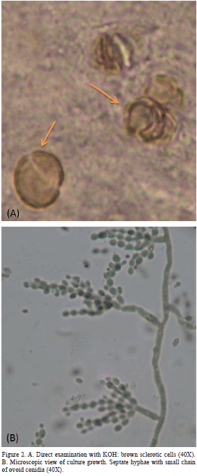
<body><![CDATA[<p style="text-autospace: none" align="justify"><font face="Verdana"><b> <span lang="EN-US" style="font-size: 10.0pt; font-family: TimesNewRomanPS-BoldMT"> Introduction</span></b></font></p>     <p style="text-autospace: none" align="justify"><font face="Verdana"> <span lang="EN-US" style="font-size: 10.0pt; font-family: TimesNewRomanPSMT"> Chromoblastomycosis (CBM) is a chronic, localized deep fungal infection of the  skin and subcutaneous tissue, caused by dematiaceous (black) fungi, living as  saprophytes on plants (</span><i><span lang="EN-US" style="font-size: 10.0pt; font-family: TimesNewRomanPS-ItalicMT">Cactaceae </span></i> <span lang="EN-US" style="font-size: 10.0pt; font-family: TimesNewRomanPSMT">spp</span><i><span lang="EN-US" style="font-size: 10.0pt; font-family: TimesNewRomanPS-ItalicMT">.</span></i><span lang="EN-US" style="font-size: 10.0pt; font-family: TimesNewRomanPSMT">)  or vegetable debris in the soil [1] All agents generate the same parasitic  forms: sclerotic cells (fumagoid cells or Medlar’s cell scleronts) [1,2].  Clinically, the infection is characterized by a slow development, and in some  cases causing irreversible deformation and partial disability of the affected  limb [1,3]. CBM is not a mandatory reportable disease, so the number of cases  reported by researchers and study groups summarizes the epidemiological data.  The description of cases in atypical anatomical areas represents an  extraordinary opportunity to update knowledge and to remind the importance of  thinking about this disease to make an appropriate diagnosis. To the best of our  knowledge, this is the first published report in Venezuela of auricular lesions  of CBM.</span></font></p>     <p style="text-autospace: none" align="justify"><font face="Verdana"><b> <span lang="EN-US" style="font-size: 10.0pt; font-family: TimesNewRomanPS-BoldMT"> Case</span></b></font></p>     <p style="text-autospace: none" align="justify"><font face="Verdana"> <span lang="EN-US" style="font-size: 10.0pt; font-family: TimesNewRomanPSMT">A  62-year-old-man, from Agua Larga, a rural town located at the south of the  “Sierra de San Luis”, Falcon state of Venezuela, with symptoms of seborrheic  dermatitis for more than 10 years. After physical examination, dandruff on the  scalp region was find, however, the characteristics of a lesion observed on the  right ear, drew significant attention. It was observe desquamative and hyperemic  areas with crusts on the right ear, and areas of black dots (<a href="#fig1">Figure 1</a>). After  patient’s acceptance through informed consent, a sample was collect by scraping  the affected areas. Direct KOH examination showed several sclerotic cells  (coffee beans like structures, of 4 to 10 </span> <span style="font-size: 10.0pt; font-family: TimesNewRomanPSMT">&#956;</span><span lang="EN-US" style="font-size: 10.0pt; font-family: TimesNewRomanPSMT">m,  brown colored, with thick walls and septum, which could be seen single or  grouped) thus confirming the clinical diagnosis (<a href="#fig2">Figure 2A</a>).</span></font></p>     <p style="text-autospace: none" align="center"><a name="fig1"> <img border="0" src="/img/fbpe/rsvm/v37n1/art08fig1.gif" width="284" height="242"></a></p>     
<p style="text-autospace: none" align="center"><a name="fig2"> <img border="0" src="/img/fbpe/rsvm/v37n1/art08fig2.gif" width="286" height="691"></a></p>     
<p style="text-autospace: none" align="justify"><font face="Verdana"> <span lang="EN-US" style="font-size: 10.0pt; font-family: TimesNewRomanPSMT"> Culture on Sabouraud Dextrose Agar (SDA), incubated at room temperature (26-28 </span><span lang="EN-US" style="font-family: TimesNewRomanPSMT"><font size="2"> o</font></span><span lang="EN-US" style="font-size: 10.0pt; font-family: TimesNewRomanPSMT">C)  after 15 days, showed velvety colonies with dark olivaceous color. Microcultures  were performing on SDA at 37 </span> <span lang="EN-US" style="font-family: TimesNewRomanPSMT"><font size="2">o</font></span><span lang="EN-US" style="font-size: 10.0pt; font-family: TimesNewRomanPSMT">C  in humid chamber, and septate hyphae with small chains of ovoid conidia were  observed (<a href="#fig2">Figure 2B</a>). Direct exam with KOH, macro and microscopic culture  characteristics, plus epidemiological and clinical findings confirmed the  diagnosis of CBM by </span><i> <span lang="EN-US" style="font-size: 10.0pt; font-family: TimesNewRomanPS-ItalicMT"> Cladophialophora carrionii</span></i><span lang="EN-US" style="font-size: 10.0pt; font-family: TimesNewRomanPSMT">.  Strain was kept on laboratory cultures as AL-2009/UNEFM. Treatment was started  with 200 mg itraconazole daily, showed improvement at the first control.  However, we could not follow evolution, as he did not attend other controls.</span></font></p>     <p style="text-autospace: none" align="justify"><font face="Verdana"><b> <span lang="EN-US" style="font-size: 10.0pt; font-family: TimesNewRomanPS-BoldMT"> Discussion</span></b></font></p>     <p style="text-autospace: none" align="justify"><font face="Verdana"> <span lang="EN-US" style="font-size: 10.0pt; font-family: TimesNewRomanPSMT"> Chromoblastomycosis is a chronic, localized deep fungal infection of the skin  and subcutaneous tissue, caused by dematiaceous (black) fungi, described over a  century ago. Usually affects individuals from rural and poverty areas.  Unfortunately, it is not considered a public health issue, and it is not among  the priorities of national/local health systems, even in endemic countries, such  as Brazil, Madagascar, Mexico, South Africa, and Venezuela. Also many cases have  been described in Australia, China, Guadalupe, India, Japan, Malaysia, United  States, other Latin American, and some European countries, extending the  classical definition of this mycosis to tropical and subtropical areas; so CBM  has a worldwide distribution [4,5]. CBM is not a mandatory reportable disease  and nor does it appear in the list of neglected diseases of the World Health  Organization (WHO), so the systematic reporting in the international  publications have clinical and epidemiological relevance and supports case  studies of working groups, which describe for each case, the typical or atypical  evolution and new species involved [3].</span></font></p>     <p style="text-autospace: none" align="justify"><font face="Verdana"> <span lang="EN-US" style="font-size: 10.0pt; font-family: TimesNewRomanPSMT"> This fungal infection usually affects asymmetrically, unilaterally and  predominantly on the lower limbs, although in some countries, it involves arms  and it might affect the thorax. The anatomical location of lesions has been  associated with exposure to trauma during work, such as goat breeding, logging,  harvesting of fruits in deciduous trees, and </span><i> <span lang="EN-US" style="font-size: 10.0pt; font-family: TimesNewRomanPS-ItalicMT"> Cactaceae </span></i> <span lang="EN-US" style="font-size: 10.0pt; font-family: TimesNewRomanPSMT"> spp. farming, without the proper use of footwear protection. These observations  suggest that CBM is also an occupational disease [1].</span></font></p>     ]]></body>
<body><![CDATA[<p style="text-autospace: none" align="justify"><font face="Verdana"> <span lang="EN-US" style="font-size: 10.0pt; font-family: TimesNewRomanPSMT"> There is little data published on auricular lesions [6-10]. Iwatsu </span><i> <span lang="EN-US" style="font-size: 10.0pt; font-family: TimesNewRomanPS-ItalicMT"> et al</span></i><span lang="EN-US" style="font-size: 10.0pt; font-family: TimesNewRomanPSMT">,  reported a case of CBM that involved the auricle in a patient from Japan and the  etiological agent was </span><i> <span lang="EN-US" style="font-size: 10.0pt; font-family: TimesNewRomanPS-ItalicMT"> Fonsecaea pedrosoi </span></i> <span lang="EN-US" style="font-size: 10.0pt; font-family: TimesNewRomanPSMT"> [6]. Bittencourt </span><i> <span lang="EN-US" style="font-size: 10.0pt; font-family: TimesNewRomanPS-ItalicMT"> et al</span></i><span lang="EN-US" style="font-size: 10.0pt; font-family: TimesNewRomanPSMT">,  presented a case of auricular CMB mimicking an eczematous lesions and the  etiologic agent was </span><i> <span lang="EN-US" style="font-size: 10.0pt; font-family: TimesNewRomanPS-ItalicMT"> Phialophora verrucosa </span></i> <span lang="EN-US" style="font-size: 10.0pt; font-family: TimesNewRomanPSMT"> [7]. Arango </span><i> <span lang="EN-US" style="font-size: 10.0pt; font-family: TimesNewRomanPS-ItalicMT"> et al</span></i><span lang="EN-US" style="font-size: 10.0pt; font-family: TimesNewRomanPSMT">,  described a 60-year-old Colombian male, urban resident, who had a dark pigmented  lesion, infiltrative and crusty in the ear for 5 years, caused by </span><i> <span lang="EN-US" style="font-size: 10.0pt; font-family: TimesNewRomanPS-ItalicMT"> Rhinocladiella aquaspersa, </span></i> <span lang="EN-US" style="font-size: 10.0pt; font-family: TimesNewRomanPSMT"> with a good response to itraconazole therapy at a dose of 200 mg/day for 7  months [8]. França </span><i> <span lang="EN-US" style="font-size: 10.0pt; font-family: TimesNewRomanPS-ItalicMT"> et al</span></i><span lang="EN-US" style="font-size: 10.0pt; font-family: TimesNewRomanPSMT">,  reported another auricular CBM in a 67-year-old man caused by </span><i> <span lang="EN-US" style="font-size: 10.0pt; font-family: TimesNewRomanPS-ItalicMT"> F</span></i><span lang="EN-US" style="font-size: 10.0pt; font-family: TimesNewRomanPSMT">. </span><i> <span lang="EN-US" style="font-size: 10.0pt; font-family: TimesNewRomanPS-ItalicMT"> pedrosoi</span></i><span lang="EN-US" style="font-size: 10.0pt; font-family: TimesNewRomanPSMT">,  the most common agent in Brazil [9], and Muñoz </span><i> <span lang="EN-US" style="font-size: 10.0pt; font-family: TimesNewRomanPS-ItalicMT"> et al</span></i><span lang="EN-US" style="font-size: 10.0pt; font-family: TimesNewRomanPSMT">,  presented a case located in the left ear of a 29-yearold male from Mexico,  caused by </span><i> <span lang="EN-US" style="font-size: 10.0pt; font-family: TimesNewRomanPS-ItalicMT"> F. pedrosoi</span></i><span lang="EN-US" style="font-size: 10.0pt; font-family: TimesNewRomanPSMT">,  treated with itraconazole 200 mg daily for 10 weeks, with complete resolution  [10].</span></font></p>     <p style="text-autospace: none" align="justify"><font face="Verdana"> <span lang="EN-US" style="font-size: 10.0pt; font-family: TimesNewRomanPSMT">In  Venezuela, the most frequent agent of CMB is </span><i> <span lang="EN-US" style="font-size: 10.0pt; font-family: TimesNewRomanPS-ItalicMT"> Cladophialophora carrionii</span></i><span lang="EN-US" style="font-size: 10.0pt; font-family: TimesNewRomanPSMT">,  reported in semi-arid regions; however, </span><i> <span lang="EN-US" style="font-size: 10.0pt; font-family: TimesNewRomanPS-ItalicMT"> F. pedrosoi</span></i><span lang="EN-US" style="font-size: 10.0pt; font-family: TimesNewRomanPSMT">, </span><i> <span lang="EN-US" style="font-size: 10.0pt; font-family: TimesNewRomanPS-ItalicMT"> P. verrucosa </span></i> <span lang="EN-US" style="font-size: 10.0pt; font-family: TimesNewRomanPSMT">and </span><i> <span lang="EN-US" style="font-size: 10.0pt; font-family: TimesNewRomanPS-ItalicMT"> R. aquaspersa </span></i> <span lang="EN-US" style="font-size: 10.0pt; font-family: TimesNewRomanPSMT">are  also found [1,2,4]. In this work, we described an auricular lesion caused by </span><i> <span lang="EN-US" style="font-size: 10.0pt; font-family: TimesNewRomanPS-ItalicMT"> C. carrionii</span></i><span lang="EN-US" style="font-size: 10.0pt; font-family: TimesNewRomanPSMT">.  The patient was a farmer and denied having suffered trauma on the ear with a  Cactaceae splinter and used a hat for sun protection. For harvesting, he used a  sort of sac made of “</span><i><span lang="EN-US" style="font-size: 10.0pt; font-family: TimesNewRomanPS-ItalicMT">cocuiza” </span></i> <span lang="EN-US" style="font-size: 10.0pt; font-family: TimesNewRomanPSMT">(</span><i><span lang="EN-US" style="font-size: 10.0pt; font-family: TimesNewRomanPS-ItalicMT">Furcraea  andina</span></i><span lang="EN-US" style="font-size: 10.0pt; font-family: TimesNewRomanPSMT">),  a rough and porous material easily allows for plants residues to adhere to the  sac, that when it is placed on the right shoulder for transporting it could rub  the ear. It is presumed that exposure may have occurred while performing this  task, because the bag was placed on the floor, dragged short stretches where  they can easily collect small pieces of plant debris containing the saprophyte  phase of the fungus, becoming a rare transmission mechanism. The initial lesion  should have been unnoticeable, without phlogosis signs, which slowly evolved  into small scaly areas, not severe and painless, so the patient felt that the  ear injured was affect by dandruff, and the CBM diagnosis was never made. The  successful treatment of CBM is often unrewarding though; therapeutic response to  itraconazole and terbinafine are good options in </span><i> <span lang="EN-US" style="font-size: 10.0pt; font-family: TimesNewRomanPS-ItalicMT"> C. carrionii </span></i> <span lang="EN-US" style="font-size: 10.0pt; font-family: TimesNewRomanPSMT"> cases [5].</span></font></p>     <p style="text-autospace: none" align="justify"><font face="Verdana"> <span lang="EN-US" style="font-size: 10.0pt; font-family: TimesNewRomanPSMT"> Unfortunately, the patient did not seek treatment, most likely because our  hospital is not easily accessible to people living in these areas. Moreover,  another situation of neglected diseases is the lack of basic, medical and  logistics services near poor and rural towns.</span></font></p>     <p style="text-autospace: none" align="justify"><font face="Verdana"> <span lang="EN-US" style="font-size: 10.0pt; font-family: TimesNewRomanPSMT">It  is very important to remember that traumatic infection by the agent does not  always has the classic presentations in limbs, therefore it is necessary to  investigate clinical, microscopic and eco-epidemiological areas.</span></font></p>     <p style="text-autospace: none" align="justify"><font face="Verdana"><b> <span style="font-size: 10.0pt; font-family: TimesNewRomanPS-BoldMT"> Acknowledgements</span></b></font></p>     <p style="text-autospace: none" align="justify"><font face="Verdana"> <span lang="EN-US" style="font-size: 10.0pt; font-family: TimesNewRomanPSMT">To  Jesús Esquivel, Dee Cocheran and Cristóbal Grau, for critically review of the  manuscript.</span></font></p>     <p style="text-autospace: none" align="justify"><font face="Verdana"><b> <span lang="EN-US" style="font-size: 10.0pt; font-family: TimesNewRomanPS-BoldMT"> Conflicts of interest</span></b></font></p>     <p style="text-autospace: none" align="justify"><font face="Verdana"> <span lang="EN-US" style="font-size: 10.0pt; font-family: TimesNewRomanPSMT">The  authors declare no conflict of interest.</span></font></p>     <p style="text-autospace: none" align="justify"><font face="Verdana"><b> <span style="font-size: 10.0pt; font-family: TimesNewRomanPS-BoldMT">References</span></b></font></p>     <!-- ref --><p style="text-autospace: none" align="justify"><font face="Verdana"> <span style="font-size: 10.0pt; font-family: TimesNewRomanPSMT">1. Yegres F,  Richard-Yegres N. </span><i> <span style="font-size: 10.0pt; font-family: TimesNewRomanPS-ItalicMT"> Cladophialophora carrionii</span></i><span style="font-size: 10.0pt; font-family: TimesNewRomanPSMT">:  aportes al conocimiento de la endemia en Venezuela durante el siglo XX. Rev Soc  Ven Microbiol. 2002; 22:153-7.</span></font>&nbsp;&nbsp;&nbsp;&nbsp;&nbsp;&nbsp;&nbsp;&nbsp;[&#160;<a href="javascript:void(0);" onclick="javascript: window.open('/scielo.php?script=sci_nlinks&ref=2025406&pid=S1315-2556201700010000800001&lng=','','width=640,height=500,resizable=yes,scrollbars=1,menubar=yes,');">Links</a>&#160;]<!-- end-ref --><!-- ref --><p style="text-autospace: none" align="justify"><font face="Verdana"> <span style="font-size: 10.0pt; font-family: TimesNewRomanPSMT">2. Pérez-Blanco  M, Fernández-Zeppenfeldt G, Hernández R, Yegres F, Borelli D. Cromomicosis por </span><i> <span style="font-size: 10.0pt; font-family: TimesNewRomanPS-ItalicMT"> Rhinocladiella aquaspera</span></i><span style="font-size: 10.0pt; font-family: TimesNewRomanPSMT">:  primer caso en Venezuela. Rev Iberoam Micol. </span> <span lang="EN-US" style="font-size: 10.0pt; font-family: TimesNewRomanPSMT"> 1998; 15:51-4.</span></font>&nbsp;&nbsp;&nbsp;&nbsp;&nbsp;&nbsp;&nbsp;&nbsp;[&#160;<a href="javascript:void(0);" onclick="javascript: window.open('/scielo.php?script=sci_nlinks&ref=2025407&pid=S1315-2556201700010000800002&lng=','','width=640,height=500,resizable=yes,scrollbars=1,menubar=yes,');">Links</a>&#160;]<!-- end-ref --><!-- ref --><p style="text-autospace: none" align="justify"><font face="Verdana"> <span lang="EN-US" style="font-size: 10.0pt; font-family: TimesNewRomanPSMT">3.  Queiroz-Telles F. Chromoblastomycosis: a neglected tropical disease. Rev Inst  Med Trop Sao Paulo. </span> <span lang="PT-BR" style="font-size: 10.0pt; font-family: TimesNewRomanPSMT"> 2015; 57(Suppl 19):S46-50.</span></font>&nbsp;&nbsp;&nbsp;&nbsp;&nbsp;&nbsp;&nbsp;&nbsp;[&#160;<a href="javascript:void(0);" onclick="javascript: window.open('/scielo.php?script=sci_nlinks&ref=2025408&pid=S1315-2556201700010000800003&lng=','','width=640,height=500,resizable=yes,scrollbars=1,menubar=yes,');">Links</a>&#160;]<!-- end-ref --><!-- ref --><p style="text-autospace: none" align="justify"><font face="Verdana"> <span style="font-size: 10.0pt; font-family: TimesNewRomanPSMT">4. Martínez  Méndez D, Hernández Valles R, Alvarado P, Mendoza M. Las micosis en Venezuela:  casuística de los Grupos de Trabajo en Micología (1984-2010). Ver Iberoam Micol. </span> <span lang="PT-BR" style="font-size: 10.0pt; font-family: TimesNewRomanPSMT"> 2013; 30:39-46.</span></font>&nbsp;&nbsp;&nbsp;&nbsp;&nbsp;&nbsp;&nbsp;&nbsp;[&#160;<a href="javascript:void(0);" onclick="javascript: window.open('/scielo.php?script=sci_nlinks&ref=2025409&pid=S1315-2556201700010000800004&lng=','','width=640,height=500,resizable=yes,scrollbars=1,menubar=yes,');">Links</a>&#160;]<!-- end-ref --><!-- ref --><p style="text-autospace: none" align="justify"><font face="Verdana"> <span lang="PT-BR" style="font-size: 10.0pt; font-family: TimesNewRomanPSMT">5.  Esterre P, Andriantsimahavandy A, Ramarcel ER, Pecarrere JL. </span> <span lang="EN-US" style="font-size: 10.0pt; font-family: TimesNewRomanPSMT"> Forty years of chromoblastomycosis in Madagascar: a review. Am J Trop Med Hyg.  1996; 55: 45-7.</span></font>&nbsp;&nbsp;&nbsp;&nbsp;&nbsp;&nbsp;&nbsp;&nbsp;[&#160;<a href="javascript:void(0);" onclick="javascript: window.open('/scielo.php?script=sci_nlinks&ref=2025410&pid=S1315-2556201700010000800005&lng=','','width=640,height=500,resizable=yes,scrollbars=1,menubar=yes,');">Links</a>&#160;]<!-- end-ref --><!-- ref --><p style="text-autospace: none" align="justify"><font face="Verdana"> <span lang="EN-US" style="font-size: 10.0pt; font-family: TimesNewRomanPSMT">6.  Iwatsu T, Takano M, Okamoto S. Auricular chromomycosis. Arch Dermatol. 1983;  119:88-9.</span></font>&nbsp;&nbsp;&nbsp;&nbsp;&nbsp;&nbsp;&nbsp;&nbsp;[&#160;<a href="javascript:void(0);" onclick="javascript: window.open('/scielo.php?script=sci_nlinks&ref=2025411&pid=S1315-2556201700010000800006&lng=','','width=640,height=500,resizable=yes,scrollbars=1,menubar=yes,');">Links</a>&#160;]<!-- end-ref --><!-- ref --><p style="text-autospace: none" align="justify"><font face="Verdana"> <span lang="EN-US" style="font-size: 10.0pt; font-family: TimesNewRomanPSMT">7.  Bittencourt AL, Londero AT, Andrade JA. Auricular chromoblastomycosis. A case  report. Rev Inst Med Trop Sao Paulo. 1994; 36:381-3.</span></font>&nbsp;&nbsp;&nbsp;&nbsp;&nbsp;&nbsp;&nbsp;&nbsp;[&#160;<a href="javascript:void(0);" onclick="javascript: window.open('/scielo.php?script=sci_nlinks&ref=2025412&pid=S1315-2556201700010000800007&lng=','','width=640,height=500,resizable=yes,scrollbars=1,menubar=yes,');">Links</a>&#160;]<!-- end-ref --><!-- ref --><p style="text-autospace: none" align="justify"><font face="Verdana"> <span lang="EN-US" style="font-size: 10.0pt; font-family: TimesNewRomanPSMT">8.  Arango M, Jaramillo C, Cortés A, Restrepo A. Auricular chromoblastomycosis  caused by </span><i> <span lang="EN-US" style="font-size: 10.0pt; font-family: TimesNewRomanPS-ItalicMT"> Rhinocladiella aquaspersa</span></i><span lang="EN-US" style="font-size: 10.0pt; font-family: TimesNewRomanPSMT">. </span> <span lang="PT-BR" style="font-size: 10.0pt; font-family: TimesNewRomanPSMT">Med  Mycol. 1998; 36:43-5.</span></font>&nbsp;&nbsp;&nbsp;&nbsp;&nbsp;&nbsp;&nbsp;&nbsp;[&#160;<a href="javascript:void(0);" onclick="javascript: window.open('/scielo.php?script=sci_nlinks&ref=2025413&pid=S1315-2556201700010000800008&lng=','','width=640,height=500,resizable=yes,scrollbars=1,menubar=yes,');">Links</a>&#160;]<!-- end-ref --><!-- ref --><p style="text-autospace: none" align="justify"><font face="Verdana"> <span lang="EN-US" style="font-size: 10.0pt; font-family: TimesNewRomanPSMT">9.  França K, Villa RT, Bastos VR, Almeida AC, Massucatti K, Fukumaru D, Bedin V.  Auricular chromoblastomycosis: a case report and review of published literature.  Mycopathologia 2011; 172:69-72.</span></font>&nbsp;&nbsp;&nbsp;&nbsp;&nbsp;&nbsp;&nbsp;&nbsp;[&#160;<a href="javascript:void(0);" onclick="javascript: window.open('/scielo.php?script=sci_nlinks&ref=2025414&pid=S1315-2556201700010000800009&lng=','','width=640,height=500,resizable=yes,scrollbars=1,menubar=yes,');">Links</a>&#160;]<!-- end-ref --><!-- ref --><p style="text-autospace: none" align="justify"><font face="Verdana"> <span style="font-size: 10.0pt; font-family: TimesNewRomanPSMT">10. Muñoz  Estrada VF, Valenzuela Paz G, Rochín Tolosa M. Cromomicosis: reporte de un caso  con topografía atípica. </span> <span lang="EN-US" style="font-size: 10.0pt; font-family: TimesNewRomanPSMT">Rev  Iberoam Micol. 2011; 28:50-2.</span></font>&nbsp;&nbsp;&nbsp;&nbsp;&nbsp;&nbsp;&nbsp;&nbsp;[&#160;<a href="javascript:void(0);" onclick="javascript: window.open('/scielo.php?script=sci_nlinks&ref=2025415&pid=S1315-2556201700010000800010&lng=','','width=640,height=500,resizable=yes,scrollbars=1,menubar=yes,');">Links</a>&#160;]<!-- end-ref --> ]]></body>
<back>
<ref-list>
<ref id="B1">
<label>1</label><nlm-citation citation-type="journal">
<person-group person-group-type="author">
<name>
<surname><![CDATA[Yegres]]></surname>
<given-names><![CDATA[F]]></given-names>
</name>
<name>
<surname><![CDATA[Richard-Yegres]]></surname>
<given-names><![CDATA[N]]></given-names>
</name>
</person-group>
<article-title xml:lang="es"><![CDATA[Cladophialophora carrionii: aportes al conocimiento de la endemia en Venezuela durante el siglo XX]]></article-title>
<source><![CDATA[Rev Soc Ven Microbiol.]]></source>
<year>2002</year>
<volume>22</volume>
<page-range>153-7</page-range></nlm-citation>
</ref>
<ref id="B2">
<label>2</label><nlm-citation citation-type="journal">
<person-group person-group-type="author">
<name>
<surname><![CDATA[Pérez-Blanco]]></surname>
<given-names><![CDATA[M]]></given-names>
</name>
<name>
<surname><![CDATA[Fernández-Zeppenfeldt]]></surname>
<given-names><![CDATA[G]]></given-names>
</name>
<name>
<surname><![CDATA[Hernández]]></surname>
<given-names><![CDATA[R]]></given-names>
</name>
<name>
<surname><![CDATA[Yegres]]></surname>
<given-names><![CDATA[F]]></given-names>
</name>
<name>
<surname><![CDATA[Borelli]]></surname>
<given-names><![CDATA[D]]></given-names>
</name>
</person-group>
<article-title xml:lang="es"><![CDATA[Cromomicosis por Rhinocladiella aquaspera: primer caso en Venezuela]]></article-title>
<source><![CDATA[Rev Iberoam Micol.]]></source>
<year>1998</year>
<volume>15</volume>
<page-range>51-4</page-range></nlm-citation>
</ref>
<ref id="B3">
<label>3</label><nlm-citation citation-type="journal">
<person-group person-group-type="author">
<name>
<surname><![CDATA[Queiroz-Telles]]></surname>
<given-names><![CDATA[F]]></given-names>
</name>
</person-group>
<article-title xml:lang="en"><![CDATA[Chromoblastomycosis: a neglected tropical disease]]></article-title>
<source><![CDATA[Rev Inst Med Trop Sao Paulo.]]></source>
<year>2015</year>
<volume>57</volume>
<numero>^s19</numero>
<issue>^s19</issue>
<supplement>19</supplement>
<page-range>S46-50</page-range></nlm-citation>
</ref>
<ref id="B4">
<label>4</label><nlm-citation citation-type="journal">
<person-group person-group-type="author">
<name>
<surname><![CDATA[Martínez Méndez]]></surname>
<given-names><![CDATA[D]]></given-names>
</name>
<name>
<surname><![CDATA[Hernández Valles]]></surname>
<given-names><![CDATA[R]]></given-names>
</name>
<name>
<surname><![CDATA[Alvarado]]></surname>
<given-names><![CDATA[P]]></given-names>
</name>
<name>
<surname><![CDATA[Mendoza]]></surname>
<given-names><![CDATA[M]]></given-names>
</name>
</person-group>
<article-title xml:lang="es"><![CDATA[Las micosis en Venezuela: casuística de los Grupos de Trabajo en Micología (1984-2010)]]></article-title>
<source><![CDATA[Ver Iberoam Micol.]]></source>
<year>2013</year>
<volume>30</volume>
<page-range>39-46</page-range></nlm-citation>
</ref>
<ref id="B5">
<label>5</label><nlm-citation citation-type="journal">
<person-group person-group-type="author">
<name>
<surname><![CDATA[Esterre]]></surname>
<given-names><![CDATA[P]]></given-names>
</name>
<name>
<surname><![CDATA[Andriantsimahavandy]]></surname>
<given-names><![CDATA[A]]></given-names>
</name>
<name>
<surname><![CDATA[Ramarcel]]></surname>
<given-names><![CDATA[ER]]></given-names>
</name>
<name>
<surname><![CDATA[Pecarrere]]></surname>
<given-names><![CDATA[JL]]></given-names>
</name>
</person-group>
<article-title xml:lang="en"><![CDATA[Forty years of chromoblastomycosis in Madagascar: a review]]></article-title>
<source><![CDATA[Am J Trop Med Hyg.]]></source>
<year>1996</year>
<volume>55</volume>
<page-range>45-7</page-range></nlm-citation>
</ref>
<ref id="B6">
<label>6</label><nlm-citation citation-type="journal">
<person-group person-group-type="author">
<name>
<surname><![CDATA[Iwatsu]]></surname>
<given-names><![CDATA[T]]></given-names>
</name>
<name>
<surname><![CDATA[Takano]]></surname>
<given-names><![CDATA[M]]></given-names>
</name>
<name>
<surname><![CDATA[Okamoto]]></surname>
<given-names><![CDATA[S]]></given-names>
</name>
</person-group>
<article-title xml:lang="en"><![CDATA[Auricular chromomycosis]]></article-title>
<source><![CDATA[Arch Dermatol.]]></source>
<year>1983</year>
<volume>119</volume>
<page-range>88-9</page-range></nlm-citation>
</ref>
<ref id="B7">
<label>7</label><nlm-citation citation-type="journal">
<person-group person-group-type="author">
<name>
<surname><![CDATA[Bittencourt]]></surname>
<given-names><![CDATA[AL]]></given-names>
</name>
<name>
<surname><![CDATA[Londero]]></surname>
<given-names><![CDATA[AT]]></given-names>
</name>
<name>
<surname><![CDATA[Andrade]]></surname>
<given-names><![CDATA[JA]]></given-names>
</name>
</person-group>
<article-title xml:lang="en"><![CDATA[Auricular chromoblastomycosis: A case report]]></article-title>
<source><![CDATA[Rev Inst Med Trop Sao Paulo.]]></source>
<year>1994</year>
<volume>36</volume>
<page-range>381-3</page-range></nlm-citation>
</ref>
<ref id="B8">
<label>8</label><nlm-citation citation-type="journal">
<person-group person-group-type="author">
<name>
<surname><![CDATA[Arango]]></surname>
<given-names><![CDATA[M]]></given-names>
</name>
<name>
<surname><![CDATA[Jaramillo]]></surname>
<given-names><![CDATA[C]]></given-names>
</name>
<name>
<surname><![CDATA[Cortés]]></surname>
<given-names><![CDATA[A]]></given-names>
</name>
<name>
<surname><![CDATA[Restrepo]]></surname>
<given-names><![CDATA[A]]></given-names>
</name>
</person-group>
<article-title xml:lang="en"><![CDATA[Auricular chromoblastomycosis caused by Rhinocladiella aquaspersa]]></article-title>
<source><![CDATA[Med Mycol.]]></source>
<year>1998</year>
<volume>36</volume>
<page-range>43-5</page-range></nlm-citation>
</ref>
<ref id="B9">
<label>9</label><nlm-citation citation-type="journal">
<person-group person-group-type="author">
<name>
<surname><![CDATA[França]]></surname>
<given-names><![CDATA[K]]></given-names>
</name>
<name>
<surname><![CDATA[Villa]]></surname>
<given-names><![CDATA[RT]]></given-names>
</name>
<name>
<surname><![CDATA[Bastos]]></surname>
<given-names><![CDATA[VR]]></given-names>
</name>
<name>
<surname><![CDATA[Almeida]]></surname>
<given-names><![CDATA[AC]]></given-names>
</name>
<name>
<surname><![CDATA[Massucatti]]></surname>
<given-names><![CDATA[K]]></given-names>
</name>
<name>
<surname><![CDATA[Fukumaru]]></surname>
<given-names><![CDATA[D]]></given-names>
</name>
<name>
<surname><![CDATA[Bedin]]></surname>
<given-names><![CDATA[V]]></given-names>
</name>
</person-group>
<article-title xml:lang="en"><![CDATA[Auricular chromoblastomycosis: a case report and review of published literature]]></article-title>
<source><![CDATA[Mycopathologia]]></source>
<year>2011</year>
<volume>172</volume>
<page-range>69-72</page-range></nlm-citation>
</ref>
<ref id="B10">
<label>10</label><nlm-citation citation-type="journal">
<person-group person-group-type="author">
<name>
<surname><![CDATA[Muñoz Estrada]]></surname>
<given-names><![CDATA[VF]]></given-names>
</name>
<name>
<surname><![CDATA[Valenzuela Paz]]></surname>
<given-names><![CDATA[G]]></given-names>
</name>
<name>
<surname><![CDATA[Rochín Tolosa]]></surname>
<given-names><![CDATA[M]]></given-names>
</name>
</person-group>
<article-title xml:lang="es"><![CDATA[Cromomicosis: reporte de un caso con topografía atípica]]></article-title>
<source><![CDATA[Rev Iberoam Micol.]]></source>
<year>2011</year>
<volume>28</volume>
<page-range>50-2</page-range></nlm-citation>
</ref>
</ref-list>
</back>
</article>