<?xml version="1.0" encoding="ISO-8859-1"?><article xmlns:mml="http://www.w3.org/1998/Math/MathML" xmlns:xlink="http://www.w3.org/1999/xlink" xmlns:xsi="http://www.w3.org/2001/XMLSchema-instance">
<front>
<journal-meta>
<journal-id>1690-4648</journal-id>
<journal-title><![CDATA[Boletín de Malariología y Salud Ambiental]]></journal-title>
<abbrev-journal-title><![CDATA[Bol Mal Salud Amb]]></abbrev-journal-title>
<issn>1690-4648</issn>
<publisher>
<publisher-name><![CDATA[Instituto de Altos Estudios en Salud Pública Dr. Arnoldo Gabaldon]]></publisher-name>
</publisher>
</journal-meta>
<article-meta>
<article-id>S1690-46482014000200007</article-id>
<title-group>
<article-title xml:lang="en"><![CDATA[Achatina fulica Bowdich, 1822 (Mollusca, Gastropoda, Achatinidae) carrier of Helminthes, Protozoa and Bacteria in northeast Venezuela]]></article-title>
<article-title xml:lang="es"><![CDATA[Achatina fulica Bowdich, 1822 (Mollusca, Gastropoda, Achatinidae) hospedador de helmintos, protozoarios y bacterias en el noreste de Venezuela]]></article-title>
</title-group>
<contrib-group>
<contrib contrib-type="author">
<name>
<surname><![CDATA[Morocoima]]></surname>
<given-names><![CDATA[Antonio]]></given-names>
</name>
<xref ref-type="aff" rid="A01"/>
</contrib>
<contrib contrib-type="author">
<name>
<surname><![CDATA[Rodríguez]]></surname>
<given-names><![CDATA[Valmore]]></given-names>
</name>
<xref ref-type="aff" rid="A02"/>
</contrib>
<contrib contrib-type="author">
<name>
<surname><![CDATA[Rivas]]></surname>
<given-names><![CDATA[René]]></given-names>
</name>
<xref ref-type="aff" rid="A03"/>
</contrib>
<contrib contrib-type="author">
<name>
<surname><![CDATA[Coriano]]></surname>
<given-names><![CDATA[Héctor]]></given-names>
</name>
<xref ref-type="aff" rid="A01"/>
</contrib>
<contrib contrib-type="author">
<name>
<surname><![CDATA[Rivero]]></surname>
<given-names><![CDATA[Sigdelis]]></given-names>
</name>
<xref ref-type="aff" rid="A01"/>
</contrib>
<contrib contrib-type="author">
<name>
<surname><![CDATA[Errante]]></surname>
<given-names><![CDATA[Rosina]]></given-names>
</name>
<xref ref-type="aff" rid="A01"/>
</contrib>
<contrib contrib-type="author">
<name>
<surname><![CDATA[Mitchell]]></surname>
<given-names><![CDATA[Makeris]]></given-names>
</name>
<xref ref-type="aff" rid="A01"/>
</contrib>
<contrib contrib-type="author">
<name>
<surname><![CDATA[Herrera]]></surname>
<given-names><![CDATA[Leidi]]></given-names>
</name>
<xref ref-type="aff" rid="A04"/>
</contrib>
<contrib contrib-type="author">
<name>
<surname><![CDATA[Urdaneta-Morales]]></surname>
<given-names><![CDATA[Servio]]></given-names>
</name>
<xref ref-type="aff" rid="A04"/>
</contrib>
</contrib-group>
<aff id="A01">
<institution><![CDATA[,Eastern Center for Tropical Medicine School of Health Sciences Eastern University]]></institution>
<addr-line><![CDATA[ Anzoátegui]]></addr-line>
<country>Venezuela</country>
</aff>
<aff id="A02">
<institution><![CDATA[,Microlab Laboratory  ]]></institution>
<addr-line><![CDATA[ Anzoátegui]]></addr-line>
<country>Venezuela</country>
</aff>
<aff id="A03">
<institution><![CDATA[,Regional Coordination of Epidemiological Monitoring  ]]></institution>
<addr-line><![CDATA[ Monagas]]></addr-line>
<country>Venezuela</country>
</aff>
<aff id="A04">
<institution><![CDATA[,Central University of Venezuela Tropical Zoology and Ecology Institute Faculty of Sciences]]></institution>
<addr-line><![CDATA[Caracas ]]></addr-line>
<country>Venezuela</country>
</aff>
<pub-date pub-type="pub">
<day>00</day>
<month>12</month>
<year>2014</year>
</pub-date>
<pub-date pub-type="epub">
<day>00</day>
<month>12</month>
<year>2014</year>
</pub-date>
<volume>54</volume>
<numero>2</numero>
<fpage>174</fpage>
<lpage>185</lpage>
<copyright-statement/>
<copyright-year/>
<self-uri xlink:href="http://ve.scielo.org/scielo.php?script=sci_arttext&amp;pid=S1690-46482014000200007&amp;lng=en&amp;nrm=iso"></self-uri><self-uri xlink:href="http://ve.scielo.org/scielo.php?script=sci_abstract&amp;pid=S1690-46482014000200007&amp;lng=en&amp;nrm=iso"></self-uri><self-uri xlink:href="http://ve.scielo.org/scielo.php?script=sci_pdf&amp;pid=S1690-46482014000200007&amp;lng=en&amp;nrm=iso"></self-uri><abstract abstract-type="short" xml:lang="es"><p><![CDATA[Por cuanto el molusco Achatina fulica nativo del África es vector de helmintos, pero su relación con protozoarios y bacterias es poco conocida, decidimos estudiar las excretas de 1.200 ejemplares capturados en los estados Anzoátegui, Monagas, Sucre y Nueva Esparta, del noreste de Venezuela. Su moco pedal y heces mostraron infección por los protozoarios Chilomastix spp., Trichomonas spp., Giardia spp., Balantidium spp., Entamoeba spp., Iodamoeba spp., Blastocystis spp. Y por los helmintos de los grupos Ascarioidea, Trichuroidea, Ancylostomatidae y Cestoda. El moco céfalopodal mostró únicamente larvas de Rhabditida. Las bacterias Citrobacter freundii, Escherichia coli, Klebsiella pneumoniae, K. azaenae, Aeromonas hydrophila, Acinetobacter baumannii, Pseudomonas aeruginosa, Campylobacter spp. infectaron a las tres excretas. Los mecanismos de transmisión y la composición de las excretas, como nichos fisiológicamente apropiados para los organismos encontrados, son discutidos en relación con el riesgo epidemiológico que el molusco representa en salud pública y veterinaria.]]></p></abstract>
<abstract abstract-type="short" xml:lang="en"><p><![CDATA[The mollusk Achatina fulica, native to Eastern Equatorial Africa, has been incriminated as a carrier or vector of helminthes. Nevertheless, information in the literature as regards its status as a carrier for bacteria is scarce, and we could find no reference at all for its relation to protozoa. We studied microscopically the excreta from 1200 snails captured in Anzoátegui, Monagas, Sucre and Nueva Esparta states, in northeast Venezuela. The pedal mucus and feces were infected by the protozoa Chilomastix spp., Trichomonas spp., Giardia spp., Balantidium spp., Entamoeba spp., Iodamoeba spp., Blastocystis spp., as well as helminthes of Ascarioidea, Trichuroidea, Ancylostomatidae and Cestoda groups. The only helminthes found in the cephalopodal mucus were Rhabditida larvae. The three excreta were also infected by the bacteria: Citrobacter freundii, Escherichia coli, Klebsiella pneumoniae, K. azaenae, Aeromonas hydrophila, Acinetobacter baumannii, Pseudomonas aeruginosa, Campylobacter spp. Risk of infection and transmission mechanisms as well as the composition of the excreta as appropriate physiological niches for the organisms mentioned, are discussed with regard to the epidemiological importance of this snail for in human and veterinary health.]]></p></abstract>
<kwd-group>
<kwd lng="es"><![CDATA[Achatina fulica]]></kwd>
<kwd lng="es"><![CDATA[carrier]]></kwd>
<kwd lng="es"><![CDATA[helmintos]]></kwd>
<kwd lng="es"><![CDATA[protozoarios]]></kwd>
<kwd lng="es"><![CDATA[bacterias]]></kwd>
<kwd lng="es"><![CDATA[Venezuela]]></kwd>
<kwd lng="en"><![CDATA[Achatina fulica carrier]]></kwd>
<kwd lng="en"><![CDATA[helminthes]]></kwd>
<kwd lng="en"><![CDATA[protozoa]]></kwd>
<kwd lng="en"><![CDATA[bacteria]]></kwd>
<kwd lng="en"><![CDATA[Venezuela]]></kwd>
</kwd-group>
</article-meta>
</front><body><![CDATA[ <p ALIGN="center"><b><i><font SIZE="2" face="Verdana">A</font><font face="Verdana">chatina  fulica </font></i><font face="Verdana">Bowdich, 1822 (Mollusca, Gastropoda,  Achatinidae) carrier of Helminthes, Protozoa and Bacteria in northeast Venezuela</p>     <p ALIGN="center">Achatina fulica <i>Bowdich, 1822 (Mollusca, Gastropoda,  Achatinidae) hospedador de helmintos, protozoarios y bacterias en el noreste de  Venezuela </p> </i> </font><font SIZE="2" face="Verdana">     <p align="center">Antonio Morocoima<sup>1</sup>, Valmore Rodríguez<sup>2</sup>,  René Rivas<sup>3</sup>, Héctor Coriano<sup>1</sup>, Sigdelis Rivero<sup>1</sup>,  Rosina Errante<sup>1</sup>, Makeris Mitchell<sup>1</sup>, Leidi Herrera<sup>4</sup>  &amp; Servio Urdaneta-Morales<sup>4</sup>*</font></p> </b>     <p ALIGN="JUSTIFY"><font size="2"><font face="Verdana">1 Eastern Center for  Tropical Medicine, School of Health Sciences, Eastern University, Anzoátegui  state, Venezuela. School of Health Sciences, Eastern University, Anzoátegui  state, Venezuela. </font></p>     <p ALIGN="JUSTIFY"><font face="Verdana">2 Microlab Laboratory, Lecheria,  Anzoátegui state, Venezuela</font></p>     <p ALIGN="JUSTIFY"><font face="Verdana">3 Regional Coordination of  Epidemiological Monitoring, Monagas state, Venezuela </font></p>     <p ALIGN="JUSTIFY"><font face="Verdana">4 Laboratory for the Biology of Vectors  and Parasites, Tropical Zoology and Ecology Institute, Faculty of Sciences,  Central University of Venezuela, Caracas, Venezuela</font></p>     <p ALIGN="JUSTIFY"><font face="Verdana">*Autor de correspondencia: <a href="mailto:tropism2006@yahoo.es">tropism2006@yahoo.es</a></font></p> </font><font SIZE="1"></font>     <p align="justify"><font SIZE="2" face="Verdana">Dedicated to Dr. Rafael  Martínez Escarbassiere for his pioneering research on Achatina fulica in America</p> </font><b>     <p ALIGN="JUSTIFY"><font size="2"><font face="Verdana">RESUMEN </font></p> </font></b><font size="2">     ]]></body>
<body><![CDATA[<p ALIGN="JUSTIFY"><font face="Verdana">Por cuanto el molusco </font></font> <font FACE="Verdana" SIZE="2"><i>Achatina fulica </i>nativo del África es vector  de helmintos, pero su relación con protozoarios y bacterias es poco conocida,  decidimos estudiar las excretas de 1.200 ejemplares capturados en los estados  Anzoátegui, Monagas, Sucre y Nueva Esparta, del noreste de Venezuela. Su moco  pedal y heces mostraron infección por los protozoarios <i>Chilomastix </i>spp., <i>Trichomonas </i>spp., <i>Giardia </i>spp., <i>Balantidium </i>spp., <i> Entamoeba </i>spp., <i>Iodamoeba </i>spp., <i>Blastocystis </i>spp. Y por los  helmintos de los grupos Ascarioidea, Trichuroidea, Ancylostomatidae y Cestoda.  El moco céfalopodal mostró únicamente larvas de Rhabditida. Las bacterias <i> Citrobacter freundii, Escherichia coli, Klebsiella pneumoniae, K. azaenae,  Aeromonas hydrophila, Acinetobacter baumannii, Pseudomonas aeruginosa,  Campylobacter </i>spp. infectaron a las tres excretas. Los mecanismos de  transmisión y la composición de las excretas, como nichos fisiológicamente  apropiados para los organismos encontrados, son discutidos en relación con el  riesgo epidemiológico que el molusco representa en salud pública y veterinaria. </p> <b>     <p align="justify">Palabras clave: </b><i>Achatina fulica</i>, carrier,  helmintos, protozoarios, bacterias, Venezuela.</p> </font><b>     <p ALIGN="JUSTIFY"><font size="2"><font face="Verdana">SUMMARY<i> </i></font> </p> </font></b><font size="2"><i>     <p ALIGN="JUSTIFY"><font face="Verdana">The mollusk </font></i></font> <font FACE="Verdana" SIZE="2">Achatina fulica</font><i><font SIZE="2" face="Verdana">,  native to Eastern Equatorial Africa, has been incriminated as a carrier or  vector of helminthes. Nevertheless, information in the literature as regards its  status as a carrier for bacteria is scarce, and we could find no reference at  all for its relation to protozoa. We studied microscopically the excreta from  1200 snails captured in Anzoátegui, Monagas, Sucre and Nueva Esparta states, in  northeast Venezuela. The pedal mucus and feces were infected by the protozoa </font></i><font FACE="Verdana" SIZE="2">Chilomastix <i>spp., </i>Trichomonas <i> spp., </i>Giardia <i>spp., </i>Balantidium <i>spp., </i>Entamoeba <i>spp., </i> Iodamoeba <i>spp., </i>Blastocystis <i>spp., as well as helminthes of  Ascarioidea, Trichuroidea, Ancylostomatidae and Cestoda groups. The only  helminthes found in the cephalopodal mucus were Rhabditida larvae. The three  excreta were also infected by the bacteria: </i>Citrobacter freundii<i>, </i> Escherichia coli<i>, </i>Klebsiella pneumoniae<i>, </i>K. azaenae<i>, </i> Aeromonas hydrophila<i>, </i>Acinetobacter baumannii<i>, </i>Pseudomonas  aeruginosa<i>, </i>Campylobacter </font><i><font SIZE="2" face="Verdana">spp.  Risk of infection and transmission mechanisms as well as the composition of the  excreta as appropriate physiological niches for the organisms mentioned, are  discussed with regard to the epidemiological importance of this snail for in  human and veterinary health.</p> </i><b>     <p align="justify">Key words<i>: </i></b></font><font FACE="Verdana" SIZE="2"> Achatina fulica <i>carrier, helminthes, protozoa, bacteria, Venezuela.</p> </i></font><b></b>     <p align="justify"><font SIZE="2" face="Verdana">Recibido el 30/05/2014 Aceptado  el 29/10/2014</font></p>     <p ALIGN="JUSTIFY"><font SIZE="2" face="Verdana"><b>INTRDUCCIÓN</b> </p> <i>     <p>Achatina fulica </i>(the Giant African snail, called in Venezuela &quot;caracol  africano&quot;) is an arboreal and terrestrial mollusk that frequently invades the  vegetated shores of tropical and subtropical water bodies. The snails were  introduced from Eastern Equatorial Africa by humans and are currently found in  Asia (India, China, Japan, Philippines), the Pacific, Madagascar, Indonesia,  Australia, the Caribbean Basin, the United States and South America (Colombia,  Venezuela, Ecuador, Brazil, Argentina) (Martínez Escarbassiere &amp; Martínez, 1997;  Correoso, 2006; USDA, 2008; Gutiérrez <i>et al.</i>, 2011). </p> <i>     <p ALIGN="JUSTIFY">Achatina fulica </i>snails are polyphagous (herbivores,  carnivores, necrophages), and along with organic detritus, their preferred food  list to date consists of over 500 known plant species, ranging from coffee and  bananas in the tropics to potatoes and tobacco in more temperate zones. The  snails are voracious eaters and competitors of native snails as well as habitat  modifiers. Their importance in agricultural systems gardens and as agents for  the reduction of biological diversity is increasing at an alarming rate making  them one of the 100 most important pests worldwide (USDA, 2007; Zanol <i>et al.</i>,  2010; Cardoso <i>et al.</i></font><font SIZE="2"><font face="Verdana">, 2012). </font></p>     <p ALIGN="JUSTIFY"><font face="Verdana">Furthermore, this species is an  intermediate host for nematode pathogens such as </font></font> <font FACE="Verdana" SIZE="2"><i>Angiostrongylus cantonensis</i>, the causal  agent of zoonotic meningoencephalitis and <i>A. costaricensis</i>, which  produces abdominal ileocolitis. They are thus also considered to represent a  potential zoonotic risk and a possible danger to public health (Martínez  Escarbassiere &amp; Martínez, 1997; Teles <i>et al.</i>, 1997). </p> <i>     ]]></body>
<body><![CDATA[<p ALIGN="JUSTIFY">Achatina fulica</i></font><font SIZE="2"><font face="Verdana">,  like all Achatinidae, needs calcium for the formation of its shell and for  reproduction. It thus prefers environments rich in calcium carbonate, such as  limestone landscapes with a pH of 7.0-8.0 and urban areas with abundant cement  or concrete. Snails are active in the early morning, late afternoon, on cloudy,  damp, or rainy days and avoid direct sunlight (USDA, 2007). </font></p>     <p><font face="Verdana">The muci of the Mollusca are produced by several glands  that secrete the mucus through pores that pass between the epidermal cells.  Pedal mucus is a gel produced when the foot gland of the snail, which can detect  vibrations, is stimulated mechanically or chemically. This gel acts as a  lubricant that permits the snails to slide over the surface of terrestrial or  aquatic habitats by reducing friction between the foot (sole) and the ground (the  slime trail). It also has strong adhesive properties that enable the snails to  crawl up vertical surfaces such as walls and roofs without falling off.  Furthermore, the mucus acts as a means by which information regarding locomotion  direction, osmoregulation and the reparation of skin lesions can be conveyed as  well as the type of food travelled over and the snail’s sexual state (Skingsley </font></font><font FACE="Verdana" SIZE="2"><i>et al.</i>, 2000). </p>     <p ALIGN="JUSTIFY">It is frequently difficult to differentiate where the head of  the snail ends and the foot begins, so they are collectively called head-foot or  cephalopodium which is a typical morphological character of all gastropods (Nordsieck,  2011) If the snail is disturbed continuously or even violently it discharges a  clear foamy mucus by the synchronised contraction of the cephalopodal gland,  located in the upper lateral region of the cephalopodium. This mucus is composed  of acidic, neutral and sulphated mucins which behave as chemical barriers and  form part of the defense mechanisms of the innate immunological system of the  snails (Skingsley <i>et al.</i>, 2000; Pinchuck &amp; Hodgson, 2009, 2012). </p> <i>     <p ALIGN="JUSTIFY">Achatina fulica </i>was captured for the first time in  Venezuela in Caracas, the capital city, and subsequently in the states of  Miranda, Lara, Portuguesa, Carabobo, Aragua, Nueva Esparta (Margarita Island),  Sucre, Monagas and the Delta Amacuro and most recently in Zulia, Anzoátegui and  Yaracuy states. The snails have been found in diverse habitats; from forests to  mountains and in both rural and strictly urban areas (Martinez Escarbassiere &amp;  Martinez, 1997; Martínez Escarbassiere <i>et al.</i>, 2008; Oletta &amp; Carvajal,  2011). In Venezuela <i>A. fulica </i>has been found infected with <i>Schistosoma  mansoni</i>, <i>Strongyloides </i>spp., <i>Trichuris </i>spp. and <i>Hymenolepis </i>spp. (Liboria <i>et al.</i>, 2010); the first case of human infection by <i> A. costaricensis </i>was reported by Incani <i>et al. </i></font><font SIZE="2"> <font face="Verdana">(2007). </font></p>     <p><font face="Verdana">To our knowledge, despite the extensive geographical  distribution and the importance of </font></font><font FACE="Verdana" SIZE="2"> <i>A. fulica </i>as a carrier or vector of helminthes, studies regarding its  status for bacteria are scarce and infrequent, and as far as we know there are  none incriminating it as a carrier for protozoa (Utomo <i>et al.</i>, 1991;  Cardoso <i>et al.</i></font><font SIZE="2"><font face="Verdana">, 2012). This,  coupled with the explosive dispersion of dense populations of the snails in  Venezuela, prompted this investigation. We chose the northeast of Venezuela as  our study area due to the lack of investigations undertaken in this region,  despite continuous complaints of the snails’ presence made to the Eastern Center  for Tropical Medicine, Eastern University, (Anzoátegui state) by the local  inhabitants. </font></p>     <p ALIGN="JUSTIFY"><font face="Verdana"><b>MATERIAL AND METHODS</b></font></p> </font><font FACE="Verdana" SIZE="2"><i>     <p ALIGN="JUSTIFY">Capture, maintenance, identification and parasitological and  microbiological analyses of the snails </p> </i></font><font SIZE="2">     <p ALIGN="JUSTIFY"><font face="Verdana">Snails were captured in the afore  mentioned habitats in the evening (USDA, 2007) from urban areas in Anzoátegui,  Monagas, Sucre and Nueva Esparta (Margarita Island) states during the months of  June to August 2011 inclusive. Adult snails (average length 18 cm, able to  copulate and oviposit) were collected by hand from plants, fruits and vegetables  in small farms and residential gardens, as well as on walls outside human  dwellings and pavements (<a href="#fig1">Fig. 1 A/F</a>). Latex gloves and safety glasses were worn  at all times to prevent any risk from infection.</font></p>     <p ALIGN="center"> <a name="fig1"> <img border="0" src="/img/fbpe/bmsa/v54n2/art07fig1.gif" width="540" height="434"></a></p>     
<p><font face="Verdana">After collection, snails were placed in plastic bags,  labeled according to date and collection site and kept in the laboratory in  darkness at a relative humidity of 70%, and temperature 24-26ºC. They were fed  with lettuce leaves (</font></font><font FACE="Verdana" SIZE="2"><i>Lactuca  sativa</i>) <i>ad libitum </i>(Liboria <i>et al.</i>, 2010) thoroughly washed  with sterile saline solution and vinegar. Species were identified according to  Martínez Escarbassiere &amp; </font><font SIZE="2"><font face="Verdana">Martínez  (1997). </font></p>     ]]></body>
<body><![CDATA[<p><font face="Verdana">For the parasitological study, three hundred snails were  collected from each of the four states mentioned (n=1200). They were separated  into groups of 150 mollusks each from two municipalities per state and placed in  plastic containers 60 x 40 x </font></font><font FACE="Verdana" SIZE="2">20 cm,  divided internally into five compartments with stiff plastic sheets and covered  with perforated lids so that the snails could breathe easily (<a href="#fig2">Fig. 2A, B</a>). Each  container was labeled according to the collection site of the snails placed in  it. Snails were washed with sterile saline solution before placing them  individually in each of the five compartments and the </font><font SIZE="2"> <font face="Verdana">different types of mucus were obtained sequentially using a  sterile pipette, as follows: pedal mucus (3 ml average) by lightly rubbing the  snails feet with a small, sterile applicator stick; cephalopodal mucus (5 ml  average) expressed by manual compression on </font> <font FACE="Verdana" SIZE="2">the cephalopodal gland and finally the feces (50  mg </font><font face="Verdana">average). In order to avoid fecal contamination,  feces released during the collection of the other excreta were immediately  recovered and both the compartment and the snail were thoroughly washed before  continuing with their extraction. Samples of the three excreta were taken with  small sterile applicator sticks and prepared as wet mounts by mixing them with  drops of sterile isotonic solution. They were examined immediately for protozoa  (trophozoites or cysts) and helminthes (eggs, larvae or adults). The material  was </font><font FACE="Verdana" SIZE="2">also stained using Lugol´s solution in  order to confirm the identification of cysts and eggs. The presence of </font> <font face="Verdana">coccidians was determined from samples stained with  Kinyoun’s solution. Fecal samples were prepared according to the Kato-Katz  method for qualitative </font><font FACE="Verdana" SIZE="2">diagnosis. All  preparations were sufficiently thin </font><font face="Verdana">so as to be  spread smoothly and evenly under the cover glass (75 x 37 mm) (Spencer &amp; Monroe,  1961). Preparations were examined immediately with an Olympus CH20 microscope  (400X; 1,000X </font><font FACE="Verdana" SIZE="2">magnification) and  micrographs were taken using a </font><font face="Verdana">digital camera (Nikon  Coolpix P550, resolution 16.1 megapixels) attached to the microscope.</font></font></p>     <p style="text-align: center"><font SIZE="2"> <a name="fig2"> <img border="0" src="/img/fbpe/bmsa/v54n2/art07fig2.gif" width="545" height="287"></a></p> <font FACE="Verdana" SIZE="2">     
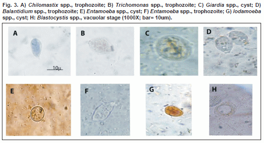
<p>The presence and identification of the </font><font face="Verdana"> <font SIZE="2">bacteria observed in the three types of excreta taken </font>from  ten snails captured from each of the four states surveyed (n = 40), was done  using Gram coloration and by inoculation in gelatin agar Thiosulfate-Citrate-Bile-  Sucrose (TCBS), Xylose-lysine-desoxycholate (XLD) agar, Salmonella-Shigella  (S-S) agar, MacConkey agar and MacConkey broth (incubated at 35ºC/24hs, under  facultative aerobic conditions); blood agar and chocolate agar (incubated at 35ºC/24hs  under microaerophilic conditions); Preston agar and chrome agar (incubated at 35ºC/48hs  under microaerophilic conditions). </font></p> </font><font FACE="Verdana" SIZE="2"><i>     <p ALIGN="JUSTIFY">Ethics Considerations </p> </i></font><font SIZE="2">     <p ALIGN="JUSTIFY"><font face="Verdana">All experiments involving animals were  conducted according to the current Bioethical Laws as laid down by the  Venezuelan Science and Technology Ministry, and were approved by the Ethics  Committee and the Committee for Animal Care of the National Fund for Science and  Technology (FONACIT, Caracas, Venezuela).</font></p>     <p ALIGN="JUSTIFY"><font face="Verdana"><b>RESULTS</b></font></p> </font><font FACE="Verdana" SIZE="2"><i>     <p ALIGN="JUSTIFY">Results of the parasitological analyses of the mollusks </p> </i>     <p>All three excreta types in every one of the 1,200 <i>A. fulica </i>specimens  examined were infected (<a href="#tab1">Tables I</a>, <a href="#tab2">II</a>). Both, the pedal mucus and the feces,  contained protozoa and helminthes taxa of human and or animal importance (<a href="#fig3">Figs.  3A/H</a>; <a href="#fig4">4A/I</a>); coccidia were not observed. The cephalopodal mucus was infected by  adult of one imputed Rhabditida. Protozoa and helminthes were common in the  pedal mucus and feces of snails collected from all the states surveyed, although  the percentages of infection did vary. In addition, these excreta contained  larval stages of Nematodes, highlighting the need for further studies for their  identification to gender or species level.</p>     <p style="text-align: center"> <a name="tab1"> <img border="0" src="/img/fbpe/bmsa/v54n2/art07tab1.gif" width="568" height="421"></a></p>     
<p style="text-align: center"> <a name="tab2"> <img border="0" src="/img/fbpe/bmsa/v54n2/art07tab2.gif" width="569" height="405"></a></p>     
]]></body>
<body><![CDATA[<p style="text-align: center"> <a name="fig3"> <img border="0" src="/img/fbpe/bmsa/v54n2/art07fig3.gif" width="544" height="294"></a></p>     
<p style="text-align: center"> <a name="fig4"> <img border="0" src="/img/fbpe/bmsa/v54n2/art07fig4.gif" width="538" height="368"></a></p> <i>     
<p ALIGN="JUSTIFY">Results of the microbiological analyses of the mollusks </p> </i>     <p ALIGN="JUSTIFY">The microbiological study (<a href="#tab3">Tables III</a>,  <a href="#tab4">IV</a>) showed that all  three types of excreta sampled from the 40 individuals examined, contained gram-negative  facultative anaerobic bacilli and coccobacilli Enterobacteriaceae and  Aeromonadaceae, as well as other bacteria whose taxonomic status is not well  defined (Murray <i>et al.</i></font><font SIZE="2"><font face="Verdana">, 2009).  All three types of excreta were negative for levaduriforme yeasts.</font></font></p>     <p ALIGN="center"><font SIZE="2"> <a name="tab3"> <img border="0" src="/img/fbpe/bmsa/v54n2/art07tab3.gif" width="537" height="497"></a></font></p>     
<p ALIGN="center"><font SIZE="2"> <a name="tab4"> <img border="0" src="/img/fbpe/bmsa/v54n2/art07tab4.gif" width="536" height="496"></a></p>     
<p ALIGN="JUSTIFY"><font face="Verdana"><b>DISCUSSION</b> </font></p>     <p ALIGN="JUSTIFY"><font face="Verdana">All the helminthes found in </font> </font><font FACE="Verdana" SIZE="2"><i>A. fulica </i>snails belong to taxa that  require soil to complete their development cycles. The Enterobacteriaceae, as  well as <i>A. hidrofila</i>, <i>P. aeruginosa</i>, <i>A. baumanni</i>, <i>E.  coli </i>and <i>Campylobacter </i>spp. also develop and proliferate in soil,  vegetation, decomposing organic material and water (Murray <i>et al.</i>, 2009),  all of which are inhabited by <i>A. fulica</i></font><font SIZE="2"><font face="Verdana">. </font></p>     <p ALIGN="JUSTIFY"><font face="Verdana">On the other hand, it has been estimated  that 5.3 billion people are at risk of contracting geohelminthiasis mainly  caused by </font></font><font FACE="Verdana" SIZE="2"><i>Ascaris</i>, <i> Trichuris</i>, <i>Necator/Ancylostoma</i>, <i>Toxocara </i>and <i>Toxascaris</i>,  with the highest prevalence reported from Africa, Asia and Latin America. These  helminthes are endemic in America and the Caribbean, with around 100, 84 and 50  million people infected with the first three helminthes listed above,  respectively. Venezuela has been reported as being among the countries with the  highest risk of infection (27% for <i>Trichuris </i>spp., indicating an urgent  need for the implementation of control measures according to the latest  recommendations drawn up by the PAHO (Pullan &amp; Brooker, 2012; Chammartin <i>et  al.</i></font><font SIZE="2"><font face="Verdana">, 2013). </font></p>     <p><font face="Verdana">Similar health risks have been linked to the presence of  the protozoa and bacteria found in the snail’s excreta during this investigation,  which taken together, are responsible for three of the greatest current global  public health problems. This is especially true for populations with a high  degree of poverty, an inadequate water supply and poor sanitation and health  education (Murray </font><font FACE="Verdana" SIZE="2"><i>et al.</i></font><font face="Verdana">,2009;  Duc </font><font FACE="Verdana" SIZE="2"><i>et al.</i></font><font face="Verdana">,  2011, Pullan &amp; Brooker, 2012); a situation typical of many tropical countries,  including Venezuela. </font></p> <font FACE="Verdana" SIZE="2">     ]]></body>
<body><![CDATA[<p ALIGN="JUSTIFY">Furthermore, two of the bacteria identified in the snails  examined, <i>E. coli </i>and <i>Campylobacter </i></font><font SIZE="2"> <font face="Verdana">spp., have been singled out as being among the seven most  important bacterial pathogens for humans (Faruque, 2012). </font></p>     <p ALIGN="JUSTIFY"><font face="Verdana">Investigations on the composition,  characterization and functions of the microbiota in the gastrointestinal tract  of the Mollusca and in particular </font></font><font FACE="Verdana" SIZE="2"> <i>A. fulica</i>, are scarce. According to Charrier <i>et al. </i>(2006) in <i> Helix </i></font><font SIZE="2"><font face="Verdana">spp. the digestive tract is  acidic in the crop but neutral or alkaline in the intestine. This highlights the  dependence of these snails on the high biochemical potential of their diverse  bacterial microbiota for the degradation and fermentation of the principal  vegetable components (soluble sugars and polymers) that make up their diet. </font></p>     <p ALIGN="JUSTIFY"><font face="Verdana">Cardoso </font></font> <font FACE="Verdana" SIZE="2"><i>et al. </i>(2012) also found diverse, abundant  and metabolically active bacterial communities that included, among others, <i> Aeromonas</i>, <i>Citrobacter</i>, <i>Klebsiella</i>, <i>Acinetobacter </i>and <i>Pseudomonas </i>in the intestinal tracts of Giant African snails starved for  72 hours in order to minimize the presence of transient bacteria. This leads us  to suppose that these bacteria are native to the intestinal tracts of <i>A.  fulica </i>making it <i>&quot;per se&quot; </i></font><font SIZE="2"><font face="Verdana"> a very effective disperser of these organisms. </font></p>     <p><font face="Verdana">The bacteria are probably acquired by </font></font> <font FACE="Verdana" SIZE="2"><i>A. fulica </i>through its diet, the polyphagous  nature of which results in the development of a structurally complex and highly  diverse microbiotic community. The impact of biotic and abiotic factors on this  bacterial community should, however, be investigated. These bacteria are  responsible for the digestion of complex polysaccharides, by converting sugars  to short-chain fatty acids and synthesizing amino acids and essential vitamins.  Cardoso <i>et al. </i>(2012) suggest that this microbiota enables the snails to  adapt to different diets. It is thus important to determine their species </font> <font face="Verdana">composition, in order to provide knowledge that could  contribute to the elaboration of strategies for snail control. With this in mind,  we would like to mention the fact that </font></font> <font FACE="Verdana" SIZE="2"><i>A. hydrophila </i>(Aeromonadaceae) the causal  agent of diseases in humans, fish and mollusks and identified in all three  excreta types collected from <i>A. fulica</i>, has been implicated in its  decline in several geographic regions (Yamada <i>et al.</i></font><font SIZE="2"><font face="Verdana">,  2011). </font></p>     <p><font face="Verdana">The polyphagous diet of </font></font> <font FACE="Verdana" SIZE="2"><i>A. fulica</i>, together with its  physiologically high environmental adaptability and capacity to proliferate in  an eclectic range of habitats over a wide geographic range (in Venezuela, snails  have been reported as having invaded 60% of the national territory; Martinez  Escarbassiere <i>et al. </i></font><font face="Verdana" SIZE="2">(2008); Oletta  &amp; Carvajal (2011)) are characteristics that make it a very important carrier and  disperser of protozoa, helminthes and bacteria. So much so that has been  reported to &quot;sow&quot; terrestrial environments and contaminate water bodies with  these organisms and has thus become an excellent indicator for their presence (Liboria </font><font FACE="Verdana" SIZE="2"><i>et al.</i></font><font SIZE="2"><font face="Verdana">,  2010). </font></p>     <p><font face="Verdana">These pathogens may be transmitted from </font></font> <font FACE="Verdana" SIZE="2"><i>A. fulica </i>to humans and animals by the  ingestion of raw plant products and water infected by their excreta (Faruque,  2012), eating raw or undercooked snails (Zanol <i>et al.</i>, 2010), or possibly  from the handling of live snails, especially if the mucous membranes of the eyes,  nose or mouth come into contact with the snails' excreta (USDA, 2008). </p>     <p>Despite the warnings given by Martinez Escarbassiere &amp; Martinez (1997) about  the risks associated with the presence of <i>A. fulica </i>in Venezuela, few  studies have determined the impact of this snail species either on the natural  environment or human and veterinary health. Thus a low priority has been given  to the proper surveillance of the zones invaded by this mollusk by the health  authorities and there have been few measures taken to restrict its invasion of  the rest of the country. There have also been a lack of control measures as  regards food security issues and little attempt to educate the population as  regards the health risks involved. Implementation of these aspects would  contribute to mitigating the number of cases of infection reported as well as  the emergence of new diseases due to the intense and rapid invasion and  proliferation of <i>A. fulica </i>in Venezuela. A greater knowledge of the snail  distribution would enable the instigation of effective measures for the control  of the protozoa, helminthes and bacteria that they harbor. We suggest that these  considerations underline the epidemiological importance of <i>A. fulica </i> </font><font SIZE="2"><font face="Verdana">for human and animal health. </font> </p>     <p><font face="Verdana">Two mucopolysaccharide groups, Glycosaminoglycans (GAGs,  heparin-, chondroitin-, dermatan sulfate and hyaluronic acid) and glycoproteins,  suspended in 80-99% water have been found ubiquitously in the animal kingdom,  from the most primitive invertebrates (Hydra, Cnidaria) to vertebrates,  demonstrating their conserved status and role in fundamental biological  processes. In the </font></font><font FACE="Verdana" SIZE="2"><i>A. fulica </i> pedal mucus, these structures are responsible for regulating cell growth,  proliferation, differentiation, morphogenesis and migration, and are also  involved in the transport of substances, thus providing energy and metabolic  resources basic to the snail´s survival (Skingsley <i>et al.</i>, 2000;  Berniyanti <i>et al.</i>, 2007; Yamada <i>et al.</i></font><font SIZE="2"><font face="Verdana">,  2011). </font></p>     <p ALIGN="JUSTIFY"><font face="Verdana">In addition, it has been demonstrated  that this mucus contains a broad spectrum antibiotic glycoprotein effective  against </font></font><font FACE="Verdana" SIZE="2"><i>E. coli</i>, <i> Streptococcus mutans</i>, <i>Staphylococus aureus </i>and <i>S. epidermidis </i> (Berniyanty <i>et al.</i>, 2007; Santana <i>et al.</i>, 2012) indicating its  importance for the mollusk immune system. With regard to this we should add that  a highly sulfated product -acharan sulfate, related to heparin/heparin sulfate  but structurally unique to <i>A. fulica</i>, may also be responsible for  protecting <i>A. fulica </i>from foreign bodies such as bacteria and virus (Yamada <i>et al.</i></font><font SIZE="2"><font face="Verdana">, 2011). </font></p>     <p ALIGN="JUSTIFY"><font face="Verdana">Mucins, a family of highly glycosylated  proteins found throughout the animal kingdom including mollusks, are basic  components of gel secretions. They have diverse functions, ranging from </font> </font><font FACE="Verdana" SIZE="2">lubrication and the regulation of the  calcification process as constituents of the snail´s shell, to the formation of  physical barriers for defence including inhibitory chemicals which bind to  parasites, commensals and predators (Skingsley <i>et al.</i></font><font SIZE="2"><font face="Verdana">,  2000, Pinchuck &amp; Hodgson, 2009, 2012). These aspects could explain the role of  the snails´ excreta as appropriate niches for the organisms mentioned.</font></p>     ]]></body>
<body><![CDATA[<p ALIGN="JUSTIFY"><font face="Verdana"><b>CONFLICT OF INTERESTS</b>: None.</font></p>     <p ALIGN="JUSTIFY"><font face="Verdana"><b>ACKNOWLEDGEMENTS </b></font></p>     <p ALIGN="JUSTIFY"><font face="Verdana">The authors would like to thank Ricardo  Guerrero and José Leonardo De Sousa for assistance in laboratory work; Hermes  Piñango (</font></font><font FACE="Verdana" SIZE="2"><i>in memoriam</i></font><font SIZE="2"><font face="Verdana">),  Frances Osborn, Wilma Urdaneta and Verónica Aguilar, for their help in the  preparation of the manuscript. </font></p>     <p ALIGN="JUSTIFY"><font face="Verdana"><b>REFERENCES</b></font></p>     <!-- ref --><p><font face="Verdana">1. Berniyanty T., Waskito E. B. &amp; Suwarno T. (2007).  Biochemical characterization of an antibacterial glycoprotein from </font> </font><font FACE="Verdana" SIZE="2"><i>Achatina fulica </i></font> <font SIZE="2"><font face="Verdana">ferussac snail mucus local isolate and their  implication on bacterial dental infection. </font><font FACE="Verdana" SIZE="2"> <i>Indonesian J. Biotech. </i><b>12: </b></font><font face="Verdana">943-951.</font>&nbsp;&nbsp;&nbsp;&nbsp;&nbsp;&nbsp;&nbsp;&nbsp;[&#160;<a href="javascript:void(0);" onclick="javascript: window.open('/scielo.php?script=sci_nlinks&ref=2853979&pid=S1690-4648201400020000700001&lng=','','width=640,height=500,resizable=yes,scrollbars=1,menubar=yes,');">Links</a>&#160;]<!-- end-ref --><!-- ref --><p ALIGN="JUSTIFY"><font face="Verdana">2. Cardoso A. M., Cavalcante J. V.,  Vieira R. P., Lima J. L., Griecol M. A., Clementino M. M., </font> <font FACE="Verdana" SIZE="2"><i>et al. </i></font><font face="Verdana">(2012).  Gut bacterial communities in the giant land snail </font> <font FACE="Verdana" SIZE="2"><i>Achatina fulica </i>and their modification </font><font face="Verdana">by sugarcane-based diet. </font> <font FACE="Verdana" SIZE="2"><i>PLos ONE </i><b>7: </b></font> <font face="Verdana">e33440.</font>&nbsp;&nbsp;&nbsp;&nbsp;&nbsp;&nbsp;&nbsp;&nbsp;[&#160;<a href="javascript:void(0);" onclick="javascript: window.open('/scielo.php?script=sci_nlinks&ref=2853980&pid=S1690-4648201400020000700002&lng=','','width=640,height=500,resizable=yes,scrollbars=1,menubar=yes,');">Links</a>&#160;]<!-- end-ref --><!-- ref --><p ALIGN="JUSTIFY"><font face="Verdana">3. Chammartin F., Scholte R., Guimarães  L. H., Tanner M., Utzinger J. &amp; Vounatsou P. (2013). Soil-transmitted helminth  infection in South America: a systematic review and geostatistical meta-analysis. </font><font FACE="Verdana" SIZE="2"><i>Lancet Infect. Dis. </i><b>13: </b> </font><font face="Verdana">507-518.</font>&nbsp;&nbsp;&nbsp;&nbsp;&nbsp;&nbsp;&nbsp;&nbsp;[&#160;<a href="javascript:void(0);" onclick="javascript: window.open('/scielo.php?script=sci_nlinks&ref=2853981&pid=S1690-4648201400020000700003&lng=','','width=640,height=500,resizable=yes,scrollbars=1,menubar=yes,');">Links</a>&#160;]<!-- end-ref --><!-- ref --><p ALIGN="JUSTIFY"><font face="Verdana">4. Charrier M. A., Fonty G., Gaillard-Martinie  B., Ainouche K. &amp; Gerard A. (2006). Isolation and characterization of cultivable  fermentative bacteria from the intestine of two edible snails, </font> <font FACE="Verdana" SIZE="2"><i>Helix pomatia </i></font><font face="Verdana"> and </font><font FACE="Verdana" SIZE="2"><i>Cornu aspersum </i></font> <font face="Verdana">(Gastropoda: Pulmonata). </font> <font FACE="Verdana" SIZE="2"><i>Biol. Res. </i><b>39: </b></font> <font face="Verdana">669-681.</font>&nbsp;&nbsp;&nbsp;&nbsp;&nbsp;&nbsp;&nbsp;&nbsp;[&#160;<a href="javascript:void(0);" onclick="javascript: window.open('/scielo.php?script=sci_nlinks&ref=2853982&pid=S1690-4648201400020000700004&lng=','','width=640,height=500,resizable=yes,scrollbars=1,menubar=yes,');">Links</a>&#160;]<!-- end-ref --><!-- ref --><p ALIGN="JUSTIFY"><font face="Verdana">5. Correoso M. (2006). Estrategia  preliminar para evaluar y erradicar </font><font FACE="Verdana" SIZE="2"><i> Achatina fulica </i></font><font face="Verdana">(Gastropoda: Achatinaceae) en  Ecuador. </font><font FACE="Verdana" SIZE="2"><i>Bol. Técnico 6, Serie  Zoológica. </i><b>2: </b></font><font face="Verdana">45-52.</font>&nbsp;&nbsp;&nbsp;&nbsp;&nbsp;&nbsp;&nbsp;&nbsp;[&#160;<a href="javascript:void(0);" onclick="javascript: window.open('/scielo.php?script=sci_nlinks&ref=2853983&pid=S1690-4648201400020000700005&lng=','','width=640,height=500,resizable=yes,scrollbars=1,menubar=yes,');">Links</a>&#160;]<!-- end-ref --><!-- ref --><p ALIGN="JUSTIFY"><font face="Verdana">6. Duc P. P., Viet H. N., Hattendorf S.,  Zinsstag J., Cam P. D. &amp; Odermatt P. (2011). Risk factors for </font> <font FACE="Verdana" SIZE="2"><i>Entamoeba histolytica </i></font> <font face="Verdana">infection in an agricultural community in Hanam province,  Vietnam. </font><font FACE="Verdana" SIZE="2"><i>Parasit. Vectors. </i><b>4: </b> </font><font face="Verdana">102-111. </font>&nbsp;&nbsp;&nbsp;&nbsp;&nbsp;&nbsp;&nbsp;&nbsp;[&#160;<a href="javascript:void(0);" onclick="javascript: window.open('/scielo.php?script=sci_nlinks&ref=2853984&pid=S1690-4648201400020000700006&lng=','','width=640,height=500,resizable=yes,scrollbars=1,menubar=yes,');">Links</a>&#160;]<!-- end-ref --><p ALIGN="JUSTIFY"><font face="Verdana">7. Faruque S. M. (2012). Introduction  3-11. In: </font><font FACE="Verdana" SIZE="2"><i>Foodborne and waterborne  bacterial pathogens: Epidemiology. </i></font><font face="Verdana">Faruque SM  (ed.) Caister Academic Press, Norfolk, UK. </font></p>     <!-- ref --><p ALIGN="JUSTIFY"><font face="Verdana">8. Gutiérrez D. E., Núñez V., Vogler R.  &amp; Rumi A. (2011). Invasion of the Argentinean Paraense rainforest by the Giant  African snail </font><font FACE="Verdana" SIZE="2"><i>Achatina fulica</i></font><font face="Verdana">. </font><font FACE="Verdana" SIZE="2"><i>Amer. Malacol. Bull. </i><b>29: </b> </font><font face="Verdana">135-137.</font>&nbsp;&nbsp;&nbsp;&nbsp;&nbsp;&nbsp;&nbsp;&nbsp;[&#160;<a href="javascript:void(0);" onclick="javascript: window.open('/scielo.php?script=sci_nlinks&ref=2853986&pid=S1690-4648201400020000700007&lng=','','width=640,height=500,resizable=yes,scrollbars=1,menubar=yes,');">Links</a>&#160;]<!-- end-ref --><!-- ref --><p ALIGN="JUSTIFY"><font face="Verdana">9. Incani R. N., Caleiras E., Martin M.  &amp; González C. (2007). Human infection by </font><font FACE="Verdana" SIZE="2"> <i>Angiostrongylus costaricensis </i>in Venezuela: first report of a confirmed  case. <i>Rev. Inst. Méd. Trop. Sâo Paulo. </i><b>49: </b></font> <font face="Verdana">197-200.</font>&nbsp;&nbsp;&nbsp;&nbsp;&nbsp;&nbsp;&nbsp;&nbsp;[&#160;<a href="javascript:void(0);" onclick="javascript: window.open('/scielo.php?script=sci_nlinks&ref=2853987&pid=S1690-4648201400020000700008&lng=','','width=640,height=500,resizable=yes,scrollbars=1,menubar=yes,');">Links</a>&#160;]<!-- end-ref --><!-- ref --><p><font face="Verdana">10. Liboria M., Morales G., Sierra C., Silva I. &amp; Luz P.  (2010). Primer hallazgo en Venezuela de huevos de </font></font> <font FACE="Verdana" SIZE="2"><i>Schistosoma mansoni </i>y de otros helmintos de  interés en salud pública, presentes en heces y secreción mucosa del molusco  terrestre <i>Achatina fulica </i>(Bowdich, 1822). <i>Zootecnia Trop. </i><b>28: </b></font><font SIZE="2"><font face="Verdana">383-394. </font>&nbsp;&nbsp;&nbsp;&nbsp;&nbsp;&nbsp;&nbsp;&nbsp;[&#160;<a href="javascript:void(0);" onclick="javascript: window.open('/scielo.php?script=sci_nlinks&ref=2853988&pid=S1690-4648201400020000700009&lng=','','width=640,height=500,resizable=yes,scrollbars=1,menubar=yes,');">Links</a>&#160;]<!-- end-ref --><!-- ref --><p ALIGN="JUSTIFY"><font face="Verdana">11. Martínez Escarbassiere R. &amp; Martínez  M. E. (1997). Nota acerca de la </font></font><font FACE="Verdana" SIZE="2"><i> Achatina (Lissachatina) fulica </i>(Bowdich, 1822), peligroso caracol africano (Pulmonata  Achatinidae) introducido en Venezuela. <i>Acta Biol. Venez. </i><b>17: </b> </font><font SIZE="2"><font face="Verdana">37-40. </font>&nbsp;&nbsp;&nbsp;&nbsp;&nbsp;&nbsp;&nbsp;&nbsp;[&#160;<a href="javascript:void(0);" onclick="javascript: window.open('/scielo.php?script=sci_nlinks&ref=2853989&pid=S1690-4648201400020000700010&lng=','','width=640,height=500,resizable=yes,scrollbars=1,menubar=yes,');">Links</a>&#160;]<!-- end-ref --><!-- ref --><p ALIGN="JUSTIFY"><font face="Verdana">12. Martínez Escarbassiere R., Martínez  M. E. &amp; </font></font><font FACE="Verdana" SIZE="2">Castillo O. (2008).  Distribución geográfica de <i>Achatina (Lissachatina) fulica </i>(Bowdich, 1882)  (Gastropoda-Stylommatophora- Achatinidae) en Venezuela. <i>Mem. Fundac. La Salle  Ciencias. </i><b>68: </b></font><font SIZE="2"><font face="Verdana">93-106. </font>&nbsp;&nbsp;&nbsp;&nbsp;&nbsp;&nbsp;&nbsp;&nbsp;[&#160;<a href="javascript:void(0);" onclick="javascript: window.open('/scielo.php?script=sci_nlinks&ref=2853990&pid=S1690-4648201400020000700011&lng=','','width=640,height=500,resizable=yes,scrollbars=1,menubar=yes,');">Links</a>&#160;]<!-- end-ref --><!-- ref --><p ALIGN="JUSTIFY"><font face="Verdana">13. Murray P. R., Rosenthal K. S. &amp;  Pfaller M. A. (2009). </font></font><font FACE="Verdana" SIZE="2"><i> Microbiología médica. </i></font><font SIZE="2"><font face="Verdana">Sexta edit.  Elsevier España, Barcelona, Spain. </font>&nbsp;&nbsp;&nbsp;&nbsp;&nbsp;&nbsp;&nbsp;&nbsp;[&#160;<a href="javascript:void(0);" onclick="javascript: window.open('/scielo.php?script=sci_nlinks&ref=2853991&pid=S1690-4648201400020000700012&lng=','','width=640,height=500,resizable=yes,scrollbars=1,menubar=yes,');">Links</a>&#160;]<!-- end-ref --><p ALIGN="JUSTIFY"><font face="Verdana">14. Nordsieck R. (2011). </font></font> <font FACE="Verdana" SIZE="2"><i>The living word of Molluscs</i></font><font SIZE="2"><font face="Verdana">.  The Locomotion of Gastropods. <a href="http://molluscs.at/gastropoda/terrestrial.html"> http://molluscs.at/gastropoda/terrestrial.html</a>. </font></font></p> <font SIZE="2">     <!-- ref --><p ALIGN="JUSTIFY"><font face="Verdana">15. Oletta J. F. &amp; Carvajal A. C.  (2011). Caracoles africanos como plaga potencial y riesgo para la salud en  Venezuela. </font></font><font FACE="Verdana" SIZE="2"><i>Red. Soc. Cient. Méd.  Venez. </i></font><font SIZE="2"><font face="Verdana">Alerta Epidemiol. No. 193:  8-15. </font>&nbsp;&nbsp;&nbsp;&nbsp;&nbsp;&nbsp;&nbsp;&nbsp;[&#160;<a href="javascript:void(0);" onclick="javascript: window.open('/scielo.php?script=sci_nlinks&ref=2853993&pid=S1690-4648201400020000700013&lng=','','width=640,height=500,resizable=yes,scrollbars=1,menubar=yes,');">Links</a>&#160;]<!-- end-ref --><!-- ref --><p ALIGN="JUSTIFY"><font face="Verdana">16. Pinchuck S. C. &amp; Hodgson A. N.  (2009). Comparative structure of the lateral pedal defensive glands of three  species of </font></font><font FACE="Verdana" SIZE="2"><i>Siphonaria </i>(Gastropoda:  Basommatophora). <i>J. Mollus. Stud. </i><b>75: </b></font><font SIZE="2"> <font face="Verdana">371-380. </font>&nbsp;&nbsp;&nbsp;&nbsp;&nbsp;&nbsp;&nbsp;&nbsp;[&#160;<a href="javascript:void(0);" onclick="javascript: window.open('/scielo.php?script=sci_nlinks&ref=2853994&pid=S1690-4648201400020000700014&lng=','','width=640,height=500,resizable=yes,scrollbars=1,menubar=yes,');">Links</a>&#160;]<!-- end-ref --><!-- ref --><p ALIGN="JUSTIFY"><font face="Verdana">17. Pinchuck S. C. &amp; Hodgson A. N.  (2012). Structure of the lateral pedal defensive glands of </font></font> <font FACE="Verdana" SIZE="2"><i>Trimusculus costatus </i>(Gastropoda:  Trimusculidae). <i>J. Mollus. Stud. </i><b>78: </b></font><font SIZE="2"> <font face="Verdana">44-51. </font>&nbsp;&nbsp;&nbsp;&nbsp;&nbsp;&nbsp;&nbsp;&nbsp;[&#160;<a href="javascript:void(0);" onclick="javascript: window.open('/scielo.php?script=sci_nlinks&ref=2853995&pid=S1690-4648201400020000700015&lng=','','width=640,height=500,resizable=yes,scrollbars=1,menubar=yes,');">Links</a>&#160;]<!-- end-ref --><!-- ref --><p><font face="Verdana">18. Pullan R. I. &amp; Brooker S. J. (2012). The global  limits and population at risk of soil-transmitted helminth infections in 2010. </font></font><font FACE="Verdana" SIZE="2"><i>Parasit. Vectors. </i><b>5: </b> </font><font SIZE="2"><font face="Verdana">81-95.</font>&nbsp;&nbsp;&nbsp;&nbsp;&nbsp;&nbsp;&nbsp;&nbsp;[&#160;<a href="javascript:void(0);" onclick="javascript: window.open('/scielo.php?script=sci_nlinks&ref=2853996&pid=S1690-4648201400020000700016&lng=','','width=640,height=500,resizable=yes,scrollbars=1,menubar=yes,');">Links</a>&#160;]<!-- end-ref --><!-- ref --><p ALIGN="JUSTIFY"><font face="Verdana">19. Santana W. A., Moura de Melo C.,  Cardoso J. C., </font></font><font FACE="Verdana" SIZE="2"><i>et al. </i>(2012).  Assessment of antimicrobial activity and healing potential of mucous secretion  of <i>Achatina fulica</i>. <i>Int. J. Morphol. </i><b>30: </b></font> <font SIZE="2"><font face="Verdana">365-373. </font>&nbsp;&nbsp;&nbsp;&nbsp;&nbsp;&nbsp;&nbsp;&nbsp;[&#160;<a href="javascript:void(0);" onclick="javascript: window.open('/scielo.php?script=sci_nlinks&ref=2853997&pid=S1690-4648201400020000700017&lng=','','width=640,height=500,resizable=yes,scrollbars=1,menubar=yes,');">Links</a>&#160;]<!-- end-ref --><!-- ref --><p ALIGN="JUSTIFY"><font face="Verdana">20. Skingsley D. R., White A. J. &amp;  Weston A. (2000). Analysis of Pulmonate mucus by infrared spectroscopy. </font> </font><font FACE="Verdana" SIZE="2"><i>J. Moll. Stud. </i><b>66: </b></font> <font SIZE="2"><font face="Verdana">363-371. </font>&nbsp;&nbsp;&nbsp;&nbsp;&nbsp;&nbsp;&nbsp;&nbsp;[&#160;<a href="javascript:void(0);" onclick="javascript: window.open('/scielo.php?script=sci_nlinks&ref=2853998&pid=S1690-4648201400020000700018&lng=','','width=640,height=500,resizable=yes,scrollbars=1,menubar=yes,');">Links</a>&#160;]<!-- end-ref --><!-- ref --><p ALIGN="JUSTIFY"><font face="Verdana">21. Spencer F. M. &amp; Monroe L. E. (1961). </font></font><font FACE="Verdana" SIZE="2"><i>The color atlas of intestinal  parasites. </i>Charles C Thomas Pub, Springfield, Illinois, USA. &nbsp;&nbsp;&nbsp;&nbsp;&nbsp;&nbsp;&nbsp;&nbsp;[&#160;<a href="javascript:void(0);" onclick="javascript: window.open('/scielo.php?script=sci_nlinks&ref=2853999&pid=S1690-4648201400020000700019&lng=','','width=640,height=500,resizable=yes,scrollbars=1,menubar=yes,');">Links</a>&#160;]<!-- end-ref --><!-- ref --><p ALIGN="JUSTIFY">22. Teles H. M., Vaz J. F., Fontes L. R. &amp; Fátima M. (1997).  Registro de <i>Achatina fulica </i>Bowdich, 1822 (Mollusca, Gastropoda) no  Brasil: caramujo hospedeiro intermediário da angiostrongilíase. <i>Rev. Saúde  Púb. </i><b>31: </b></font><font SIZE="2"><font face="Verdana">310-312. </font> &nbsp;&nbsp;&nbsp;&nbsp;&nbsp;&nbsp;&nbsp;&nbsp;[&#160;<a href="javascript:void(0);" onclick="javascript: window.open('/scielo.php?script=sci_nlinks&ref=2854000&pid=S1690-4648201400020000700020&lng=','','width=640,height=500,resizable=yes,scrollbars=1,menubar=yes,');">Links</a>&#160;]<!-- end-ref --><p ALIGN="JUSTIFY"><font face="Verdana">23. USDA. United States Department of  Agriculture. National Invasive Species Information Center (2007). New Pest  Response Guidelines. Giant African Snails: Snail Pests in the Family Achatinidae,  New York, USA.</font></p>     <p ALIGN="JUSTIFY"><font face="Verdana">24. USDA. United States Department of  Agriculture. National Invasive Species Information Center (2008). </font></font> <font FACE="Verdana" SIZE="2"><i>Achatina fulica </i></font><font SIZE="2"> <font face="Verdana">Bowdich; Giant African Land Snail). Available from: <a href="http://www.invasivespeciesinfo.gov/animals/africansnail.shtml"> http://www.invasivespeciesinfo.gov/animals/africansnail.shtml</a>.</font></font></p> <font SIZE="2">     <!-- ref --><p ALIGN="JUSTIFY"><font face="Verdana">25. Utomo B. N., Poernomo S.,  Hardjoutomo S., </font></font><font FACE="Verdana" SIZE="2"><i>et al. </i> (1991). A bacteriological study on earth snails (<i>Achatina fulica</i>). <i> Penyakit Hewan. </i><b>23: </b></font><font SIZE="2"><font face="Verdana">15-17.</font>&nbsp;&nbsp;&nbsp;&nbsp;&nbsp;&nbsp;&nbsp;&nbsp;[&#160;<a href="javascript:void(0);" onclick="javascript: window.open('/scielo.php?script=sci_nlinks&ref=2854003&pid=S1690-4648201400020000700021&lng=','','width=640,height=500,resizable=yes,scrollbars=1,menubar=yes,');">Links</a>&#160;]<!-- end-ref --><!-- ref --><p ALIGN="JUSTIFY"><font face="Verdana">26. Yamada S., Sugahara K. &amp; Özbek S.  (2011). Evolution of glycosaminoglycans Comparative biochemical study. </font> </font><font FACE="Verdana" SIZE="2"><i>Commun. Integr. Biol. </i><b>4: </b> </font><font SIZE="2"><font face="Verdana">150-158.</font>&nbsp;&nbsp;&nbsp;&nbsp;&nbsp;&nbsp;&nbsp;&nbsp;[&#160;<a href="javascript:void(0);" onclick="javascript: window.open('/scielo.php?script=sci_nlinks&ref=2854004&pid=S1690-4648201400020000700022&lng=','','width=640,height=500,resizable=yes,scrollbars=1,menubar=yes,');">Links</a>&#160;]<!-- end-ref --><!-- ref --><p><font face="Verdana">27. Zanol J., Fernandez M. A., de Oliveira A. P. M., </font></font><font FACE="Verdana" SIZE="2"><i>et al. </i>(2010). O caramujo  exótico invasor <i>Achatina fulica </i>(Stylommatophora, Mollusca) no Estado de  Rio de Janeiro (Brasil): situação atual. <i>Biota Neurotrop. </i><b>10: </b> 448-451. &nbsp;&nbsp;&nbsp;&nbsp;&nbsp;&nbsp;&nbsp;&nbsp;[&#160;<a href="javascript:void(0);" onclick="javascript: window.open('/scielo.php?script=sci_nlinks&ref=2854005&pid=S1690-4648201400020000700023&lng=','','width=640,height=500,resizable=yes,scrollbars=1,menubar=yes,');">Links</a>&#160;]<!-- end-ref --> ]]></body>
<back>
<ref-list>
<ref id="B1">
<label>1</label><nlm-citation citation-type="journal">
<person-group person-group-type="author">
<name>
<surname><![CDATA[Berniyanty]]></surname>
<given-names><![CDATA[T.]]></given-names>
</name>
<name>
<surname><![CDATA[Waskito]]></surname>
<given-names><![CDATA[E. B.]]></given-names>
</name>
<name>
<surname><![CDATA[Suwarno]]></surname>
<given-names><![CDATA[T]]></given-names>
</name>
</person-group>
<article-title xml:lang="en"><![CDATA[Biochemical characterization of an antibacterial glycoprotein from Achatina fulica ferussac snail mucus local isolate and their implication on bacterial dental infection]]></article-title>
<source><![CDATA[Indonesian J. Biotech.]]></source>
<year>2007</year>
<volume>12</volume>
<page-range>943-951</page-range></nlm-citation>
</ref>
<ref id="B2">
<label>2</label><nlm-citation citation-type="journal">
<person-group person-group-type="author">
<name>
<surname><![CDATA[Cardoso]]></surname>
<given-names><![CDATA[A. M.]]></given-names>
</name>
<name>
<surname><![CDATA[Cavalcante]]></surname>
<given-names><![CDATA[J. V.]]></given-names>
</name>
<name>
<surname><![CDATA[Vieira]]></surname>
<given-names><![CDATA[R. P.]]></given-names>
</name>
<name>
<surname><![CDATA[Lima]]></surname>
<given-names><![CDATA[J. L.]]></given-names>
</name>
<name>
<surname><![CDATA[Griecol]]></surname>
<given-names><![CDATA[M. A.]]></given-names>
</name>
<name>
<surname><![CDATA[Clementino]]></surname>
<given-names><![CDATA[M. M.]]></given-names>
</name>
</person-group>
<article-title xml:lang="en"><![CDATA[Gut bacterial communities in the giant land snail Achatina fulica and their modification by sugarcane-based diet]]></article-title>
<source><![CDATA[PLos ONE]]></source>
<year>2012</year>
<volume>7</volume>
<page-range>e33440</page-range></nlm-citation>
</ref>
<ref id="B3">
<label>3</label><nlm-citation citation-type="journal">
<person-group person-group-type="author">
<name>
<surname><![CDATA[Chammartin]]></surname>
<given-names><![CDATA[F.]]></given-names>
</name>
<name>
<surname><![CDATA[Scholte]]></surname>
<given-names><![CDATA[R.]]></given-names>
</name>
<name>
<surname><![CDATA[Guimarães]]></surname>
<given-names><![CDATA[L. H.]]></given-names>
</name>
<name>
<surname><![CDATA[Tanner]]></surname>
<given-names><![CDATA[M.]]></given-names>
</name>
<name>
<surname><![CDATA[Utzinger]]></surname>
<given-names><![CDATA[J.]]></given-names>
</name>
<name>
<surname><![CDATA[Vounatsou]]></surname>
<given-names><![CDATA[P]]></given-names>
</name>
</person-group>
<article-title xml:lang="en"><![CDATA[Soil-transmitted helminth infection in South America: a systematic review and geostatistical meta-analysis]]></article-title>
<source><![CDATA[Lancet Infect. Dis.]]></source>
<year>2013</year>
<volume>13</volume>
<page-range>507-518</page-range></nlm-citation>
</ref>
<ref id="B4">
<label>4</label><nlm-citation citation-type="journal">
<person-group person-group-type="author">
<name>
<surname><![CDATA[Charrier]]></surname>
<given-names><![CDATA[M. A.]]></given-names>
</name>
<name>
<surname><![CDATA[Fonty]]></surname>
<given-names><![CDATA[G.]]></given-names>
</name>
<name>
<surname><![CDATA[Gaillard-Martinie]]></surname>
<given-names><![CDATA[B.]]></given-names>
</name>
<name>
<surname><![CDATA[Ainouche]]></surname>
<given-names><![CDATA[K.]]></given-names>
</name>
<name>
<surname><![CDATA[Gerard]]></surname>
<given-names><![CDATA[A]]></given-names>
</name>
</person-group>
<article-title xml:lang="en"><![CDATA[Isolation and characterization of cultivable fermentative bacteria from the intestine of two edible snails, Helix pomatia and Cornu aspersum (Gastropoda: Pulmonata)]]></article-title>
<source><![CDATA[Biol. Res.]]></source>
<year>2006</year>
<volume>39</volume>
<page-range>669-681</page-range></nlm-citation>
</ref>
<ref id="B5">
<label>5</label><nlm-citation citation-type="journal">
<person-group person-group-type="author">
<name>
<surname><![CDATA[Correoso]]></surname>
<given-names><![CDATA[M]]></given-names>
</name>
</person-group>
<article-title xml:lang="es"><![CDATA[Estrategia preliminar para evaluar y erradicar Achatina fulica (Gastropoda: Achatinaceae) en Ecuador]]></article-title>
<source><![CDATA[Bol. Técnico 6, Serie Zoológica]]></source>
<year>2006</year>
<volume>2</volume>
<page-range>45-52</page-range></nlm-citation>
</ref>
<ref id="B6">
<label>6</label><nlm-citation citation-type="journal">
<person-group person-group-type="author">
<name>
<surname><![CDATA[Duc]]></surname>
<given-names><![CDATA[P. P.]]></given-names>
</name>
<name>
<surname><![CDATA[Viet]]></surname>
<given-names><![CDATA[H. N.]]></given-names>
</name>
<name>
<surname><![CDATA[Hattendorf]]></surname>
<given-names><![CDATA[S.]]></given-names>
</name>
<name>
<surname><![CDATA[Zinsstag]]></surname>
<given-names><![CDATA[J.]]></given-names>
</name>
<name>
<surname><![CDATA[Cam]]></surname>
<given-names><![CDATA[P. D.]]></given-names>
</name>
<name>
<surname><![CDATA[Odermatt]]></surname>
<given-names><![CDATA[P]]></given-names>
</name>
</person-group>
<article-title xml:lang="en"><![CDATA[Risk factors for Entamoeba histolytica infection in an agricultural community in Hanam province, Vietnam]]></article-title>
<source><![CDATA[Parasit. Vectors.]]></source>
<year>2011</year>
<volume>4</volume>
<page-range>102-111</page-range></nlm-citation>
</ref>
<ref id="B7">
<label>8</label><nlm-citation citation-type="journal">
<person-group person-group-type="author">
<name>
<surname><![CDATA[Gutiérrez]]></surname>
<given-names><![CDATA[D. E.]]></given-names>
</name>
<name>
<surname><![CDATA[Núñez]]></surname>
<given-names><![CDATA[V.]]></given-names>
</name>
<name>
<surname><![CDATA[Vogler]]></surname>
<given-names><![CDATA[R.]]></given-names>
</name>
<name>
<surname><![CDATA[Rumi]]></surname>
<given-names><![CDATA[A]]></given-names>
</name>
</person-group>
<article-title xml:lang="en"><![CDATA[Invasion of the Argentinean Paraense rainforest by the Giant African snail Achatina fulica]]></article-title>
<source><![CDATA[Amer. Malacol. Bull.]]></source>
<year>2011</year>
<volume>29</volume>
<page-range>135-137</page-range></nlm-citation>
</ref>
<ref id="B8">
<label>9</label><nlm-citation citation-type="journal">
<person-group person-group-type="author">
<name>
<surname><![CDATA[Incani]]></surname>
<given-names><![CDATA[R. N.]]></given-names>
</name>
<name>
<surname><![CDATA[Caleiras]]></surname>
<given-names><![CDATA[E.]]></given-names>
</name>
<name>
<surname><![CDATA[Martin]]></surname>
<given-names><![CDATA[M.]]></given-names>
</name>
<name>
<surname><![CDATA[González]]></surname>
<given-names><![CDATA[C]]></given-names>
</name>
</person-group>
<article-title xml:lang="en"><![CDATA[Human infection by Angiostrongylus costaricensis in Venezuela: first report of a confirmed case]]></article-title>
<source><![CDATA[Rev. Inst. Méd. Trop. Sâo Paulo.]]></source>
<year>2007</year>
<volume>49</volume>
<page-range>197-200</page-range></nlm-citation>
</ref>
<ref id="B9">
<label>10</label><nlm-citation citation-type="journal">
<person-group person-group-type="author">
<name>
<surname><![CDATA[Liboria]]></surname>
<given-names><![CDATA[M.]]></given-names>
</name>
<name>
<surname><![CDATA[Morales]]></surname>
<given-names><![CDATA[G.]]></given-names>
</name>
<name>
<surname><![CDATA[Sierra]]></surname>
<given-names><![CDATA[C.]]></given-names>
</name>
<name>
<surname><![CDATA[Silva]]></surname>
<given-names><![CDATA[I.]]></given-names>
</name>
<name>
<surname><![CDATA[Luz]]></surname>
<given-names><![CDATA[P]]></given-names>
</name>
</person-group>
<article-title xml:lang="es"><![CDATA[Primer hallazgo en Venezuela de huevos de Schistosoma mansoni y de otros helmintos de interés en salud pública, presentes en heces y secreción mucosa del molusco terrestre Achatina fulica (Bowdich, 1822)]]></article-title>
<source><![CDATA[Zootecnia Trop.]]></source>
<year>2010</year>
<volume>28</volume>
<page-range>383-394</page-range></nlm-citation>
</ref>
<ref id="B10">
<label>11</label><nlm-citation citation-type="journal">
<person-group person-group-type="author">
<name>
<surname><![CDATA[Martínez Escarbassiere]]></surname>
<given-names><![CDATA[R.]]></given-names>
</name>
<name>
<surname><![CDATA[Martínez]]></surname>
<given-names><![CDATA[M. E]]></given-names>
</name>
</person-group>
<article-title xml:lang="es"><![CDATA[Nota acerca de la Achatina (Lissachatina) fulica (Bowdich, 1822), peligroso caracol africano (Pulmonata Achatinidae) introducido en Venezuela]]></article-title>
<source><![CDATA[Acta Biol. Venez.]]></source>
<year>1997</year>
<volume>17</volume>
<page-range>37-40</page-range></nlm-citation>
</ref>
<ref id="B11">
<label>12</label><nlm-citation citation-type="journal">
<person-group person-group-type="author">
<name>
<surname><![CDATA[Martínez Escarbassiere]]></surname>
<given-names><![CDATA[R.]]></given-names>
</name>
<name>
<surname><![CDATA[Martínez]]></surname>
<given-names><![CDATA[M. E.]]></given-names>
</name>
<name>
<surname><![CDATA[Castillo]]></surname>
<given-names><![CDATA[O]]></given-names>
</name>
</person-group>
<article-title xml:lang="es"><![CDATA[Distribución geográfica de Achatina (Lissachatina) fulica (Bowdich, 1882) (Gastropoda-Stylommatophora- Achatinidae) en Venezuela]]></article-title>
<source><![CDATA[Mem. Fundac. La Salle Ciencias.]]></source>
<year>2008</year>
<volume>68</volume>
<page-range>93-106</page-range></nlm-citation>
</ref>
<ref id="B12">
<label>13</label><nlm-citation citation-type="book">
<person-group person-group-type="author">
<name>
<surname><![CDATA[Murray]]></surname>
<given-names><![CDATA[P. R.]]></given-names>
</name>
<name>
<surname><![CDATA[Rosenthal]]></surname>
<given-names><![CDATA[K. S.]]></given-names>
</name>
<name>
<surname><![CDATA[Pfaller]]></surname>
<given-names><![CDATA[M. A]]></given-names>
</name>
</person-group>
<source><![CDATA[Microbiología médica]]></source>
<year>2009</year>
<edition>Sexta</edition>
<publisher-loc><![CDATA[Barcelona ]]></publisher-loc>
<publisher-name><![CDATA[Elsevier España]]></publisher-name>
</nlm-citation>
</ref>
<ref id="B13">
<label>15</label><nlm-citation citation-type="journal">
<person-group person-group-type="author">
<name>
<surname><![CDATA[Oletta]]></surname>
<given-names><![CDATA[J. F.]]></given-names>
</name>
<name>
<surname><![CDATA[Carvajal]]></surname>
<given-names><![CDATA[A. C]]></given-names>
</name>
</person-group>
<article-title xml:lang="es"><![CDATA[Caracoles africanos como plaga potencial y riesgo para la salud en Venezuela]]></article-title>
<source><![CDATA[Red. Soc. Cient. Méd. Venez. Alerta Epidemiol]]></source>
<year>2011</year>
<volume>193</volume>
<page-range>8-15</page-range></nlm-citation>
</ref>
<ref id="B14">
<label>16</label><nlm-citation citation-type="journal">
<person-group person-group-type="author">
<name>
<surname><![CDATA[Pinchuck]]></surname>
<given-names><![CDATA[S. C.]]></given-names>
</name>
<name>
<surname><![CDATA[Hodgson]]></surname>
<given-names><![CDATA[A. N]]></given-names>
</name>
</person-group>
<article-title xml:lang="en"><![CDATA[Comparative structure of the lateral pedal defensive glands of three species of Siphonaria (Gastropoda: Basommatophora)]]></article-title>
<source><![CDATA[J. Mollus. Stud.]]></source>
<year>2009</year>
<volume>75</volume>
<page-range>371-380</page-range></nlm-citation>
</ref>
<ref id="B15">
<label>17</label><nlm-citation citation-type="journal">
<person-group person-group-type="author">
<name>
<surname><![CDATA[Pinchuck]]></surname>
<given-names><![CDATA[S. C.]]></given-names>
</name>
<name>
<surname><![CDATA[Hodgson]]></surname>
<given-names><![CDATA[A. N]]></given-names>
</name>
</person-group>
<article-title xml:lang="en"><![CDATA[Structure of the lateral pedal defensive glands of Trimusculus costatus (Gastropoda: Trimusculidae)]]></article-title>
<source><![CDATA[J. Mollus. Stud.]]></source>
<year>2012</year>
<volume>78</volume>
<page-range>44-51</page-range></nlm-citation>
</ref>
<ref id="B16">
<label>18</label><nlm-citation citation-type="journal">
<person-group person-group-type="author">
<name>
<surname><![CDATA[Pullan]]></surname>
<given-names><![CDATA[R. I.]]></given-names>
</name>
<name>
<surname><![CDATA[Brooker]]></surname>
<given-names><![CDATA[S. J]]></given-names>
</name>
</person-group>
<article-title xml:lang="en"><![CDATA[The global limits and population at risk of soil-transmitted helminth infections in 2010]]></article-title>
<source><![CDATA[Parasit. Vectors.]]></source>
<year>2012</year>
<volume>5</volume>
<page-range>81-95</page-range></nlm-citation>
</ref>
<ref id="B17">
<label>19</label><nlm-citation citation-type="journal">
<person-group person-group-type="author">
<name>
<surname><![CDATA[Santana]]></surname>
<given-names><![CDATA[W. A.]]></given-names>
</name>
<name>
<surname><![CDATA[Moura de Melo]]></surname>
<given-names><![CDATA[C.]]></given-names>
</name>
<name>
<surname><![CDATA[Cardoso]]></surname>
<given-names><![CDATA[J. C.]]></given-names>
</name>
</person-group>
<article-title xml:lang="en"><![CDATA[Assessment of antimicrobial activity and healing potential of mucous secretion of Achatina fulica]]></article-title>
<source><![CDATA[Int. J. Morphol.]]></source>
<year>2012</year>
<volume>30</volume>
<page-range>365-373</page-range></nlm-citation>
</ref>
<ref id="B18">
<label>20</label><nlm-citation citation-type="journal">
<person-group person-group-type="author">
<name>
<surname><![CDATA[Skingsley]]></surname>
<given-names><![CDATA[D. R.]]></given-names>
</name>
<name>
<surname><![CDATA[White]]></surname>
<given-names><![CDATA[A. J.]]></given-names>
</name>
<name>
<surname><![CDATA[Weston]]></surname>
<given-names><![CDATA[A]]></given-names>
</name>
</person-group>
<article-title xml:lang="en"><![CDATA[Analysis of Pulmonate mucus by infrared spectroscopy]]></article-title>
<source><![CDATA[J. Moll. Stud.]]></source>
<year>2000</year>
<volume>66</volume>
<page-range>363-371</page-range></nlm-citation>
</ref>
<ref id="B19">
<label>21</label><nlm-citation citation-type="book">
<person-group person-group-type="author">
<name>
<surname><![CDATA[Spencer]]></surname>
<given-names><![CDATA[F. M.]]></given-names>
</name>
<name>
<surname><![CDATA[Monroe]]></surname>
<given-names><![CDATA[L. E]]></given-names>
</name>
</person-group>
<source><![CDATA[The color atlas of intestinal parasites]]></source>
<year>1961</year>
<publisher-loc><![CDATA[Illinois ]]></publisher-loc>
<publisher-name><![CDATA[Charles C Thomas Pub, Springfield]]></publisher-name>
</nlm-citation>
</ref>
<ref id="B20">
<label>22</label><nlm-citation citation-type="journal">
<person-group person-group-type="author">
<name>
<surname><![CDATA[Teles]]></surname>
<given-names><![CDATA[H. M.]]></given-names>
</name>
<name>
<surname><![CDATA[Vaz]]></surname>
<given-names><![CDATA[J. F.]]></given-names>
</name>
<name>
<surname><![CDATA[Fontes]]></surname>
<given-names><![CDATA[L. R.]]></given-names>
</name>
<name>
<surname><![CDATA[Fátima]]></surname>
<given-names><![CDATA[M]]></given-names>
</name>
</person-group>
<article-title xml:lang="en"><![CDATA[Registro de Achatina fulica Bowdich, 1822 (Mollusca, Gastropoda) no Brasil: caramujo hospedeiro intermediário da angiostrongilíase]]></article-title>
<source><![CDATA[Rev. Saúde Púb.]]></source>
<year>1997</year>
<volume>31</volume>
<page-range>310-312</page-range></nlm-citation>
</ref>
<ref id="B21">
<label>25</label><nlm-citation citation-type="journal">
<person-group person-group-type="author">
<name>
<surname><![CDATA[Utomo]]></surname>
<given-names><![CDATA[B. N.]]></given-names>
</name>
<name>
<surname><![CDATA[Poernomo]]></surname>
<given-names><![CDATA[S.]]></given-names>
</name>
<name>
<surname><![CDATA[Hardjoutomo]]></surname>
<given-names><![CDATA[S.]]></given-names>
</name>
</person-group>
<article-title xml:lang="en"><![CDATA[A bacteriological study on earth snails (Achatina fulica)]]></article-title>
<source><![CDATA[Penyakit Hewan.]]></source>
<year>1991</year>
<volume>23</volume>
<page-range>15-17</page-range></nlm-citation>
</ref>
<ref id="B22">
<label>26</label><nlm-citation citation-type="journal">
<person-group person-group-type="author">
<name>
<surname><![CDATA[Yamada]]></surname>
<given-names><![CDATA[S.]]></given-names>
</name>
<name>
<surname><![CDATA[Sugahara]]></surname>
<given-names><![CDATA[K.]]></given-names>
</name>
<name>
<surname><![CDATA[Özbek]]></surname>
<given-names><![CDATA[S]]></given-names>
</name>
</person-group>
<article-title xml:lang="en"><![CDATA[Evolution of glycosaminoglycans Comparative biochemical study]]></article-title>
<source><![CDATA[Commun. Integr. Biol.]]></source>
<year>2011</year>
<volume>4</volume>
<page-range>150-158</page-range></nlm-citation>
</ref>
<ref id="B23">
<label>27</label><nlm-citation citation-type="journal">
<person-group person-group-type="author">
<name>
<surname><![CDATA[Zanol]]></surname>
<given-names><![CDATA[J.]]></given-names>
</name>
<name>
<surname><![CDATA[Fernandez]]></surname>
<given-names><![CDATA[M. A.]]></given-names>
</name>
<name>
<surname><![CDATA[de Oliveira]]></surname>
<given-names><![CDATA[A. P. M.]]></given-names>
</name>
</person-group>
<article-title xml:lang="pt"><![CDATA[O caramujo exótico invasor Achatina fulica (Stylommatophora, Mollusca) no Estado de Rio de Janeiro (Brasil): situação atual]]></article-title>
<source><![CDATA[Biota Neurotrop.]]></source>
<year>2010</year>
<volume>10</volume>
<page-range>448-451</page-range></nlm-citation>
</ref>
</ref-list>
</back>
</article>