


Servicios Personalizados
Revista
Articulo
 Español (pdf)
 Articulo en XML
 Referencias del artículo
 Enviar articulo por email
Indicadores
-
 Citado por SciELO
 -
 Accesos
Links relacionados
-
 Similares en
SciELO
Similares en
SciELO
Compartir

 Permalink
Investigación Clínica
versión impresa ISSN 0535-5133
Invest. clín vol.56 no.3 Maracaibo set. 2015
Duodenal gossypiboma: a case report and literature review.
Stephany Velasco-Mata, Marialy Díaz-Gómez, Tamarys Cova-Bianco, Evelyn Hopp-Mora, Roselin Rodriguez-Rojas, Yeirama Chirinos-Malave y Manuel Carreiro-Rodriguez.
Stephany Velasco-Mata, Marialy Díaz-Gómez, Tamarys Cova-Bianco, Evelyn Hopp-Mora, Roselin Rodriguez-Rojas, Yeirama Chirinos-Malave y Manuel Carreiro-Rodriguez.
Gastroenterology Division. “Dr Domingo Luciani General Hospital”. Venezuelan Institute of Social Security (IVSS). Caracas, Venezuela.
Abstract. Gossypiboma is a retained surgical cotton matrix material in the body after a surgical procedure. Cases are rarely reported due to medico-legal concerns. Although infrequent, it causes serious morbidity and even mortality if left undiagnosed. We present the case of a trans-mural migration of gossypiboma and a review of the literature. Gossypiboma’s trans-duodenal migration is a rare complication of retained gauzes. Cases reported in the literature were easy to diagnose based on clinical grounds and endoscopic studies.
Palabras clave: Gossypiboma; duodenum; foreign body.
Gossypiboma duodenal: reporte de un caso y revisión de la literatura
Resumen. La palabra gossypiboma define una gasa o matriz de algodón retenida en el organismo después de un procedimiento quirúrgico. Se reportan con poca frecuencia debido a las implicaciones médico-legales. A pesar de ser poco frecuentes, pueden ser causa de morbilidad si no se diagnostican. En el presente trabajo se reporta un caso de un gossypiboma con migración transduodenal. La migración transduodenal de un gossypiboma es una complicación rara que suele diagnosticarse sin dificultad con base a la clínica y a la endoscopía.
Keywords: Gossypiboma; duodeno; cuerpo extraño.
Recibido: 26-01-2014 Aceptado: 06-11-2014
INTRODUCTION
Items left behind unintentionally in patients following surgery are commonly referred as retained foreign bodies. These objects include needles, knife blades, surgical instruments and above all surgical sponges. Gossypiboma (textiloma, gauzeoma, muslinoma, cottonbaloma, cottonoid) is the term used to describe a retained “mass of cotton”. It is derived from the latin word gossypium” (cotton) and the Swahili voice “boma” (place of concealment) (1) and makes reference to the cotton matrix and surrounding inflammatory foreign body reaction or granuloma (2). It replaced the former Latin expression corpus alienum intraabdominalis (3). The retention of surgical sponges can result in significant morbidity and even mortality if left undiagnosed and, once diagnosed. serious medico-legal consequences to the surgeon and the hospital where the procedure was performed (res ipsa loquitur= the thing itself ¿speaks and in common law is a synonymous of negligence). It mandates a direct apology to the patient, hospital payments for all costs incurred as a result of the event and costly legal proceedings if the patient claims for any compensation (4,5).
CASE REPORT
A 34-year-old female patient, who had undergone open cholecystectomy four months earlier, was brought to the emergency room of our hospital with a one-month history of diffuse, mild to moderate colicky abdominal pain that worsened by the ingestion of most foods. Two weeks before admission, she developed immediate post-prandial non-bilious vomiting, marked halitosis, anorexia, flatulence and a progressive body weight loss of more than 10 kg over this period. On admission she appeared chronically ill. The abdomen was soft and not tender without distension or organomegaly. A 5-cm right subcostal surgical scar was noted. Bowel sounds were present and normal. Findings of the remainder of her examination were unremarkable. Laboratory tests revealed a mild thrombocytosis (578.000) and slightly elevated alkaline phosphatase; 172 U/L (37-100 U/L). A plain abdominal radiograph showed a radioopaque string as well as a whorl-like opacity (Fig. 1). A diagnosis of upper intestinal obstruction was made and based on the presence of this finding, a retained surgical sponge was highly suspected. Upper digestive endoscopy confirmed the suspicion and disclosed a gauze between the bulb and the second part of the duodenum, as well as a Forrest III ulcer (Fig. 2). Attempts were made to retrieve the foreign body with the biopsy forceps and a polypectomy snare without success. The patient went to the operating room and a laparotomy was performed. The retained sponge was dissected out with no intestinal resection. The duodenal defect was closed. The patient did well postoperative and was successfully discharged on the sixth day.
Fig. 1. Plain abdominal radiograph showed a radio-opaque string as well as a whorllike opacity
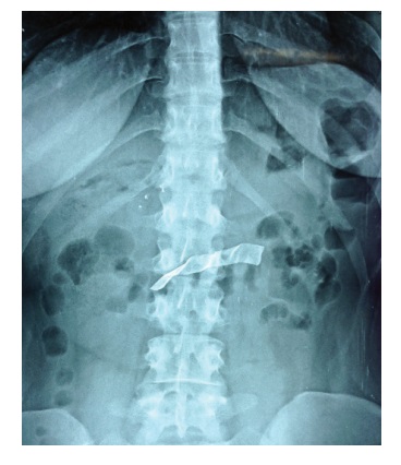
Fig. 2. Endoscopy disclosed a gauze between the bulb and the second part of the duodenum, as well as a Forrest III ulcer

Fig. 2. Endoscopy disclosed a gauze between the bulb and the second part of the duodenum, as well as a Forrest III ulcer
DISCUSSION
Gossypiboma is an important and preventable iatrogenic complication whose delay in diagnosis and treatment can lead to serious morbidity and mortality (6). The magnitude of the problem is not entirely known because cases are rarely reported due to medico-legal concerns and adverse publicity (6) A PubMed search using the terms “gossypiboma” and “gossypiboma and duodenum” were performed for English articles. Latin American articles written in Spanish or Portuguese were also reviewed with LILACS’s database search engine with the terms “gossypiboma” and “gossypiboma y duodeno” used in the search. Articles were selected by the authors based on their experience and clinical relevance. The cases reported were summarized.
The first medical case of gossypiboma was reported by Wilson in 1897 (7). The first malpractice suit related to it occurred in 1933 [5]. The incidence of retained foreign bodies following surgery has a reported rate of 0.01% to 0.001% of which gossypibomas account for 80 % of the cases (2). It has been classically estimated as 1 in 1000 to 1500 intra-abdominal open surgeries and in 1 in 3000 in all surgical interventions (6). More recently, studies based on malpractice claims found a 1 in 8801 to 1 in 18760 (4)]. However these figures omit large numbers of patients that go underreported. The retained foreign bodies induce two types of inflammatory reactions. The exudative type leads to abscess formation and chronic fistulae and usually become symptomatic in the early post-operative period.
The fibrotic type produces an aseptic fibrous reaction that creates adhesions and encapsulation, leading finally to foreign body formation and pseudotumors (6) The time interval between the surgical procedure and clinical presentation ranges from the immediate postoperative period to decades after surgery (11 days to 28 years) (5), with approximately 50 % of retained gauze pieces being discovered at least five years after surgery and one third of all patients were symptom free (8). These patterns of inflammatory reactions and the location of the sponge are responsible for the two classical forms of clinical presentation (9). Symptoms usually are nonspecific, ranging from none (incidental finding) to fatal, depending on the site and type of complication resulting from the retained foreign body (10). The acute presentation is characterized by a septic course with abscess generation and intestinal obstruction secondary to granuloma formation and adhesions. The delayed or chronic form (months or years after surgery) is the result of sponge adhesion and encapsulation to the surrounded tissues. Expressed clinically, depending on the location of the foreign body, as sub-acute intestinal obstruction, tumor, fistula, free perforation, erosion into the bowel or vessels, extrusion of the sponge via rectum and migration into the bladder (11,12). Sponge retention is classically associated with emergency surgery, gynecological or upper abdominal After surgeries, unexpected changes in the surgical procedure, disorganization, long procedures, unstable patient condition, inexperienced staff and obesity (9).
The diagnosis may be suspected based on the antecedent of a previous surgical procedure and confirmed by the presence of a radio-opaque marker in the plain abdominal films. These markers however, slowly disintegrate and fragment over time and as time passes since surgery, while clinical concern for this condition slowly declines to the point to rule out the possibility (11). After the gossypiboma’s diagnosis is made, the removal of the retained sponge either by open surgery, laparoscopy or endoscopy depending of clinical presentation and facilities available, is highly recommended to prevent severe complications that may lead even to death (15-22 %) (13,14). There are however some reports of conservative management of these patients (15). Migration of the gossypiboma may occur externally (extrusion) through a fistulous tract or internally, when it is adjacent to a hollow viscus (rectum, vagina, bladder, or intestinal lumen) (10).
The small intestine is the most commonly affected site due to its large surface (jejunal and ileal segments) and thin wall which offers the least resistance (16) Post cholecystectomy gossypibomas are rare and even more rare the reports of transduodenal migration (11). Endoscopy may be a diagnostic useful tool when the gauze migrates to the lumen of the digestive tube, and in some cases allows the retrieval of the foreign body with a snare, biopsy forceps or a basket. In selected cases, endoscopicultrasound-guided transmural drainage can be the first approach, followed by endoscopic removal if it succeeds (17). Clinical presentation includes the presence of an abdominal mass, intestinal obstruction, discharging sinus, intra-abdominal abscesses and peritonitis (6). A correct preoperative diagnosis is made in about one-third of the patients (18). The presence of a foreign body should be kept in mind in the differential diagnosis of any postoperative patient who presents with pain, infection or a palpable mass (19). The practice of counting surgical gauzes pre- and post-operatively, and the use of radiopaque markers have significantly reduced the incidence of this complication (20). As a conclusion, gossypiboma is a diagnostic dilemma due to its nonspecific symptoms and variable radiological findings (6). Trans-duodenal migration is rare and generally, presents with signs and symptoms of upper gastrointestinal obstruction. Endoscopy may be helpful in achieving a rapid diagnosis and in some cases in retrieving the foreign body.
REFERENCES
1. Shibi M, Mahesh V, Mukunda M, Noronha S, Krishnadas D, Kattoor V, Nair R. Intraduodenal gossypiboma as an unusual cause of gastric outlet obstruction. J Dig Endosc 2010; 1(2):63-65. [ Links ]
2. Rizwan F, Swaleh A, Ahmed F, Iqbal Z, Ahmed R. Gastric gossypiboma. PJR 2011; 21(1):37-39. [ Links ]
3. Borraez OA, Borraez BA, Orozco M, Matzalik G. Cuerpos extraños en abdomen: presentación de casos y revisión bibliográfica. Rev Colomb Cir 2009; 24:114-122. [ Links ]
4. McIntyre LK, Jurkovich GJ, Gunn ML, Maier RV. Gossypiboma: tales of lost sponges and lessons learned. Arch Surg 2010; 145(8):770-775. [ Links ]
5. Garg M, Aggarwa MG. A review of medicolegal consequences of gossypiboma. J Indian Acad Forensic Med 2010; 32(4):358-361. [ Links ]
6. Olnick HM, Weens HS, Rogers JR Jr. Radiological diagnosis of retained surgical sponges. JAMA 1955;159(16):1525-1527. [ Links ]
7. Lauwers PR, Van Hee RH. Intraperitoneal gossypibomas:the need to count sponges. World J Surg 2000; 24(5):521-527. [ Links ]
8. Nizamuddin S: “Gossypiboma” an operative team’s dilemma. Pak J Surg 2008; 3(24):159162.
9. Lata I, Kapoor D, Sahu S. Gossypiboma, a rare cause of acute abdomen: A case report and review of literature. Int J Crit Ill Inj Sci 2011; 1(2):157-160. [ Links ]
10. Manzella A, Filho PB, Albuquerque E, Farias F, Kaercher J. Imaging of gossypibomas: pictorial review. AJR 2009; 193(6 Suppl):S94-101. [ Links ]
11. Asuquo ME, Ogbu N, Udosen J, Ekpo R, Agbor C, Ozinko M, Emelike K. Acute abdomen from gossypiboma: A case series and review of literature. Nig J Surg Res 2006; 8(34):174-176. [ Links ]
12. Moyle H, Hines OJ, McFadden DW. Gossypiboma of the abdomen. Arch Surg 1996; 131(5):566-568. [ Links ]
13. Hung-Shun S, Sung-Lang C, Chia-Cheng K, Shao-Chuan W, Yu-Lin K. Gossypiboma— retained surgical sponge. J Chin Med Ass : JCMA 2007; 70(11):511-513.
14. Kansakar R, Hamal BK. Cystoscopic removal of an intravesical gossypiboma mimicking a bladder mass: a case report. J Med Case Rep 2011; 5(1):579. [ Links ]
15. Alis H, Soylu A, Dolay K, Kalayci M, Ciltas A: Surgical intervention may not always be required in gossypiboma with intraluminal migration. World J Gastroenterol : WJG 2007, 13(48):6605-6607. [ Links ]
16. Erbay G, Koc Z, Caliskan K, Araz F, Ulusan S. Imaging and clinical findings of a gossypiboma migrated into the stomach. Turk J Gastroenterol 2012; 23(1):54-57. [ Links ]
17. Matsumoto K, Katanuma A, Maguchi H, Takahashi K, Osanai M, Yane K, Kin T, Takaki R, Matsumori T, Gon K, Tomonari A. Gossypiboma successfully removed by endoscopy after endoscopic ultrasound-guided transmural drainage. Endoscopy 2013; 45 Suppl 2 UCTN:E212-213. [ Links ]
18. Kopka L, Fischer U, Gross AJ, Funke M, Oestmann JW, Grabbe E. CT of retained surgical sponges (textilomas): pitfalls in detection and evaluation. J Comput Assist Tomo 1996; 20(6):919-923. [ Links ]
19. Malhotra MK. Migratory surgical gossypiboma-cause of iatrogenic perforation: case report with review of literature. Nig J Surg 2012; 18(1):27-29. [ Links ]
20. Malot R, Meena DS: Gossypiboma of the thigh mimicking soft tissue sarcoma: case report and review of literature. JOCR 2012, 2(3):21-24. [ Links ]
21. Sharma R, Sachdev A, Gupta S, Kaushik R, Attri A. Gossypiboma eroding into the duodenum. Indian J Surg 2006; 68(5):279. [ Links ]
22. Sinha SK, Udawat HP, Yadav TD, Lal A, Rana SS, Bhasin DK. Gossypiboma diagnosed by upper-GI endoscopy. Gastrointestinal Endosc 2007; 65(2):347-349. [ Links ]
23. Peyrin-Biroulet L, Oliver A, Bigard MA. Gossypiboma successfully removed by upperGI endoscopy. Gastrointest Endosc 2007; 66(6):1251-1252. [ Links ]
24. Kalkan IH, Etik DO, Oztas E, Sayilir A, Disibeyaz S, Kuran SO. A rare cause of upper GI hemorrhage: an uncorrupted sponge migrating into the duodenal bulb (with video). Gastrointest Endosc 2012; 76(6):1246.
