


 Articulo en XML
 Referencias del artículo
 Enviar articulo por email
 Citado por SciELO
  Similares en
SciELO
Similares en
SciELO  uBio
uBio 
 Permalink
Investigación Clínica
versión impresa ISSN 0535-5133
Invest. clín vol.57 no.3 Maracaibo set. 2016
Lip projection analysis in brachycephalic, dolichocephalic and mesocephalic individuals of a Mexican population.
Análisis de la proyección labial en pacientes braquiocefálicos, mesocefálicos y dolicocefálicos de una población mexicana.
Wulfrano Sánchez Meraz1, Raymundo Arredondo Hérnandez1, Diana Avalos Sanchez2, Humberto Mariel Murga1, Francisco Javier Gutiérrez Cantu1 and Jairo Mariel Cárdenas1.
1 Deparment of Orthodontics, Faculty of Stomatology, Autonomous University of San Luis Potosí, México. Email: llairo@yahoo.com.mx
2 School of Dentistry, University of San Nicolas de Hidalgo, Michoacán, México.
Abstract.
The aim of this study was to examine the lip projection of brachycephalic, dolichocephalic and mesocephalic Mexican patients and to analyze the relationship between dental protrusion and the lip position, to identify if a labial soft tissue projection exists and to determine what features predominated in this study. A total of 120 lateral radiographs of the skull were randomly selected from patients aged 16-25 years. The linear and angular data were measured, the values of the upper and lower lip projection relative to Ricketts E-line, Steiner S-line and Arnett TV-line were collected. The angles to be analyzed were the nasola bial, mandibular plane and interincisal, and the mentolabial groove depth. The statistical sig nificance was determined by the Student t-test. A significant difference between mesocephalic and brachycephalic patients was observed measuring the angle between the Frankfurt plane and the mandibular plane (P<0.001). Between dolichocephalic and mesocephalic individuals significant differences were observed in the lower lip to E-line (P<0.031), lower lip to S-line (P<0.010), the interincisal angle (P<0.032) and the mandibular ́s plane (P<0.001). A statistical significant difference was shown between brachycephalic and dolichocephalic individuals: lower lip to E-line (P<0.001), upper lip to S-line (P<0.037), lower lip to S-line (P<0.001), interincisal angle (P<0.034) and the angle between the mandibular ́s plane (P<0.001). Lip soft tissue projection will depend on the population studied; we found some significant differences when compared with the cephalometric norms.
Key words: cephalometrics; lip projection; soft tissue.
Corresponding author: Jairo Mariel Cárdenas. Department of Orthodontics. Faculty of Stomatology, Autonomous University of San Luis Potosí, Mexico. Address: Av. Manuel Nava 2, San Luis Potosí, S.L.P. México, Postal Code: 78290. Tel 00524448262357 ext. 5122, Fax: 00524448139743. Email: llairo@yahoo.com.mx
Resumen.
El objetivo del presente estudio fue examinar la proyección labial en pacien tes braquicéfalos, mesocéfalos y dolicocéfalos mexicanos al igual que analizar la relación entre la protrusión dental y la posición labial, identificar si existe proyección labial de los tejidos blandos y determinar las características predominantes en el estudio. Un total de 120 radio grafías laterales de cráneo se seleccionaron de manera aleatoria de pacientes entre 16-25 años. Los datos angulares y lineales se identificaron, los valores de la proyección labial superior e inferior con respecto a línea línea-E de Ricketts, línea-S de Steiner y línea-VV de Arnett fueron recolectados, los valores angulares a analizar fueron el ángulo nasolabial, el plano mandibular y el ángulo interincisal al igual que el surco mentolabial. Se usó la prueba T de Student para obtener la significancia. Una diferencia significativa se observó entre pacientes mesocéfalos y braquicéfalos en el plano mandibular con Frankfort (P <0.001). Entre dolicocéfalos y meso céfalos diferencias significativas se observaron en el labio inferior a línea-E (P <0.031), labio inferior a S (P <0.010), ángulo interincisal (P <0.032) y el plano mandibular (P <0.001). Dife rencias significativas se observaron entre pacientes braquicéfalos y dolicocéfalos: labio inferior a línea-E (P <0.001), labio superior a línea-S (P <0.037), labio inferior a línea-S (P <0.001), ángulo interincisal (P < 0.034) y el plano mandibular (P <0.001). La proyección labial depende de la población y se pueden observar algunas diferencias significativas al ser comparadas con la norma cefalométrica.
Palabras clave: cefalometría; proyección labial; tejidos blandos.
Recibido: 22-09-2015 . Aceptado: 14-04-2016
INTRODUCTION
The concept of beauty has changed over the centuries and differs from one population to another, but it has always been a topic of inte rest and importance to humankind. Throughout history different authors as Ricketts, Steiner, Al temus and Arnett have conducted cephalometric studies to determine which features determine facial beauty being one of them the lip projec tion. Over time, beauty parameters have been modified, however still had an important role in society. Cephalometrics is a study in which a number of records of points, lines and measure ments are obtained from lateral radiographs of the skull in which certain anatomical structures are analyzed allowing the prediction of the patient ́s growth pattern (1-7).
The importance of facial aesthetics and rela tionships of the soft tissues in orthodontic treat ment was emphasized by Ricketts. He observed that the ideal for one patient might not be for another, for which the results of the cephalome tric analysis vary by race or ethnic group, be cause they have different growth and physical characteristics due to genetic influences, therefore orthodontic treatment́s needs are different and must be adapted to the characteristics of each person (8-13).
Dentofacial features of many ethnic groups have been reviewed by several researchers for orthodontic purposes. Some of these show similarities, but others showed significant differences regarding the projection of the lips. Given that every population has different dentoskeletal characteristics, it is difficult to apply a general cephalometric standard.
The aim of this study was to examine the lip projection of brachycephalic, dolichocepha lic and mesocephalic Mexican patients and to analyze the relationship between dental protrusion and the lip position, to identify if a labial soft tissue projection exists and to determine what features predominated in this study.
MATERIALS AND METHODS
A sample of 120 digital lateral skull cepha lometric radiographs, that showed good defini tion of the hard and soft tissues, were randomly selected from Mexican patients with Mexican grandparents, aged 16-25 years, who attended the Clinic of Orthodontics and Dentomaxilofa cial Orthopedics of the Autonomous University of San Luis Potosi. Permits for the use of the material were given, and the H. Research Ethics Committee of the Faculty of Dentistry revised informed and approved consents.
Each individual ́s lateral cephalometric ra diographs of the skull where taken without any previous orthodontic treatment, using a Digital Panoramic and Cephalometric System (Kodak 8000c, Germany) at 68.0 kV, 5.0 mA for 17 s at 18.5 mGy/cm2, at the Faculty of Dentistry of the Autonomous University of San Luis Potosi. Inclusion criteria were healthy individuals, with no facial anomalies and lack of skeletal abnor malities. These radiographs were manually tra ced on 0.003-mm matte acetate sheets. All radiographs were traced by the third author (DAS) and then reviewed by the second author (RAH), for accurate landmark identification.
Ten measurements (seven linear and three an gular) were traced according to Steiner, Ricketts and Arnett ́s soft tissue analysis.
The lines used in this study to determine the lip projection where the S-line described by Steiner, ranging from soft tissue pogonion to half of columnela ́s nose, which should touch both lips. Ricketts ́ E-line goes from pogonion soft tissue to the tip of the nose; the lips should pass behind or nearly touching the line.
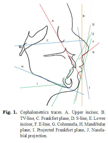
Arnett ́s True Vertical Line (TV-line) passing through the subnasale and perpendicu lar to the ground, both upper and lower lips must pass ahead of the line. The angles to be analyzed were: the nasolabial angle, formed by the nose columnela and the nasolabial projection; the interincisal angle conformed by the upper and lower incisors; the mentolabial groove depth, that gave the length of depth between the lower lip and the chin; and the mandibular plane an gle, which divide the sample, due to its degree of divergence with Frankfort ́s plane, into bra chycephalic (≥ 25°), mesocephalic (≥ 30°) and dolichocephalic (≥ 35°) (Fig. 1) individuals.
A single operator with blinding measured the linear and angular data. Statistical analysis was performed using the Minitab́s 17 version software by a specialist with data blinding, to analyze the normality of variables. The Shapiro Wilk test was applied and Student t-test statis tical significance was determined. Confidence intervals were determined at 95 % and the sta tistical significance of p< 0.05 value were de termined.
RESULTS
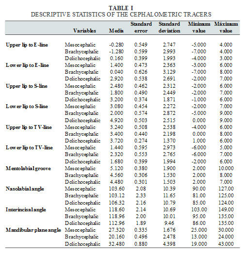
The mean, mean error, standard deviation, minimum and maximum values were determi ned (Table I).
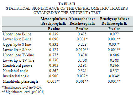
Significant differences between mesocephalic and brachycephalic patients were observed measuring the angle between the Frankfurt plane and the mandibular plane (P<0.001) but no other significant measure ments differences were identified. When compa ring between dolichocephalic and mesocepha lic, significant differences were observed in the lower lip to E-line (P<0.031), lower lip to S-line (P<0.010), the interincisal angle (P<0.032) and the mandibular ́s plane (P<0.001). Statistical significant differences were shown between bra chycephalic and dolichocephalic: lower lip to E-line (P<0.001), upper lip to S-line (P<0.037), lower lip to S-line (P<0.001), interincisal angle (P<0.034) and the angle between the mandi bular ́s plane (P<0.001).
These findings show no significant differences between upper lip to E-line in all the groups as well as upper lip to TV-line, lower lip to TV-line, the mentolabial groove, the nasolabial angle. Comparing toge ther brachycephalic-mesocephalic and meso cephalic-dolichocephalic groups no significant difference was present from the upper lip to S. (Table II).
DISCUSSION
All facial features vary from one population to another, and within the same population. This is due to miscegenation, as it allows an infinite combination of characteristics causing difficulties to establish a standard to help achieve facial harmony in all of them. However, investigations based on the cephalometric norms, show that there are similarities in some data and a greater or lesser degree depending on the population.
Sinojiya et al. (14) compared a population of Mahabubnagar in the south of India, and found some differences with the standard of Arnett, thicker soft tissue, acute nasolabial angle, increased facial length, increased deficiency midface, a more convex profile and less verti cal lower incisors, this show that some people have more similarity than others regarding the standard, upper lip thickness (P<0.000), lower lip thickness (P<0.000), Pog- Pog (P<0.0459) nasolabial angle (P<0.0090) and upper lip angle (P<0.0011). We agree with the authors, as our studies showed differences in most of the data obtained; the facial growth and their characteristics directly influence on the proportions of the head, as seen in the results showed between brachycephalic and dolichocephalic, as well as mesocephalic and dolichocephalic, in measures of the interincisal angle, lower lip to E-li ne (p<0.001) and lower lip to S-line (p<0.001), which shows that the incisal inclination has a direct effect on the lip position, allowing us to observe lip protrusion. All factors relating to Arnett́s norms were similar in the study of Uysal et al. (15), which analyzed a population of Turkey and found features like thin upper and lower lips and retruded incisors, resulting that the majority of Turks have harmony and the values obtained are within the norm of Arnett. In the present study, the results show significant differences, and when we compared mesocephalic with dolichocephalic and brachycephalic with dolichocephalic, statistical significant differences were identified in lower lip to S-line and E-line, which may be due to the difference in labial thickness and incisal protrusion causing a more pronounced lip projection in our population.
In various studies based on the Steineŕs standard such as the case of Ikenna et al. (16), a Nigerian population was analyzed against the standards established by Ricketts and Steiner, differences were shown in most of the recorded measurements, obtained as a greater lip protrusion against the standard. Our study agrees with this. No significant differences in both upper lip and lower lip were found, mainly on the com parison between the Mexican brachycephalic and dolichocephalic populations, so it may be assumed that both lips are protruded, but the lower lip which is greater and it may be due to the position of the chin, the nosés projection or excessive protrusion of the lower incisors to compensate for the lack of growth in the jaw.
Gupta et al. (17) showed that similarity be tween the soft tissue of a northern Indian popu lation and the norm, except for nasal prominence and the thickness of the upper lip, the results that are shown to be significant were: thickness of the upper lip (P<0.000), increased lower lip thickness (P<0.000), lower lip to E-line (P<0.009) and thickness of the chin (P<0.27). They concluded that individuals with a relati vely minor nose, lip protrusion and slightly convex profile are more aesthetic. The present results show significant differences: lower lip to S-line (P<0.001), lower lip to E-line (P<0.001) between brachycephalic and dolichocephalic, as well as significant differences with the E-line, a more pronounced projection of the lower lip this, may be due, to the difference in genetic characteristics.
Both, Lahlou et al. (18) and Erbay et al. (19,20) found that soft tissue analyses differ according to the population. Every race has its own nose and chin characteristics. We observe major facial features differences of the Mexi can population against the standard, identifying greater projection of the lower lips, perhaps caused by the interincisal position, being the incisors in a protruding position and making the lips look more protruded, also features of the chin will have a direct effect on the aesthetic characteristics.
Cephalometric standards aim to maintain harmony and balance in the facial features applied despite the obvious variations that exist depending on the population. Due to this, implementation of various cephalometric studies are recommended considering the characteristics of each population in order to develop an appropriate treatment plan. The results show both similarities and differences compared to diffe rent standards, this may be due to the racial mix through the history generating variables; howe ver, the standards are only general guidelines to achieve better orthodontic results and always taking into account the particular facial features of each population.
ACKNOWLEDGEMENT
To the clinic of Orthodontics and Dentomaxilofacial Orthopedics of the Autonomous University of San Luis Potosi for the equipment and installations to perform this research.
REFERENCES
1. Mejia-Maidl M, Evans CA, Viana G, Anderson KN, Giddon BD. Preferences for facial profiles between Mexican Americans and Caucasians. Angle Orthod 2005; 75(6): 953-958. [ Links ]
2. Joshi M, Wu LP, Maharjan, S, Reg mi MR. Sagittal lip positions in different skeletal malocclusions: a cephalometric analysis. Prog Orthod 2015; 16(1), 1-8. [ Links ]
3. Al Zain T, Ferguson DJ. Cephalometric characterization of an adult Emirati sam ple with Class I malocclusion. J Orthod Sci 2012; 1(1), 11. [ Links ]
4. Prabu NM, Kohila K, Sivaraj S, Pra bu PS. Appraisal of the cephalometric norms for the upper and lower lips of the South Indian ethnic population. J Pharm Bioallied Sci 2012; 4(2), S136. [ Links ]
5. Bergman RT, Waschakb J, Borzabadi FA, Murphy NC. Longitudinal study of cephalometric soft tissue profile traits be tween the ages of 6 and 18 years. Angle Orthod 2014; 84: 48-55. [ Links ]
6. Shindoi JM, Matsumoto Y, Sato Y, Ono T, Harada K. Soft tissue cephalometric norms for orthognathic and cosmetic sur gery. J Oral Maxillofac Surg 2013; 71(1), e24-e30. [ Links ]
7. Mariel Cárdenas J, Arredondo Hernán dez R, Sánchez Meraz W, Mariel Murga H, Oliva Rodriguez R, Gutierrez Cantú FJ. Análisis morfológico del grosor labial en individuos mesofaciales y braquifacia les en una población mexicana. Int J Mor phol 2015; 33(4), 1282-1286. [ Links ]
8. Parikakis KA, Moberg S, Hellsing E. Evaluation of the variable anchorage strai ghtwire technique using Ricketts growth prediction. Eur J Orthod 2009; 31: 76-83. [ Links ]
9. Uchikurab K, Shimookae S, Ishidac K, Shundoc I, Sakaedad K. Thresholds for clinically significant tooth-size discrepan cy. Angle Orthod 2009; 79: 740-746. [ Links ]
10. Eun-ju B, Hye-jin K, Oh-won K. Chan ges in longitudinal craniofacial growth in subjects with normal occlusions using the Ricketts analysis. Korean J Orthod 2014; 44(2): 77-87. [ Links ]
11. Filiaci F, Ramieri V, Fatone FMG, Gen naro P, Arangio P, Rinna C, Vellone V, Agrillo A, Ungari C, Cascone O. New parameter for the evaluation of disgnathic patients surgical planning: a preliminary report. Eur Rev Med Pharmacol Sci 2012; 16: 1430-1432. [ Links ]
12. Aparna P, Kumar DN, Prasad M, Sham nur N, Kumar AG, Sridhar KR, Krishna BRG, Gupta N. Comparative assessment of sagittal skeletal discrepancy: a cephalo metric study. J Clin Diagn Res 2015; 9(4): 38-41. [ Links ]
13. Kavitha L, Karthik K. Comparison of cephalometric norms of caucasians and non-caucasians: A forensic aid in ethnic determination. J Forensic Dent Sci 2012; 4(1): 53-55. [ Links ]
14. Sinojiya J, Kaladhar RA, Madhukar RR, Jaipal RP, Vankre M, Manjunatha RC. Soft tissue esthetic norms for Maha bubnagar population of southern India. J Clin Diagn Res 2014; 8(1): 255-259. [ Links ] 15.
Uysal T, Yagci A, Ayhan BF, Sisman Y. Standards of soft tissue Arnett analysis for surgical planning in Turkish adults. Eur J Orthod 2009; 31: 449-456.
16. Ikenna IG, Olatokunbo DO, Chukwudi IM. A cephalometric investigation of ho rizontal lip position in adult Niguerians. J Orthod 2012; 39: 160-169. [ Links ]
17. Gupta A, Garg J, Anand N, Hegde M, Parashar S. Establishment of soft tissue norms for the north indian population ba sed on laymen perception. J Maxillofac Oral Surg 2014; 13(1): 22-28. [ Links ]
18. Lahlou K, Bahoum A, Makhoukhi MB, Aalloula EH. Comparison of dentoalveo lar protrusion values in Moroccans and other populations. Eur J Orthod 2010; 32: 430–434. [ Links ]
19. Erbay EF, Caniklioglu CM, Erbay SK. Soft tissue profile in Anatolian Turkish adults. Part I: Evaluation of horizontal lip position using different soft tissue analy sis. Am J Orthod Dentofacial Orthop 2002; 121: 57-64. [ Links ]
20. Erbay EF, Caniklioglu CM, Erbay SK. Soft tissue profile in Anatolian Turkish adults. Part II: Evaluation of horizontal lip position using different soft tissue analysis. Am J Orthod Dentofacial Orthop 2002; 121: 65-72. [ Links ]
