


Servicios Personalizados
Revista
Articulo
 Articulo en XML
 Referencias del artículo
 Enviar articulo por email
Indicadores
-
 Citado por SciELO
 -
 Accesos
Links relacionados
-
 Similares en
SciELO
Similares en
SciELO
Compartir

 Permalink
Archivos Venezolanos de Farmacología y Terapéutica
versión impresa ISSN 0798-0264
AVFT vol.34 no.3 Caracas set. 2015
Cardiomiopatía hipertrófica y hemorragia pulmonar inducida por el ejercicio un estudio post mortem en caballos pura sangre
Hypertrophic cardiopatia e induzida pelo exercício pulmonar hemorragia eqüinos da raça um estudo de pós-morte
Abelardo Morales1, Aniceto Mendez1, Kimberly Brewer2, Thomas Tobin2, María Morales3, Cesar Castillo3.
1 Department of Anatomy and Comparative Anatomic Pathology, College of Veterinary Medicine. University of Cordoba, Spain. 2 Maxwell H. Gluck Equine Research Center, University of Kentucky, Lexington USA. 3 Private Practice
Autor corresponsal: Abelardo Morales Briceño Department of Anatomy and Comparative Anatomic Pathology, College of Veterinary Medicine. University of Cordoba, Spain. Edificio de Sanidad Animal, Campus de Rabanales Ctra. de Madrid km 396, 14071, Cordoba, Spain. +0034619307223 Email: aamorales13@gmail.com
Abstract
The aim of this study was to describe hypertrophic cardiomyopathy occurring in conjunction with exercise induced pulmonary hemorrhage in Thoroughbred horses in the national racetrack La Rinconada, Caracas Venezuela. The study included 26 equine Thoroughbreds, of which 20 were male and six were female, within 2-4 years old. All horses had previous episodes of pulmonary hemorrhage induced by exercise and had no clinical signs consistent with cardiomyopathy, and had a routine diagnostic necropsy. Samples of tissue heart and lungs and other organs were collected, fixed, prepared and stained with Hematoxylin & Eosin (H&E) and special stain Prussian Blue, for light microscopy. On necropsy, all cases revealed hypertrophic cardiomyopathy, specifically left ventricular concentric hypertrophy, with severe obliteration of the left ventricular lumen. There were no valve diseases or mural or valve endocarditis. The average diameter of left ventricular wall was approximately ±9.20 cm (DS: ±0.925). The thickness of the wall of the left ventricle was on average ±6.19 cm (DS: ±0.409). Weight heart was to ±5.11 (DS: ±0.530). Histological lesions showed congestion, edema, and acute pulmonary intra-alveolar and interstitial hemorrhage due to rupture of focal bronchial arterioles, replete with red blood effusion. The Prussian blue special stain was positive for hemosiderophages in all cases studied. Microscopically there is fiber disarray, hypertrophic cardiomyocytes and some fibrosis. The cardiac fibers increase in width, nuclei increase in size. In conclusion these results suggest a close relationship between the developments of exercise-induced pulmonary hemorrhage associated with ventricular concentric hypertrophic cardiomyopathy in Thoroughbred horses.
Keywords: Cardiomyopathy, EIPH, HCV, Pathology, Thoroughbreds.
Resumen
El objetivo de este estudio fue describir miocardiopatía hipertrófica en conjunción con hemorragia pulmonar inducida por el ejercicio en caballos Pura Sangre en el hipódromo La Rinconada, Caracas, Venezuela. El estudio incluyó a 26 equinos Pura Sangre, 20 machos y 6 hembras, con edades entre 2-4 años. Todos los caballos presentaron episodios previos de hemorragia pulmonar inducida por el ejercicio y no tenía signos clínicos compatibles con la cardiomiopatía, se practicó necropsia de diagnóstico. Se colectaron muestras de corazón y pulmones y de otros órganos, las cuales se fijaron en formol y procesadas por los métodos convencionales histológicos, fueron coloreadas con hematoxilina y eosina (H&E) y la tinción especial azul de Prusia, y observadas por en el microscopio óptico. En todos los casos la necropsia reveló una miocardiopatía hipertrófica, específicamente hipertrofia ventricular izquierda concéntrica, con obliteración grave de la luz ventricular izquierda. No hubo enfermedades de la válvula o válvulas murales o endocarditis. El diámetro promedio de la pared ventricular izquierda fue aproximadamente ± 9,20 cm (DS: ± 0,925). El espesor de la pared del ventrículo izquierdo fue en promedio ± 6,19 cm (DS: ± 0,409). El peso del corazón fue ± 5,11 (DS: ± 0,530). Histológicamente se observo congestión, edema y hemorragia intra-alveolar e intersticial pulmonar aguda debido a la ruptura de las arteriolas bronquiales focales, repletos con derrame de sangre roja. La coloración Azul de Prusia fue positiva para hemosiderófagos en todos los casos estudiados. Microscópicamente se observo desorganización de las fibras, los cardiomiocitos hipertróficos y algunos focos de fibrosis. Las fibras cardíacas aumentan en anchura, núcleos aumento de tamaño. En conclusión, estos resultados sugieren una estrecha relación entre la evolución de la hemorragia pulmonar inducida por el ejercicio asociados a miocardiopatía hipertrófica concéntrica ventricular en caballos pura sangre.
Palabras clave: EIPH, Miocardiopatia, Patología, pura sangre, VHC.
Resumo
O objetivo deste estudo foi descrever a cardiomiopatia hipertrófica ocorre em conjunto com o exercício hemorragia pulmonar induzida em eqüinos da raça na pista de corrida nacional La Rinconada, Caracas, Venezuela. O estudo incluiu 26 eqüinos puro-sangue, dos quais 20 eram do sexo masculino e seis do sexo feminino, com 2-4 anos de idade. Todos os cavalos tiveram episódios anteriores de hemorragia pulmonar induzida por exercício e não tinham sinais clínicos compatíveis com cardiomiopatia, e tinha uma necropsia de diagnóstico de rotina. Amostras de tecido do coração e os pulmões e outros órgãos foram fixadas, preparadas e coradas com Hematoxilina e Eosina (H & E) e corante especial Prussian Blue, por microscopia de luz. Em todos os casos de necropsia revelou cardiomiopatia hipertrófica, hipertrofia concêntrica do ventrículo esquerdo, especificamente, com obliteração grave do lúmen do ventrículo esquerdo. Não havia doenças valvares ou endocardite mural ou válvulas. O diâmetro médio da parede do ventrículo esquerdo foi de aproximadamente ± 9,20 centímetros (DS: ± 0,925). A espessura da parede do ventrículo esquerdo foi de, em média, ± 6,19 centímetros (DS: ± 0,409). Coração de peso foi de ± 5,11 (DS: ± 0,530). Lesão histológica revelou congestão, edema e hemorragia intra-alveolar e intersticial pulmonar agudo por ruptura das arteríolas brônquica focais, repleto de efusão de sangue vermelha. O corante especial azul da Prússia foi positivo para hemosiderophages em todos os casos estudados. Microscopicamente há desorganização das fibras, cardiomiócitos hipertrofiados e alguns fibrose. As fibras cardíacas aumentam em largura, aumento de núcleos de tamanho. Em conclusão estes resultados sugerem uma estreita relação entre a evolução da hemorragia pulmonar induzida por exercício associadas a cardiomiopatia hipertrófica concêntrica ventricular em cavalos puro-sangue.
Palavras-chave: Cardiomiopatia EIPH, HCV, patologia, puro- sangue.
Recibido: 20/10/2015 Aceptado: 21/11/2015
Introduction
The term cardiomyopathy was coined for a group of myocardial diseases in humans which were at the time unknown or obscure cause5. Hypertrophic cardiopathy is much less common than the dilated form. Associated clinical syndromes include sudden death, death during anesthesia, and congestion heart failure5. A recent review of the literature of cases of cardiomyopathy reported in horses showed: hypertrophic cardiomyopathy (HCM) is an uncommon genetic cardiovascular disease that affects the left ventricle2,4,7. Chronic administrations of beta2-agonists have been shown to have toxic effects on the heart; however, no data exist on cardiac function after chronic clenbuterol administration9. In 2013 a previous study described hypertensive cardiomyopathy in 5 horses (1995-2011)3. A case of Clostridial myonecrosis, hemolytic anemia, hepatopathy, osteitis, and transient hypertrophic cardiomyopathy after intramuscular injection in a Thoroughbred gelding were reported in 20131. Multiple factors have been previously described as associated with the development of epistaxis, exercised induced pulmonary hemorrhage (EIPH), and sudden death in horses, which include hypertrophic cardiomyopathy. However, congenital cardiomyopathy can be a factor in the development of pulmonary hemorrhage and its consequences have been poorly studied. The aim of this study was to describe hypertrophic cardiomyopathy occurring in conjunction with exercise induced pulmonary hemorrhage in Thoroughbreds horses in the national racetrack La Rinconada, Caracas Venezuela.
Material and methods
Study horses included 26 Thoroughbreds (20 male and 6 female), between 2-4 years old, in the National Racetrack La Rinconada, Caracas-Venezuela. All horses had previous episodes of pulmonary hemorrhage induced by exercise during racing with recurrent epistaxis in previous training and were treated at the time and their athletic activity continued with no clinical signs consistent with cardiomyopathy. The rules of racing in Venezuela allowed administration of furosemide four hours prior to race time, at a maximum dose of 250 mg (Regulation National Horse Racing Venezuela, 1995). However it is common to use a clenbuterol dose of 2ml/100lbs body weight for these cases. However, it was not possible to conduct a toxicological study to determine the presence of clenbuterol. These horses were followed for 12 months, died of various causes: colic, euthanasia, and for catastrophic skeletal muscle injury, all of which had a routine diagnostic necropsy. Additionally 5 Thoroughbred horses were included in the study (3 male and 2 female), that died of other causes not associated with epistaxis and EIPH and who had normal hearts. Macroscopic and morphological heart dissection was performed to record measurements according to standard protocols previously described in the literature14. Samples o heart and lung tissues and other organs were collected and fixed in 10% buffered formalin. Tissue sections were prepared and stained with Hematoxylin & Eosin (H & E) and special stain Prussian blue, for light microscopy.
Results
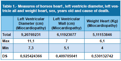
On necropsy all cases revealed hypertrophic cardiomyopathy, specifically in the left ventricle concentric hypertrophy, with severe obliteration of the left ventricle lumen (or chamber) (Figures:1,2,3,4).
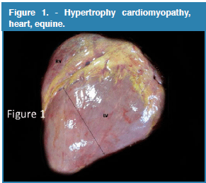
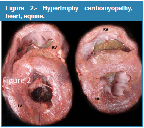
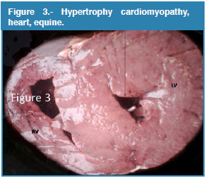
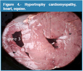
There were no valve diseases or mural or valve endocarditis. The average diameter of left ventricle wall was approximately ±9.20 cm (DS: ±0.925). The thickness of the wall of the left ventricle was on average ±6.19 cm (DS: ±0.409). Average heart weight was ±5.11 (DS: ±0.530). The results are shown in the table 1. The right ventricle wall thickness averaged 4 cm, while the left and right atria were ±3.5 cm on average (Table 1). In Venezuela, there are no previous reports of average Thoroughbred heart dimensions, so measures were taken randomly on 5 horses that died of other causes unrelated to heart disease and EIPH. The mean diameter of these hearts was 6.12, SD: ±0.52, left ventricular wall mean was 5.76, SD: ±0.35 and mean heart weight was 4.04, SD: ±0.11. Echocardiography measurement of normal hearts in this study were: Left ventricle free wall in diastole (FWd) 2.4 cm +/- 0.3 and left ventricle free wall in systole (FWs) 4.0 cm +/- 0.9 5,8. The right ventricular wall thickness averaged 4 cm, while the left and right atria were ±3 cm on average.
Edema, congestion, pulmonary hemorrhage pulmonary and fibrosis in the caudal lobe was observed in all cases. Histological lesions showed congestion, edema, and acute pulmonary intra-alveolar and interstitial hemorrhage due to rupture of focal bronchial arterioles, replete with red blood effusion. The Prussian blue special stain was positive for hemosiderophages in all cases studied. Microscopically the myocytes, normally arranged in parallel bundles, were found at oblique and perpendicular angles to each other, often in an interlacing or basket weave pattern; an indication of cardiac fiber disarray, hypertrophic cardiomyocytes and some fibrosis. The cardiac fibers were increased in width and nuclei were diameter in size.
Discussion
The lesion observed in the heart at necropsy of horses has traditionally been given little attention. Recently cases of sudden death in horses in international competition have attracted much interest in the lesions of the heart as the primary cause of sudden death. Cardiac hypertrophy occurs in the left ventricle, right ventricle, or bi-ventricle6. Hypertrophy can be primary (idiopathic cardiomyopathy) or secondary to another underlying condition that increases workload demand. Primary hypertrophy is rare and irreversible6. Secondary hypertrophy is a physiologic and partially reversible increase in cardiac mass that results from an attempt to meet increased work demand. Concentric cardiac hypertrophy results in increased myocardial mass with thickened walls and reduced ventricular chamber volume. Cardiac hypertrophy has three sequential cellular stages: 1. Initiation (increase in cell size by increasing the number of sarcomeres and mitochondria). 2. Compensation: stable hyper function of the heart; absence or minimal clinical signs of heart failure. 3. Deterioration: degeneration of hypertrophied cardiomyocytes, loss of myocardial contractility and frank evidence of heart failure6. In horses previous reports highlight the complications that can arise from clostridial myonecrosis, including the effect on the myocardium with hypertrophic cardiomyopathy after intramuscular injection1. Clenbuterol is a β 2 -adrenergic receptor agonist licensed for veterinary use as a bronchodilator. At doses ≥ 102 μg/kg (4.5 μg/lb), in excess of those normally prescribed, β-adrenergic stimulation by clenbuterol may cause sustained tachycardia, muscle tremors, hyperglycemia, and cardiac and skeletal muscle necrosis11. Laminitis, acute renal failure, rabdomiolisys, and cardiomyopathy were fatal complications with clenbuterol overdose in 2 horses in the present report11. Other study after treatment, CLENEX and CLEN demonstrated significantly higher left ventricular internal dimension (LVD) at end diastole (+23.7 +/- 4.8%; +25.6 +/- 4.1%), LVD at end systole (+29.2 +/- 8.7%; +40.1 +/- 7.9%), interventricular septum wall thickness (IVS) at end diastole (+28.9 +/- 11.0%; +30.7 +/- 7.0%), IVS at end systole (+29.2 +/- 8.7%; +40.1 +/- 7.9%), and left ventricular posterior wall systolic thickness (+43.1 +/- 14.%; +45.8 +/- 14.1%). CLENEX and CLEN had significantly increased aortic root dimensions (+29.9 +/- 6.1%; +24.0 +/- 1.7%)9. Taken together, these data indicate that chronic clenbuterol administration may negatively alter cardiac function9. Recently there have been a significant number of cases of sudden death during exercise or after exercise in California USA. A detailed report to the California Horse Racing Board at its Nov. 21 meeting concluded that the cluster of sudden deaths and the medications routinely dispensed in these horses were Ventipulmin (clenbuterol) and Thyro-L (levothyroxine), both of which were administered legally. In the Horse Racing Venezuela,is common to use a clenbuterol dose of 2ml/100lbs. Left ventricular hypertrophy is the most consistently reported effect of clenbuterol in humans and laboratory animals, and increased mural thickness may be due to a combination of true hypertrophy and myocardial fibrosis. In horses receiving clenbuterol at 2.4μg/kg twice daily for 8 weeks, no echocardiography alterations were evident at rest, but all parameters measured were significantly different in treated horses following strenuous exercise, except for end-diastolic thickness of the left ventricular free wall12. Another recent report in horses concludes a clinical relevance-hypertensive cardiomyopathy should be considered as a comorbid diagnosis in horses with laminitis or chronic renal failure3. Congestive heart failure could be unilateral or bilateral, and acute or chronic. Fluid retention, edema, venous congestion and, in some cases, cyanosis are the most common signs of heart failure. Left heart failure: pulmonary congestion and edema (acute); intra-alveolar hemorrhage, alveolar hemosiderophages known as heart failure cells and pulmonary fibrosis in chronic cases. Fluid accumulation in lungs caused by left heart failure is clinically referred to as cardiogenic pulmonary edema6. This may increase the risk of developing hypertension and pulmonary hemorrhage induced by exercise in the athletic horse. Remodeling of pulmonary veins (VR) in equine EIPH was recently described, suggesting that it contributes to the pathogenesis of the disease. The cause of VR is unknown, we tested the hypothesis that the development of VR follows pulmonary blood flow distribution, preferentially occurring in the caudal-dorsal lung region13. Similarity of the distribution of EIPH lesions and the reported fractal distribution of pulmonary blood flow suggests that VR develops in regions of high blood flow13. In summary, concentric left ventricular hypertrophy generates a decrease of end systole volume and an increase in blood volume in a left atrial retrograde stagnation of blood in the pulmonary veins. This causes an increase in blood volume in the lungs with increased pulmonary vascular pressure. Moreover systemic hypertension secondarily impacting the system with increased venous pressure in the vena cava, right atrium, right ventricle and pulmonary arteries can explain the development of pulmonary hemorrhage induced by exercise in the athletic horse. In conclusion, this study reported hypertrophic cardiomyopathy, specifically left ventricular concentric hypertrophy and the development of pulmonary hemorrhage induced by exercise in Thoroughbreds horses. However further immunohistochemical and genetic studies are required. The lesions observed in the heart of these horses were necropsy findings however, given the previous history of exercise-induced pulmonary hemorrhage, epistaxis and a pathophysiological association between pulmonary hemorrhage, epistaxis and cardiomyopathy is established.
Acknowledgments: The authors acknowledge the technical assistance of Raul Kobe Briceño Senges in image editing, Luis Leal for technical assistance during necropsies, Francisco Garcia and Emilio Suniaga by histological processing.
References
1. Anderson FL, Secombe CJ, Lester GD. Clostridial myonecrosis, haemolytic anaemia, hepatopathy, osteitis and transient hypertrophic cardiomyopathy after intramuscular injection in a Thoroughbred gelding. Aust Vet J. 2013 May;91(5):204-8. [ Links ]
2. Buergelt CD. Equine cardiovascular pathology: an overview. Animal Health Res Rev. 2003; 4(2):109-29. [ Links ]
3. De Solis CN, Slack J, Boston RC, Reef VB. Hypertensive cardiomyopathy in horses: 5 cases (1995-2011). J Am Vet Med Assoc. 2013 Jul 1;243(1):126-30. [ Links ]
4. Dudan F, Luginbühl H. Cardiovascular study of the horse: correlations between vascular and myocardial tissue changes. Schweiz Arch Tierheilkd. 1984; 126(6):277-86. [ Links ]
5. Jubb K., Palmer N. Pathology of Domestic Animals. Fifth Edition. Volume 3. Elsiever, Philadelphia, USA. 2007 pp 1–107. [ Links ]
6. Long KJ, Bonagura JD, Darke PG. Standardised imaging technique for guided M-mode and Doppler echocardiography in the horse. Equine Vet J 1992; 24:226-235. [ Links ]
7. Lopez A. Pathology of the Cardiovascular system. Pathology and Microbiology Atlantic Veterinary College. 2013. [ Links ]
8. Martin BB JR, Reef VB, Parente EJ, Sage AD. Causes of poor performance of horses during training, racing, or showing: 348 cases (1992-1996). J Am Vet Med Assoc. 2000. 15;216(4):554-8. [ Links ]
9. Patteson MW, Gibbs C, Wotton PR, Cripps PJ. Echocardiographic measurements of cardiac dimensions and indices of cardiac function in normal adult Thoroughbred horses. Equine Vet J 1995; Suppl 19:18-27. [ Links ]
10. Sleeper MM, Kearns CF, Mckeever KH. Chronic clenbuterol administration negatively alters cardiac function. Med Sci Sports Exerc. 2002 Apr;34(4):643-50. [ Links ]
11. Thomas B. The Equine Heart. Horse Report 2006. v. 24, n 4. [ Links ]
12. Thompson JA, Mirza MH, Barker SA, Morgan TW, Bauer RW, Mcconnico RS. Clenbuterol toxicosis in three Quarter Horse racehorses after administration of a compounded product. J Am Vet Med Assoc. 2011 Sep 15;239(6):842-9. [ Links ]
13. Thompson JA. Effects of clenbuterol on skeletal and cardiac muscle in horses. A Thesis Submitted to the Graduate Faculty of the Louisiana State University and Agricultural and Mechanical College.Texas, USA. August, 2009. [ Links ]
14. Williams KJ, Robinson NE, Defeijter-Rupp H, Millerick-May M, Stack A, Hauptman J, Derksen FJ. Distribution of venous remodeling in exercise-induced pulmonary hemorrhage of horses follows reported blood flow distribution in the equine lung. J Appl Physiol. 2013; 114(7):869-78. [ Links ]
